Anticorps Polyclonal de lapin anti-GFP tag
GFP tag Polyclonal Antibody for WB, IF/ICC, IF-P, IP, ELISA
Hôte / Isotype
Lapin / IgG
Réactivité testée
Protéine recombinante, Aequorea Victoria et plus (6)
Applications
WB, IHC, IF/ICC, IF-P, IP, CoIP, ChIP, RIP, IP-MS, ELISA
Conjugaison
Non conjugué
N° de cat : 50430-2-AP
Synonymes
Galerie de données de validation
 at various dilutions.")
 at dilution of 1:1000 incubated at room temperature for 1.5 hours.")
 with Transfected HEK-293T cells lysate 400 ug.")
 fixed paraffin-embedded transgenic mouse brain tissue using GFP tag antibody (50430-2-AP) at dilution of 1:200 and CoraLite®594-Conjugated AffiniPure Goat Anti-Rabbit IgG(H+L) (SA00013-2). Heat mediated antigen retrieval with Tris-EDTA buffer (pH 9.0).")
 fixed Transfected HEK-293 cells using 50430-2-AP (GFP tag antibody) at dilution of 1:100 and Alexa Fluor 594-conjugated AffiniPure Goat Anti-Rabbit IgG(H+L).")
 fixed Transfected HEK-293 cells using GFP tag antibody (50430-2-AP) at dilution of 1:200 and CoraLite®594-Conjugated Goat Anti-Rabbit IgG(H+L) (SA00013-4).")
Applications testées
| Résultats positifs en WB | Protéine recombinante, cellules HEK-293 transfectées |
| Résultats positifs en IP | Transfected HEK-293T cells, |
| Résultats positifs en IF-P | transgenic mouse brain tissue |
| Résultats positifs en IF/ICC | cellules HEK-293 transfectées, |
Dilution recommandée
| Application | Dilution |
|---|---|
| Western Blot (WB) | WB : 1:1000-1:4000 |
| Immunoprécipitation (IP) | IP : 0.5-4.0 ug for 1.0-3.0 mg of total protein lysate |
| Immunofluorescence (IF)-P | IF-P : 1:50-1:500 |
| Immunofluorescence (IF)/ICC | IF/ICC : 1:50-1:500 |
| It is recommended that this reagent should be titrated in each testing system to obtain optimal results. | |
| Sample-dependent, check data in validation data gallery | |
Informations sur le produit
50430-2-AP cible GFP tag dans les applications de WB, IHC, IF/ICC, IF-P, IP, CoIP, ChIP, RIP, IP-MS, ELISA et montre une réactivité avec des échantillons Protéine recombinante, Aequorea Victoria
| Réactivité | Protéine recombinante, Aequorea Victoria |
| Réactivité citée | rat, canin, levure, porc, souris, Ver à soie |
| Hôte / Isotype | Lapin / IgG |
| Clonalité | Polyclonal |
| Type | Anticorps |
| Immunogène | GFP tag Protéine recombinante Ag2128 |
| Nom complet | GFP tag |
| Masse moléculaire calculée | 26 kDa |
| Numéro d’acquisition GenBank | M62653 |
| Symbole du gène | |
| Identification du gène (NCBI) | |
| Conjugaison | Non conjugué |
| Forme | Liquide |
| Méthode de purification | Purification par affinité contre l'antigène |
| Tampon de stockage | PBS with 0.02% sodium azide and 50% glycerol |
| Conditions de stockage | Stocker à -20°C. Stable pendant un an après l'expédition. L'aliquotage n'est pas nécessaire pour le stockage à -20oC Les 20ul contiennent 0,1% de BSA. |
Informations générales
Green Fluorescent Proteins (GFPs) encompass a diverse range of proteins carrying a green chromophore, originating from various species and forming different protein lineages.
Wildtype GFP consists of 238 amino acid residues (26.9 kDa). GFP was first identified in the jellyfish Aequorea victoria. It emits green light with a peak wavelength of 509 nm upon excitation by blue light at 395 nm.
When fused with other proteins, GFP serves as a versatile reporter protein e.g. for quantifying expression levels or facilitates visualization of subcellular localization through fluorescence microscopy.
This antibody is a rabbit polyclonal antibody, generated against the full-length eGFP protein. It exhibits reactivity towards variants of Aequorea victoria GFP, including S65T-GFP, RS-GFP, YFP, CFP, and eGFP.
Protocole
| Product Specific Protocols | |
|---|---|
| WB protocol for GFP tag antibody 50430-2-AP | Download protocol |
| IF protocol for GFP tag antibody 50430-2-AP | Download protocol |
| IP protocol for GFP tag antibody 50430-2-AP | Download protocol |
| Standard Protocols | |
|---|---|
| Click here to view our Standard Protocols |
Publications
| Species | Application | Title |
|---|---|---|
Signal Transduct Target Ther Circulating tumor cells shielded with extracellular vesicle-derived CD45 evade T cell attack to enable metastasis | ||
Gastroenterology PTEN deficiency facilitates exosome secretion and metastasis in cholangiocarcinoma by impairing TFEB-mediated lysosome biogenesis | ||
Nat Genet Pathogenic SPTBN1 variants cause an autosomal dominant neurodevelopmental syndrome. | ||
Mol Plant A TT1-SCE1 module integrates ubiquitination and SUMOylation to regulate heat tolerance in rice |
Avis
The reviews below have been submitted by verified Proteintech customers who received an incentive for providing their feedback.
FH Manon (Verified Customer) (09-25-2025) | It works well
|
FH Ioana (Verified Customer) (08-27-2025) | I have used this antibody for a lot of different applications and it has performed well throughout. I was particularly impressed with it's efficiency in doing IP experiments.
|
FH Yi (Verified Customer) (07-23-2025) | This GFP antibody performs reliably in both Western blot and co-IP applications. It shows strong specificity and sensitivity for GFP-tagged proteins, with clean results and minimal background for detecting and pulling down GFP fusion proteins.
|
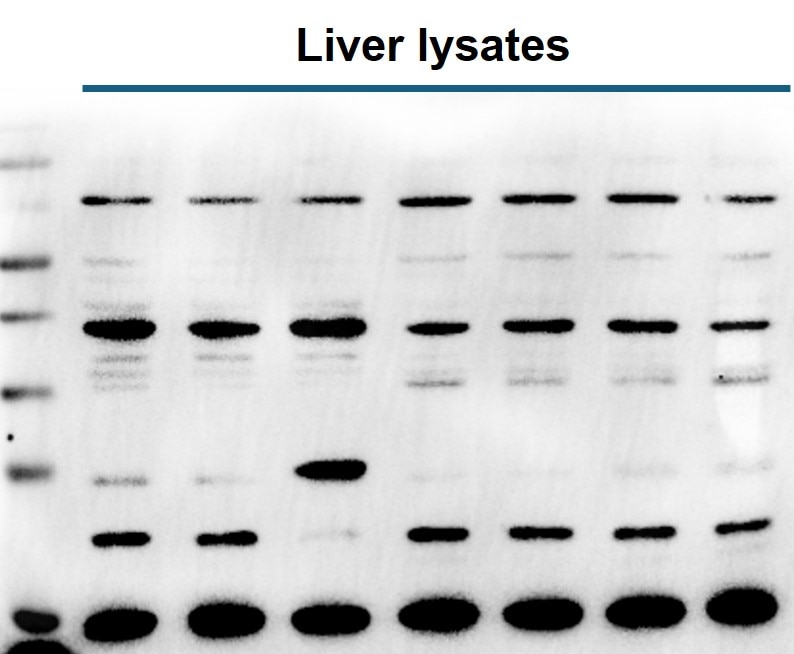
FH Kamal (Verified Customer) (02-27-2025) | Liver lysates were subjected to SDS-PAGE and immunoblotted with GFP antibody. Multiple non-specific protein bands were detected in liver tissue lysates.
 |
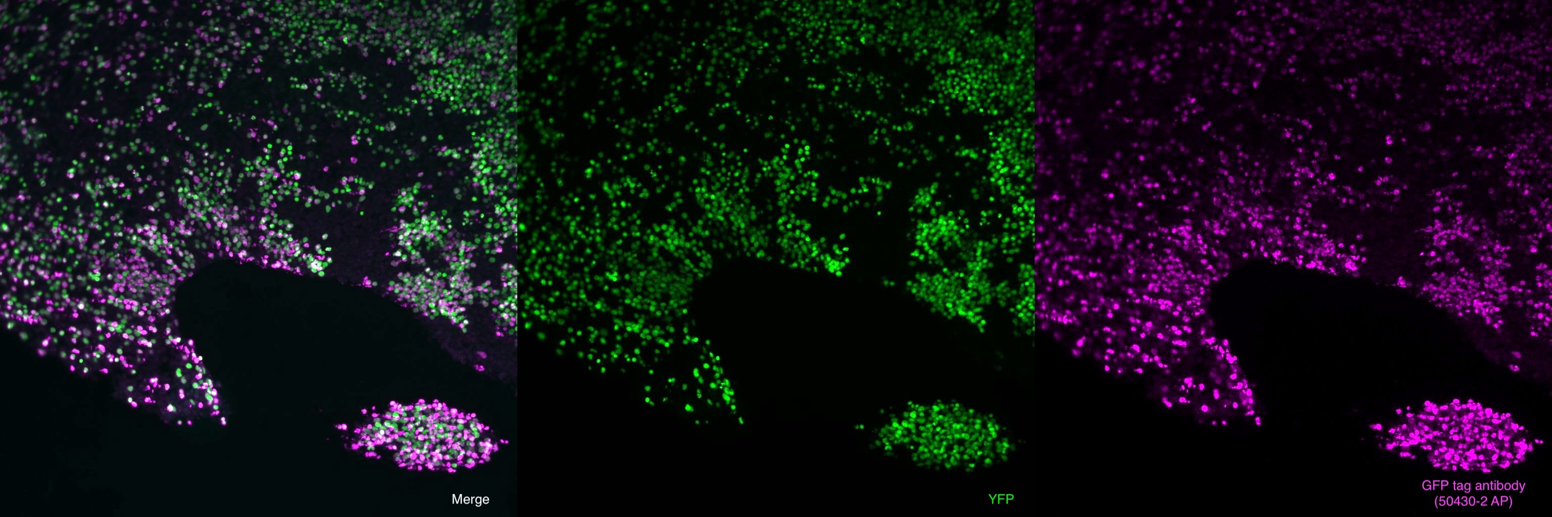
FH Raquel (Verified Customer) (07-12-2024) | Immunofluorescence analysis in cryostat sections of 4% PFA fixed human brain organoid derived from iPSCs expressing YFP with 50430-2-AP GFP tag antibody at dilution of 1:100 (under 20x lens). (Green:YFP; Magenta: GFP tag antibody)
 |
FH S (Verified Customer) (05-26-2023) | Excellent
 |
FH PK (Verified Customer) (03-20-2023) | Very Good
 |
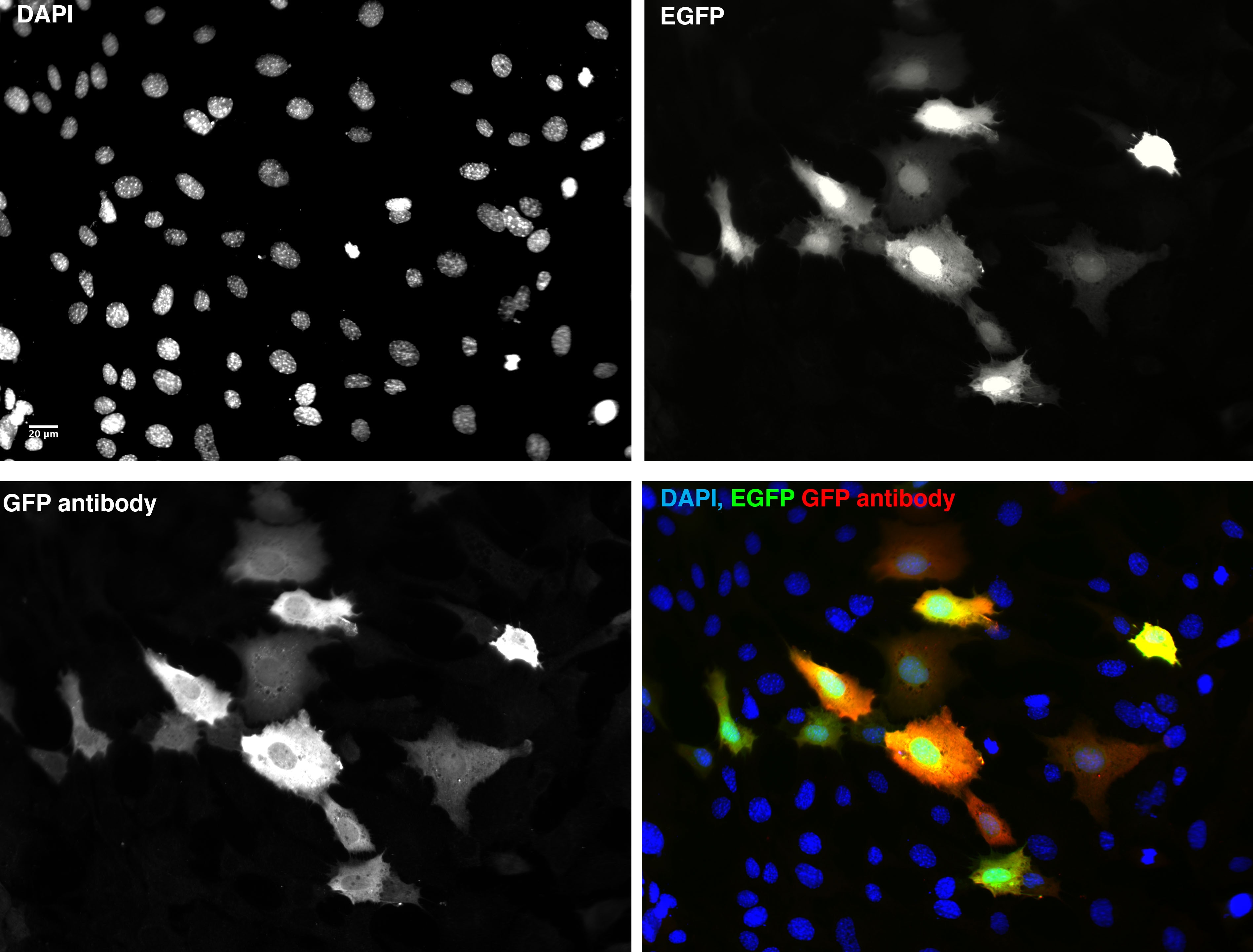
FH Stephen (Verified Customer) (08-02-2022) | HeLa cells transfected with plasmid expressing EGFP fixed with 4% PFA for 15 mins and immunostained with rabbit GFP antibody at 4 degrees incubated for 20h washed then incubated secondary alexa 594 and DAPI for 1 hour room temp imaged on wide field fluorescence microscope.
 |
FH A (Verified Customer) (01-04-2022) | Works for IP
|
FH Rinalda (Verified Customer) (06-21-2021) | Great product
|
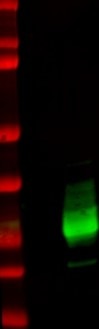
FH Lana (Verified Customer) (12-22-2020) | SDS-PAGE: 15 ug/ul RIPA protein lysate, 4-12% Bis-Tris gradient gel.Transfer: Immobilon-FL transfer membranes (Millipore) for 2h at 80V, 4C.Blocking: SEA Block Blocking Buffer 1h, room T.Primary Ab: O/N incubation at 4C, 1:2500.Secondary Ab: IRDye 800CW Goat anti-Rabbit, 1:15000.Lines of WB image: 1 – protein ladder, 2 – HEK293 whole cell lysate, negative transfection, 3 – whole cell lysate of cells transfected with eGFP.
 |
FH Paul (Verified Customer) (01-15-2020) | Works well for WB.
|
FH Laura (Verified Customer) (01-14-2020) | Good sensitivity for Western blot.
|
FH Aamir (Verified Customer) (01-08-2020) | Works well for WB and IF
|
FH Erica (Verified Customer) (09-26-2019) | This antibody works very well with both western blots and IP. We previously used GFP antibody from another company and it didn't work well. We then switched to Proteinntech's and the signal was very strong! Highly recommend!
|
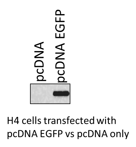
FH Mayur (Verified Customer) (04-30-2019) | The H4 cells were transfected with pcDNA only and pcDNA EGFP . The expression of the EGFP was measured using the GFP antibody from ProteinTech. Great antibody.
 |

