Anticorps Polyclonal de lapin anti-Ki-67
Ki-67 Polyclonal Antibody for IF/ICC, IF-P, FC (Intra)
Hôte / Isotype
Lapin / IgG
Réactivité testée
souris
Applications
IF/ICC, IF-P, FC (Intra)
Conjugaison
CoraLite® Plus 647 Fluorescent Dye
N° de cat : CL647-28074
Synonymes
Galerie de données de validation
Applications testées
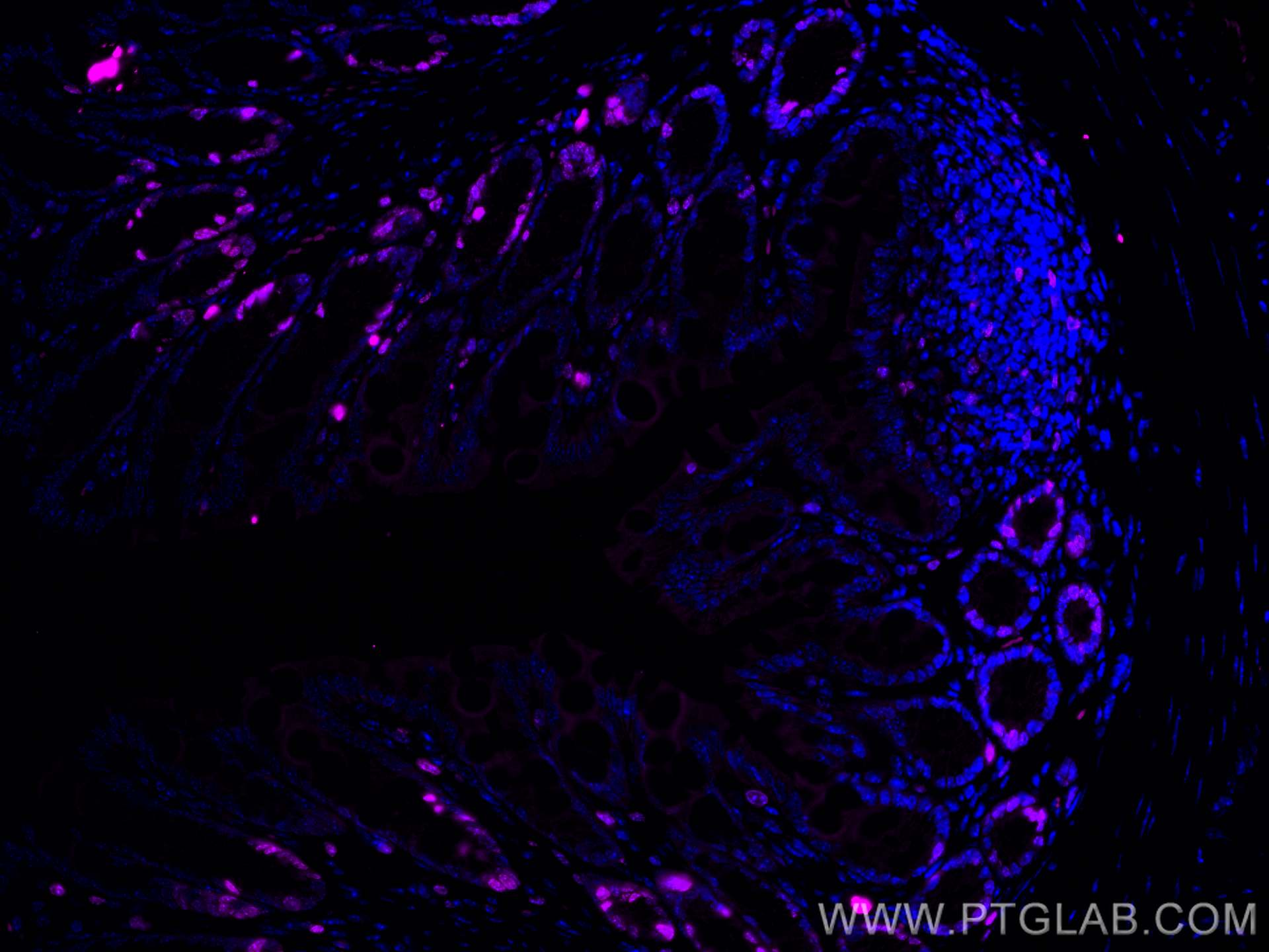
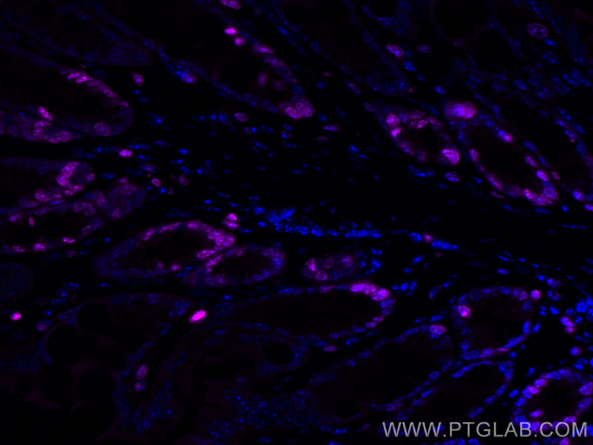
| Résultats positifs en IF-P | tissu de côlon de souris, |
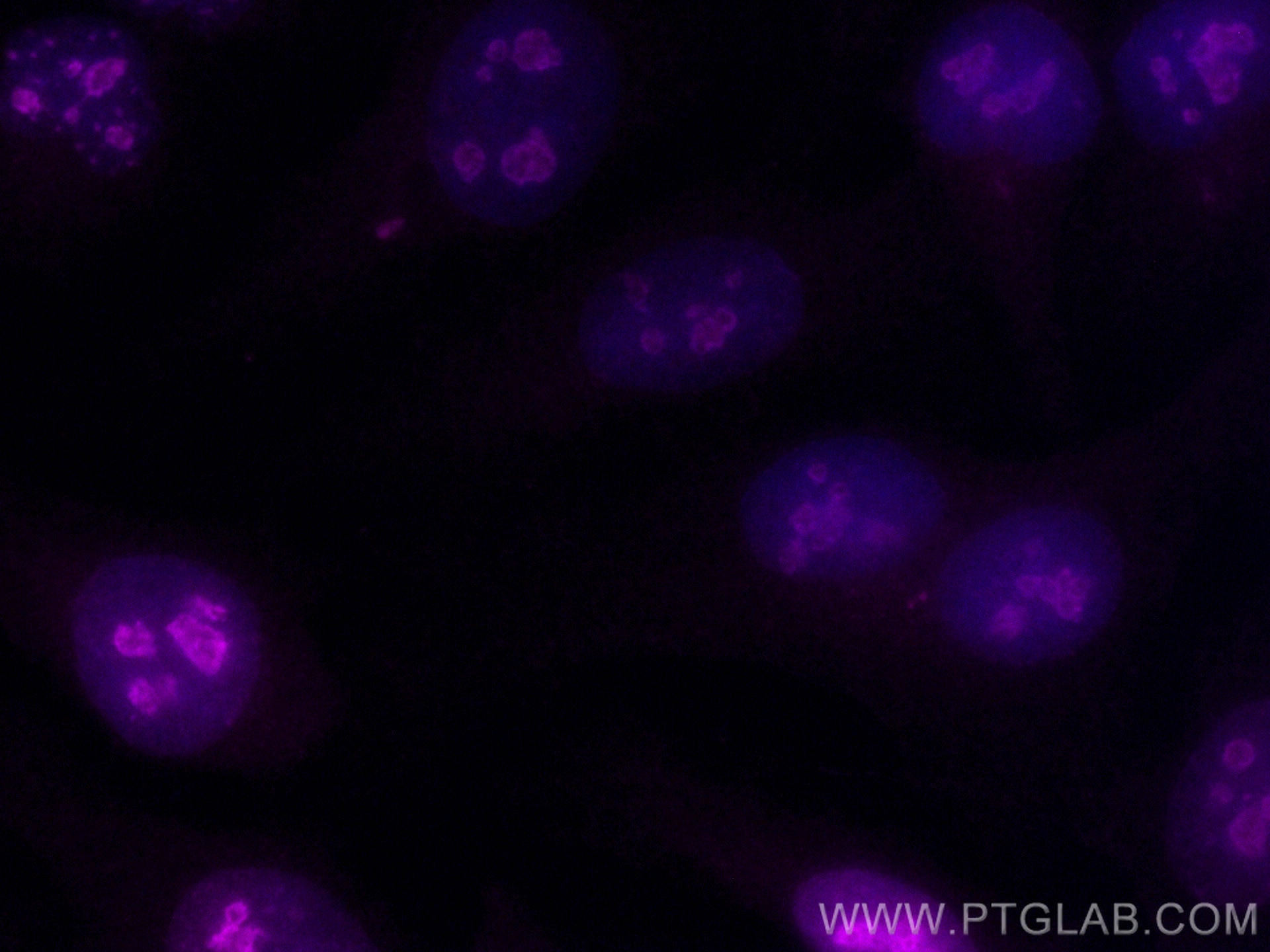
| Résultats positifs en IF/ICC | cellules HeLa, |
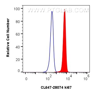
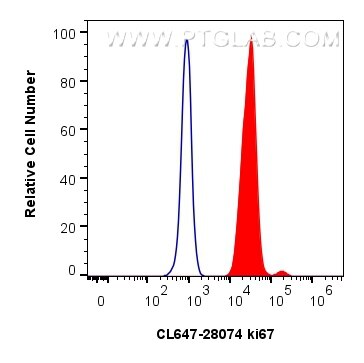
| Résultats positifs en FC (Intra) | cellules NIH/3T3, cellules Jurkat |
Dilution recommandée
| Application | Dilution |
|---|---|
| Immunofluorescence (IF)-P | IF-P : 1:50-1:500 |
| Immunofluorescence (IF)/ICC | IF/ICC : 1:50-1:500 |
| Flow Cytometry (FC) (INTRA) | FC (INTRA) : 0.40 ug per 10^6 cells in a 100 µl suspension |
| It is recommended that this reagent should be titrated in each testing system to obtain optimal results. | |
| Sample-dependent, check data in validation data gallery | |
Informations sur le produit
CL647-28074 cible Ki-67 dans les applications de IF/ICC, IF-P, FC (Intra) et montre une réactivité avec des échantillons souris
| Réactivité | souris |
| Hôte / Isotype | Lapin / IgG |
| Clonalité | Polyclonal |
| Type | Anticorps |
| Immunogène | Ki-67 Protéine recombinante Ag27894 |
| Nom complet | antigen identified by monoclonal antibody Ki 67 |
| Masse moléculaire calculée | 351 kDa |
| Numéro d’acquisition GenBank | NM_001081117 |
| Symbole du gène | ki67 |
| Identification du gène (NCBI) | 17345 |
| Conjugaison | CoraLite® Plus 647 Fluorescent Dye |
| Excitation/Emission maxima wavelengths | 654 nm / 674 nm |
| Forme | Liquide |
| Méthode de purification | Purification par affinité contre l'antigène |
| Tampon de stockage | PBS with 50% glycerol, 0.05% Proclin300, 0.5% BSA |
| Conditions de stockage | Stocker à -20 °C. Éviter toute exposition à la lumière. Stable pendant un an après l'expédition. L'aliquotage n'est pas nécessaire pour le stockage à -20oC Les 20ul contiennent 0,1% de BSA. |
Informations générales
The Ki-67 protein (also known as MKI67) is a cellular marker for proliferation. Ki67 is present during all active phases of the cell cycle (G1, S, G2 and M), but is absent in resting cells (G0). Cellular content of Ki-67 protein markedly increases during cell progression through S phase of the cell cycle. Therefore, the nuclear expression of Ki67 can be evaluated to assess tumor proliferation by immunohistochemistry.
Protocole
| Product Specific Protocols | |
|---|---|
| IF protocol for CL Plus 647 Ki-67 antibody CL647-28074 | Download protocol |
| Standard Protocols | |
|---|---|
| Click here to view our Standard Protocols |
