Anticorps Recombinant de lapin anti-Phospho-TDP43 (Ser409/410)
Phospho-TDP43 (Ser409/410) Recombinant Antibody for WB, IHC, ELISA
Hôte / Isotype
Lapin / IgG
Réactivité testée
Humain, souris et plus (1)
Applications
WB, IHC, IF, ELISA
Conjugaison
Non conjugué
CloneNo.
6M10
N° de cat : 80007-1-RR
Synonymes
Galerie de données de validation
 antibody) at dilution of 1:10000 incubated at room temperature for 1.5 hours.The membrane was stripped and re-blotted with Alpha Tubulin (66031-1-Ig) antibody as a loading control.")
 rabbit recombinant antibody (80007-1-RR, 1000) with the frontal cortex from FTLD-TDP type B patients. IHC experiment was done with Ventana automatic staining system and Optiview DAB detection kit with heat-induced epitope retrieval (boiling for 32 min in Tris-EDTA based solution CC1 buffer, Ventana). Fig from the lab of Dr. Neumann.")
 and temporal cortex (medium-mag) from subject with limbic-predominant age-related TDP-43 encephalopathy neuropathologic change (LATE-NC). Staining provided by Pete Nelson and Ela Patel, U. Kentucky AD Research Center Neuropathology Core.")
Applications testées
| Résultats positifs en WB | cellules HeLa, Calyculin A treated cells |
| Résultats positifs en IHC | cortex frontal d'un cas de DLFT-TDP de type B, tissu cérébral humain il est suggéré de démasquer l'antigène avec un tampon de TE buffer pH 9.0; (*) À défaut, 'le démasquage de l'antigène peut être 'effectué avec un tampon citrate pH 6,0. |
Dilution recommandée
| Application | Dilution |
|---|---|
| Western Blot (WB) | WB : 1:5000-1:50000 |
| Immunohistochimie (IHC) | IHC : 1:500-1:2000 |
| It is recommended that this reagent should be titrated in each testing system to obtain optimal results. | |
| Sample-dependent, check data in validation data gallery | |
Applications publiées
| WB | See 7 publications below |
| IHC | See 8 publications below |
| IF | See 10 publications below |
Informations sur le produit
80007-1-RR cible Phospho-TDP43 (Ser409/410) dans les applications de WB, IHC, IF, ELISA et montre une réactivité avec des échantillons Humain, souris
| Réactivité | Humain, souris |
| Réactivité citée | Humain, poisson-zèbre, souris |
| Hôte / Isotype | Lapin / IgG |
| Clonalité | Recombinant |
| Type | Anticorps |
| Immunogène | Peptide |
| Nom complet | TAR DNA binding protein |
| Masse moléculaire calculée | 43 kDa |
| Poids moléculaire observé | 45-50 kDa |
| Numéro d’acquisition GenBank | NM_007375 |
| Symbole du gène | TDP-43 |
| Identification du gène (NCBI) | 23435 |
| Conjugaison | Non conjugué |
| Forme | Liquide |
| Méthode de purification | Purification par protéine A |
| Tampon de stockage | PBS with 0.02% sodium azide and 50% glycerol |
| Conditions de stockage | Stocker à -20°C. Stable pendant un an après l'expédition. L'aliquotage n'est pas nécessaire pour le stockage à -20oC Les 20ul contiennent 0,1% de BSA. |
Informations générales
Transactivation response (TAR) DNA-binding protein of 43 kDa (also known as TARDBP or TDP-43) was first isolated as a transcriptional inactivator binding to the TAR DNA element of the HIV-1 virus. Neumann et al. (2006) found that a hyperphosphorylated, ubiquitinated, and cleaved form of TARDBP, known as pathologic TDP-43, is the major component of the tau-negative and ubiquitin-positive inclusions that characterize amyotrophic lateral sclerosis (ALS) and the most common pathological subtype of frontotemporal lobar degeneration (FTLD-U). Various forms of TDP-43 exist, including 18-35 kDa of cleaved C-terminal fragments, 45-50 kDa phospho-protein, 55 kDa glycosylated form, 75 kDa hyperphosphorylated form, and 90-300 kDa cross-linked form. (PMID: 17023659,19823856, 21666678, 22193176). 80007-1-RR is a recombinant rabbit monoclonal antibody recognizing TDP-43 only when phosphorylated at 409/410. Immunohistochemical analyses using this antibody only stain the insoluble inclusions in pathologic tissues without normal diffuse nuclear staining.
Protocole
| Product Specific Protocols | |
|---|---|
| WB protocol for Phospho-TDP43 (Ser409/410) antibody 80007-1-RR | Download protocol |
| Standard Protocols | |
|---|---|
| Click here to view our Standard Protocols |
Publications
| Species | Application | Title |
|---|---|---|
J Biol Chem Regulation of TAR DNA binding protein 43 (TDP-43) homeostasis by cytosolic DNA accumulation | ||
Biochim Biophys Acta Mol Basis Dis Phosphorylated TAR DNA-Binding Protein-43: Aggregation and Antibody-Based Inhibition. | ||
bioRxiv TDP-43 pathology links innate and adaptive immunity in amyotrophic lateral sclerosis | ||
bioRxiv Fructose-2,6-bisphosphate restores TDP-43 pathology-driven genome repair deficiency in motor neuron diseases | ||
Ecotoxicol Environ Saf Arsenic exposure activates microglia, inducing neuroinflammation and promoting the occurrence and development of Alzheimer's disease-like neurodegeneration in mice | ||
Res Sq Endogenous TDP-43 mislocalization in a novel knock-in mouse model reveals DNA repair impairment, inflammation, and neuronal senescence |
Avis
The reviews below have been submitted by verified Proteintech customers who received an incentive for providing their feedback.
FH Rashmi (Verified Customer) (02-14-2025) | used for Western blot, IHC, highly recommended
|
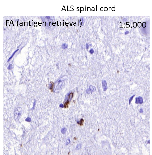
FH Silvia (Verified Customer) (08-21-2020) | Excellent phospho-specific TDP-43 antibody. Tested by IHC in ethanol and NBF fixed FTLD-TDP brains and ALS spinal cord. The antibody works well with citrate buffer and FA as antigen retrieval. The dilutions that worked better showing high specificity and low background ranged from 1,000-5,000.Works great also by western blot at dilutions 1:1,000-1:2,000.
 |

