

Tested Applications
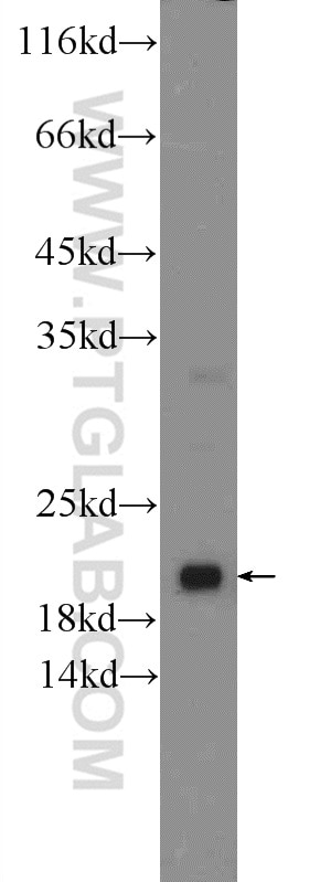
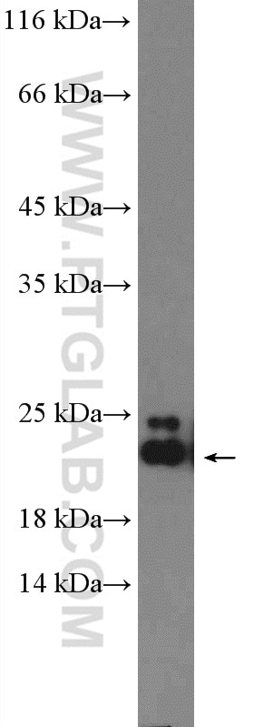
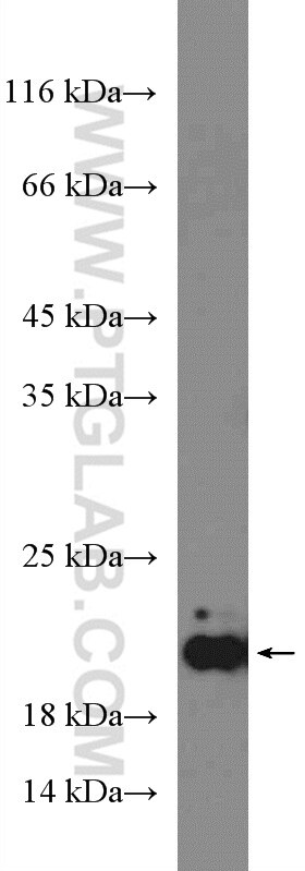
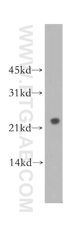
| Positive WB detected in | fetal human brain tissue, human brain tissue, mouse brain tissue, NIH/3T3 cells |
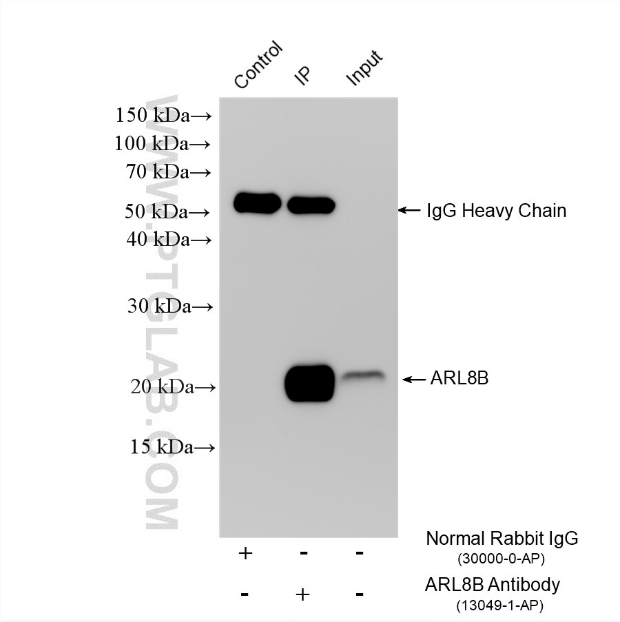
| Positive IP detected in | A431 cells |
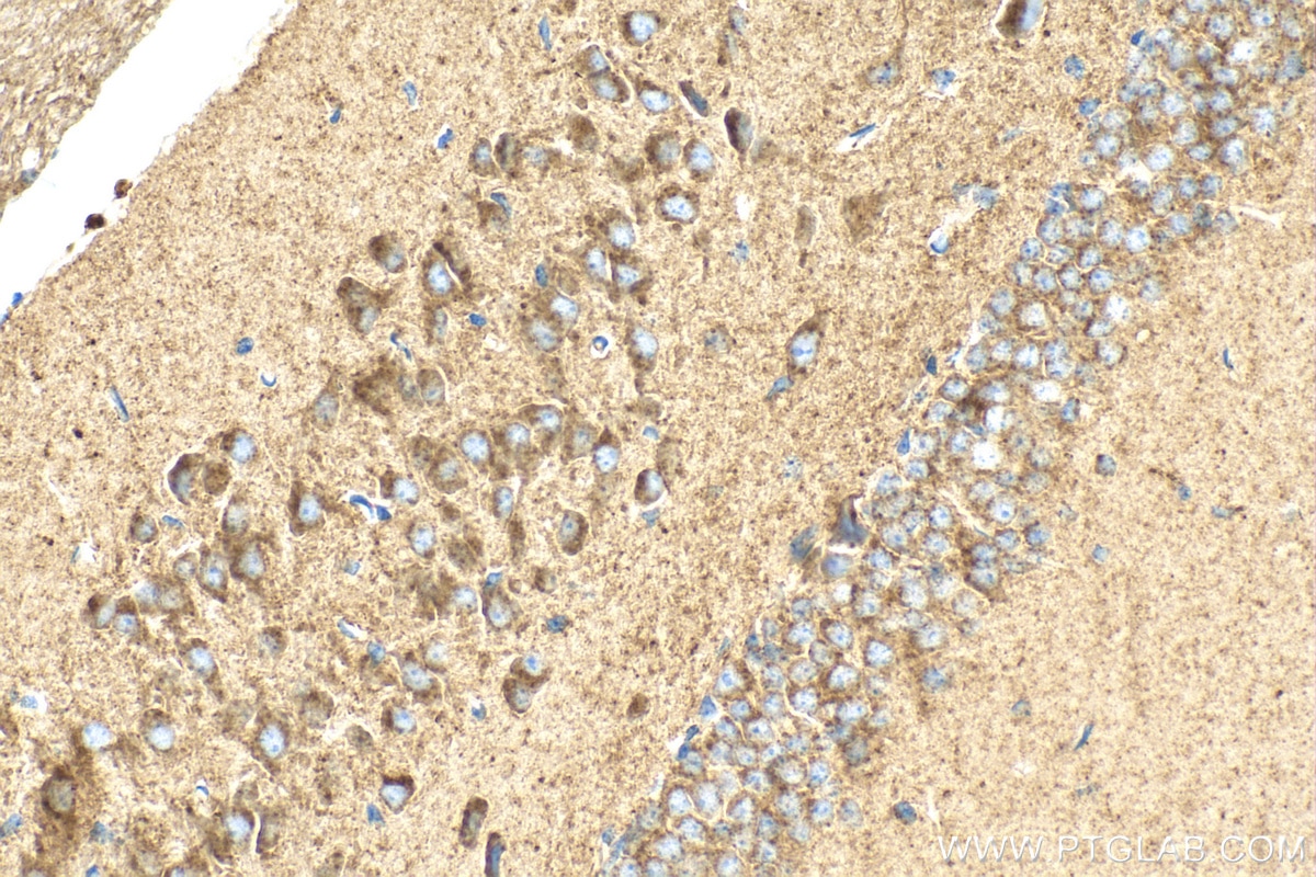
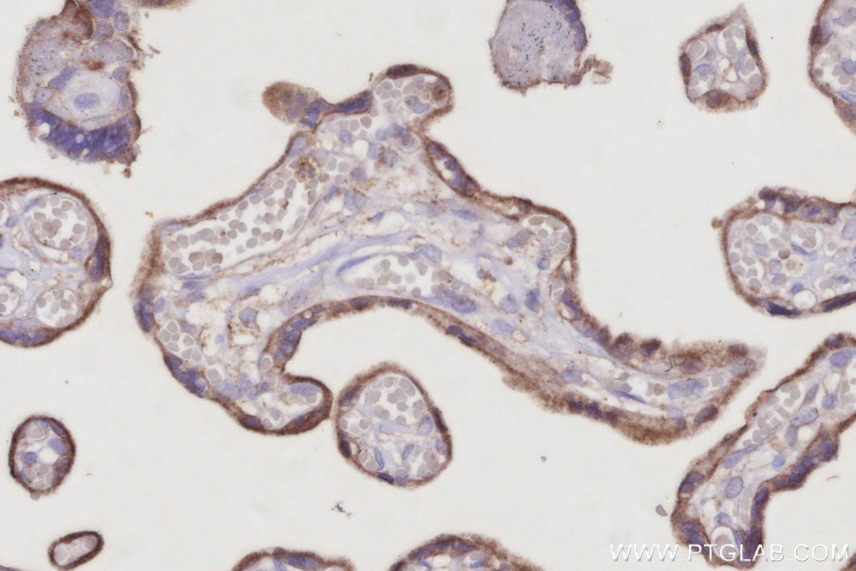
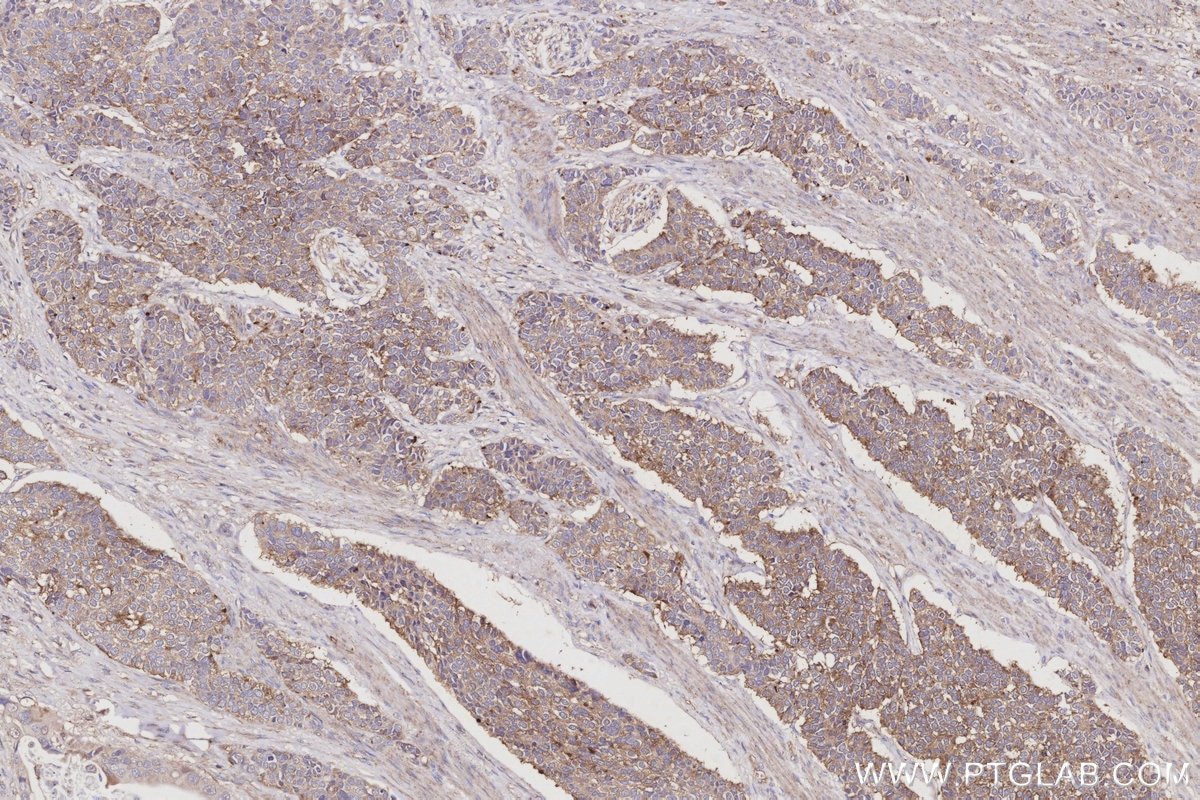
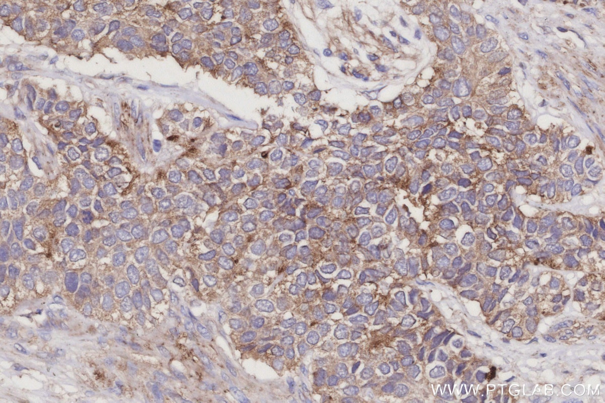
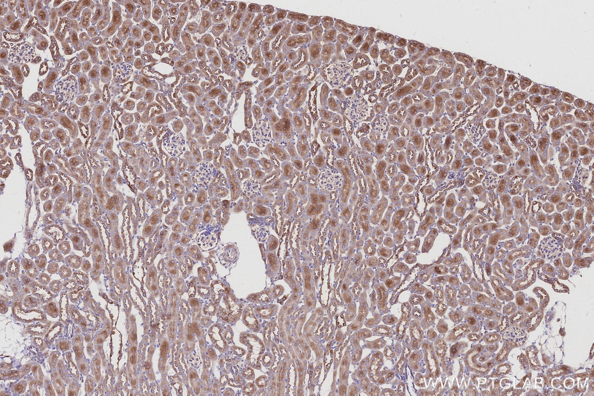
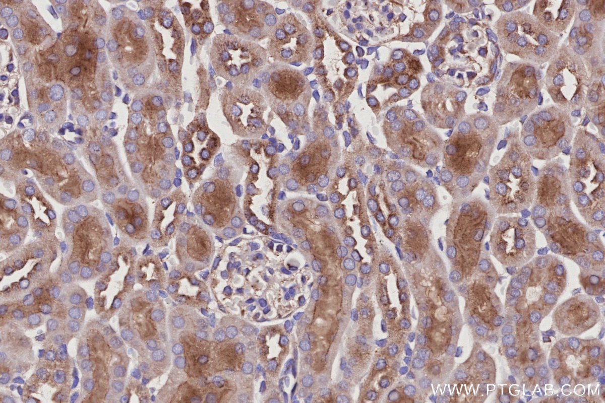
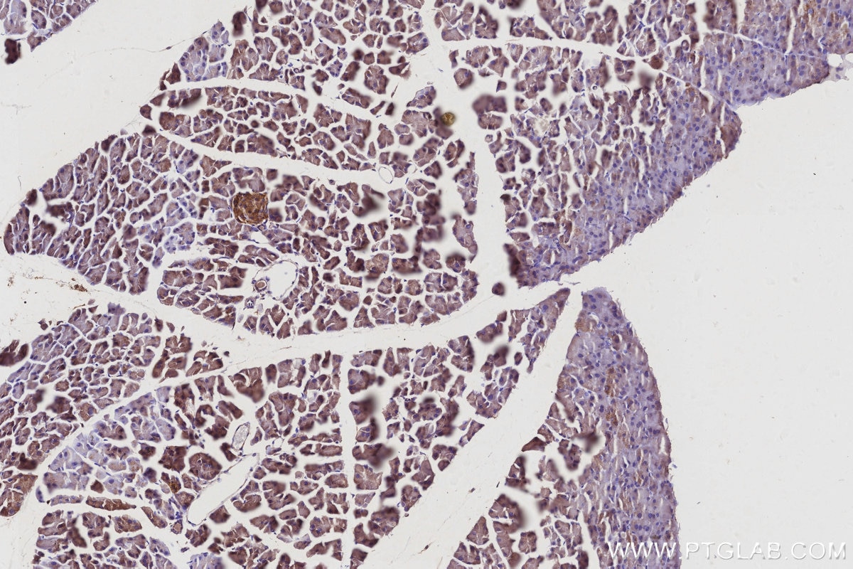
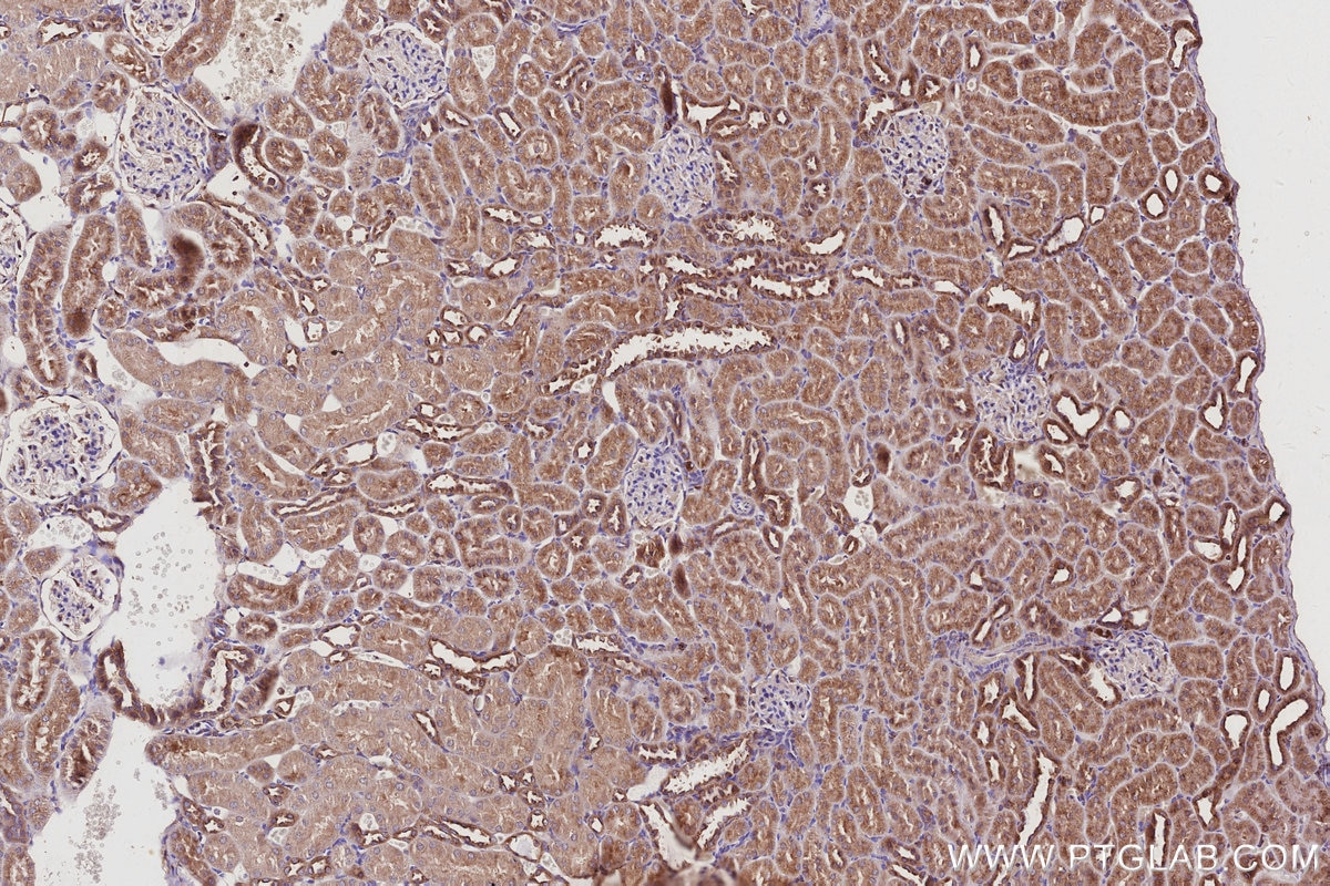
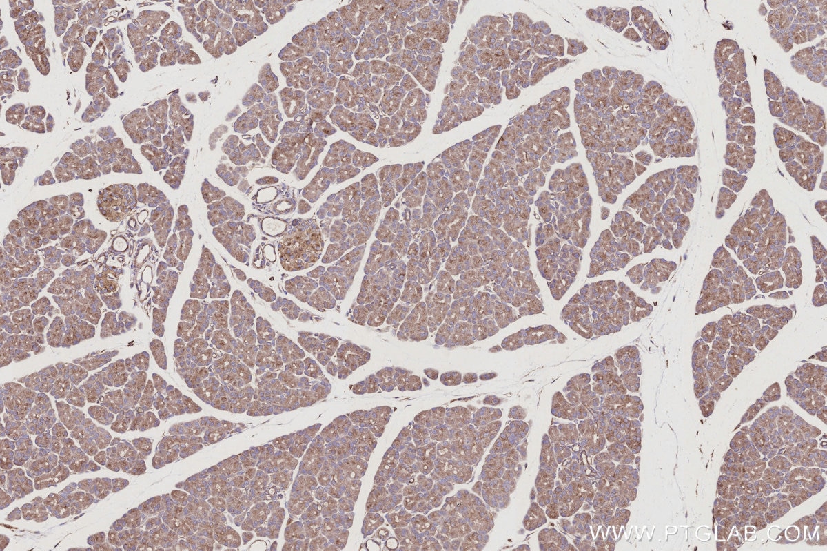
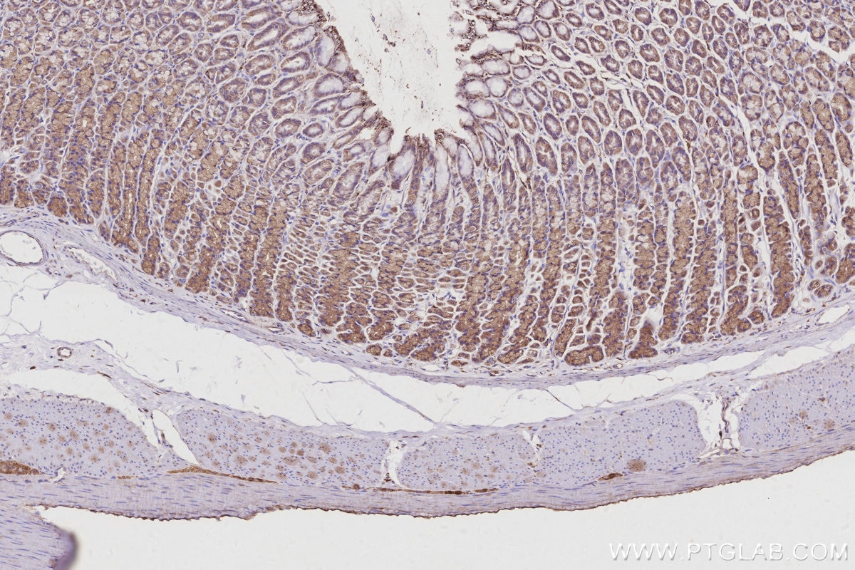
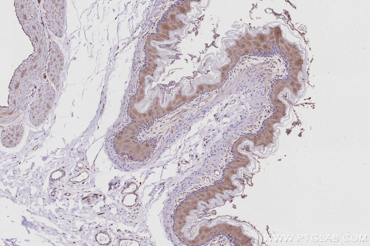
| Positive IHC detected in | mouse brain tissue, rat kidney tissue, rat stomach tissue, human placenta tissue, human stomach cancer tissue, mouse kidney tissue, rat pancreas tissue, mouse pancreas tissue Note: suggested antigen retrieval with TE buffer pH 9.0; (*) Alternatively, antigen retrieval may be performed with citrate buffer pH 6.0 |
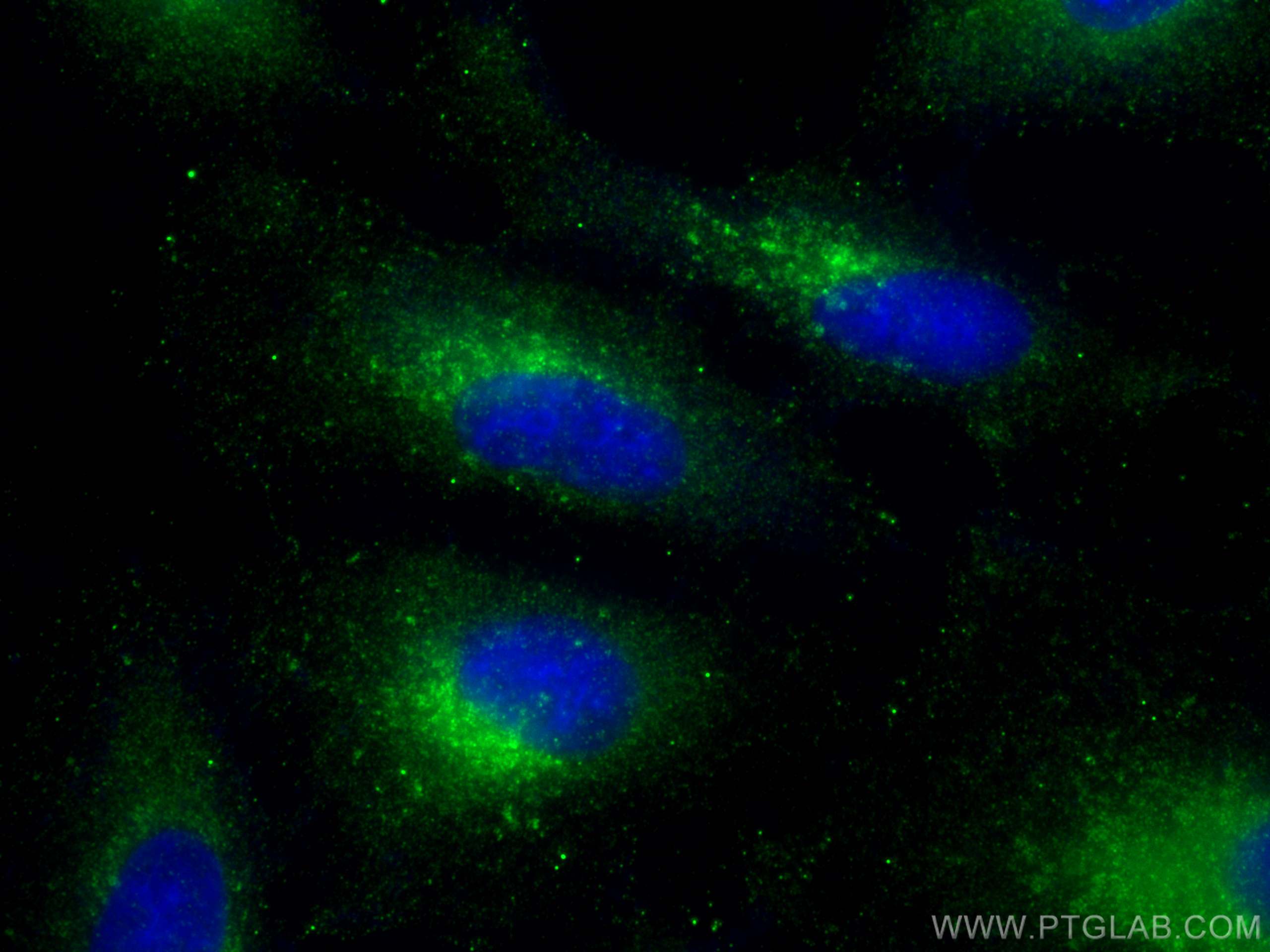
| Positive IF/ICC detected in | A549 cells |
Recommended dilution
| Application | Dilution |
|---|---|
| Western Blot (WB) | WB : 1:500-1:1000 |
| Immunoprecipitation (IP) | IP : 0.5-4.0 ug for 1.0-3.0 mg of total protein lysate |
| Immunohistochemistry (IHC) | IHC : 1:50-1:500 |
| Immunofluorescence (IF)/ICC | IF/ICC : 1:50-1:500 |
| It is recommended that this reagent should be titrated in each testing system to obtain optimal results. | |
| Sample-dependent, Check data in validation data gallery. | |
Product Information
13049-1-AP targets ARL8B in WB, IHC, IF/ICC, IP, CoIP, ELISA applications and shows reactivity with human, mouse, rat samples.
| Tested Reactivity | human, mouse, rat |
| Cited Reactivity | human, mouse, rat, monkey, chicken |
| Host / Isotype | Rabbit / IgG |
| Class | Polyclonal |
| Type | Antibody |
| Immunogen |
CatNo: Ag3711 Product name: Recombinant human ARL8B protein Source: e coli.-derived, PGEX-4T Tag: GST Domain: 1-186 aa of BC013131 Sequence: MLALISRLLDWFRSLFWKEEMELTLVGLQYSGKTTFVNVIASGQFSEDMIPTVGFNMRKVTKGNVTIKIWDIGGQPRFRSMWERYCRGVNAIVYMIDAADREKIEASRNELHNLLDKPQLQGIPVLVLGNKRDLPNALDEKQLIEKMNLSAIQDREICCYSISCKEKDNIDITLQWLIQHSKSRRS Predict reactive species |
| Full Name | ADP-ribosylation factor-like 8B |
| Calculated Molecular Weight | 186 aa, 22 kDa |
| Observed Molecular Weight | 22 kDa |
| GenBank Accession Number | BC013131 |
| Gene Symbol | ARL8B |
| Gene ID (NCBI) | 55207 |
| RRID | AB_2059000 |
| Conjugate | Unconjugated |
| Form | Liquid |
| Purification Method | Antigen affinity purification |
| UNIPROT ID | Q9NVJ2 |
| Storage Buffer | PBS with 0.02% sodium azide and 50% glycerol, pH 7.3. |
| Storage Conditions | Store at -20°C. Stable for one year after shipment. Aliquoting is unnecessary for -20oC storage. 20ul sizes contain 0.1% BSA. |
Background Information
ARL8B ( ADP-ribosylation factor-like 8B) , also known as ARL10C or GIE1, is a small GTPase of the Arl-family, that is associated with the lysosome. ARL8A interacts with Tubulin, and plays a role in lysosome motility, as well as in chromosomal segregation. ARL8B plays a role in the spatial distribution and positioning of lysosomes. ARL8B is regarded as a regulator of endosome to lysosome trafficking pathways of special significance for host defense.
Protocols
| Product Specific Protocols | |
|---|---|
| IF protocol for ARL8B antibody 13049-1-AP | Download protocol |
| IHC protocol for ARL8B antibody 13049-1-AP | Download protocol |
| IP protocol for ARL8B antibody 13049-1-AP | Download protocol |
| WB protocol for ARL8B antibody 13049-1-AP | Download protocol |
| Standard Protocols | |
|---|---|
| Click here to view our Standard Protocols |
Publications
What published studies show
The ARL8B polyclonal antibody (Cat# 13049-1-AP) has 36 published citations appearing in journals including Nature Cell Biology, Nature Communications, Science Advances, Cell Reports, Current Biology, Journal of Cell Biology, PNAS, and Developmental Cell. Research fields encompass lysosomal positioning and transport, autophagy, endocytosis, intracellular trafficking, neurodegeneration, cell migration, viral infection, and cancer biology.
| Species | Application | Title |
|---|---|---|
Nat Commun Spatiotemporal proteomics reveals the biosynthetic lysosomal membrane protein interactome in neurons
| ||
Nat Commun Lysosomal NKG7 restrains mTORC1 activity to promote CD8+ T cell durability and tumor control | ||
Nat Commun Control of lysosome function by the GTPase-activating protein TBC1D9B and its binding partner TMEM55B. | ||
Nat Commun DENND6A links Arl8b to a Rab34/RILP/dynein complex, regulating lysosomal positioning and autophagy |
Reviews
What customers say
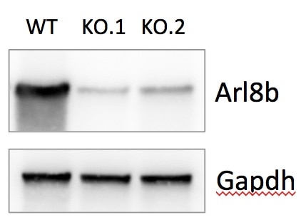
One reviewer (Olivia M.) rated this Arl8b antibody 4/5 stars in January 2020. She used it at a 1:500 dilution on mouse bone marrow-derived macrophages for both Western Blot and Immunofluorescence. For WB, she probed lysates via SDS-PAGE with overnight incubation at 4°C. For IF, cells were fixed in 4% PFA, permeabilized with 0.5% saponin, incubated with the primary antibody overnight at 4°C, then detected with anti-rabbit Alexa Fluor 568 at room temperature for 1 hour. Overall, the antibody performed well in both applications.
The reviews below have been submitted by verified Proteintech customers who received an incentive for providing their feedback.
Olivia (Verified Customer) (01-08-2020) | WB: Lysates from mouse bone marrow-derived macrophages were subjected to SDS PAGE followed by western blot with 13049-1-AP(Arl8b antibody) at a dilution of 1:500 overnight at 4°C.IF: Mouse bone marrow-derived macrophages were fixed in 4% PFA and permeabilized with 0.5% saponin before incubation with 1:500 Arl8b antibody (in 1% BSA/0.1% saponin/PBS) overnight at 4°C, followed by incubation with anti-rabbit Alexa Fluor 568 for 1h at room temperature.
 |


















