

Tested Applications
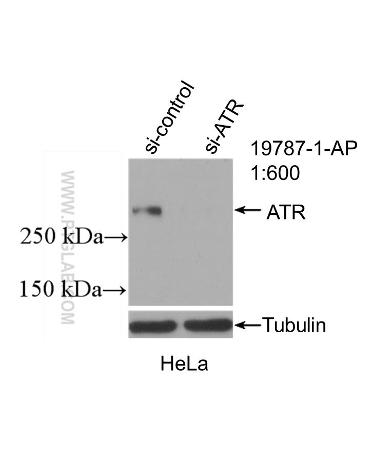
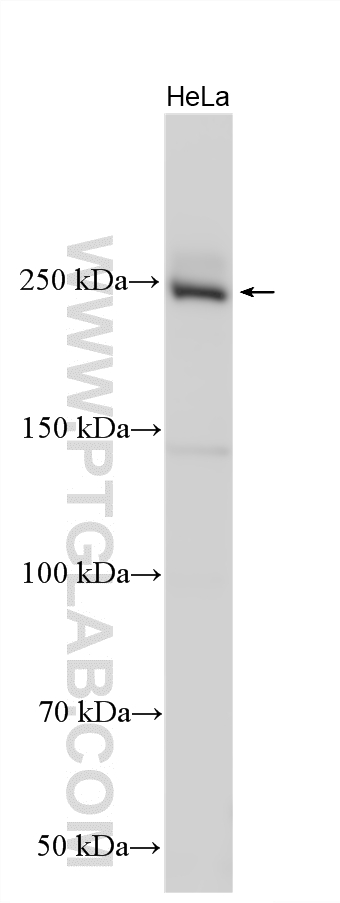
| Positive WB detected in | HeLa cells |
Recommended dilution
| Application | Dilution |
|---|---|
| Western Blot (WB) | WB : 1:1000-1:4000 |
| It is recommended that this reagent should be titrated in each testing system to obtain optimal results. | |
| Sample-dependent, Check data in validation data gallery. | |
Product Information
19787-1-AP targets ATR in WB, IHC, IP, CoIP, ELISA applications and shows reactivity with human samples.
| Tested Reactivity | human |
| Cited Reactivity | human, mouse, rat |
| Host / Isotype | Rabbit / IgG |
| Class | Polyclonal |
| Type | Antibody |
| Immunogen |
Peptide Predict reactive species |
| Full Name | ataxia telangiectasia and Rad3 related |
| Calculated Molecular Weight | 301 kDa |
| Observed Molecular Weight | 250-290 kDa |
| GenBank Accession Number | NM_001184 |
| Gene Symbol | ATR |
| Gene ID (NCBI) | 545 |
| RRID | AB_10639516 |
| Conjugate | Unconjugated |
| Form | Liquid |
| Purification Method | Antigen affinity purification |
| UNIPROT ID | Q13535 |
| Storage Buffer | PBS with 0.02% sodium azide and 50% glycerol, pH 7.3. |
| Storage Conditions | Store at -20°C. Stable for one year after shipment. Aliquoting is unnecessary for -20oC storage. 20ul sizes contain 0.1% BSA. |
Background Information
ATR, also named as FRP1, belongs to the PI3/PI4-kinase family and ATM subfamily. ATR is a serine/threonine protein kinase which activates checkpoint signaling upon genotoxic stresses such as ionizing radiation (IR), ultraviolet light (UV), or DNA replication stalling, thereby acting as a DNA damage sensor. ATR recognizes the substrate consensus sequence [ST]-Q. ATR phosphorylates BRCA1, CHEK1, MCM2, RAD17, RPA2, SMC1 and TP53/p53, which collectively inhibit DNA replication and mitosis and promote DNA repair, recombination and apoptosis. ATR phosphorylates 'Ser-139' of histone variant H2AX/H2AFX at sites of DNA damage, thereby regulating DNA damage response mechanism. It is required for FANCD2 ubiquitination. It is critical for maintenance of fragile site stability and efficient regulation of centrosome duplication. ATR catalyze the reaction: ATP + a protein = ADP + a phosphoprotein. Defects in ATR are a cause of Seckel syndrome type 1 (SCKL1) which is a rare autosomal recessive disorder characterized by growth retardation, microcephaly with mental retardation, and a characteristic 'bird-headed' facial appearance. The antibody can recognize all the isoforms of ATR.
Protocols
| Product Specific Protocols | |
|---|---|
| WB protocol for ATR antibody 19787-1-AP | Download protocol |
| Standard Protocols | |
|---|---|
| Click here to view our Standard Protocols |
Publications
What published studies show
ATR Polyclonal antibody (Cat# 19787-1-AP) has 57 citations in journals including Cell, Nature Communications, Cancer Research, Nucleic Acids Research, Oncogene, Cell Death & Disease, ACS Nano, Journal of Experimental & Clinical Cancer Research, Advanced Science, Drug Resistance Updates, and Brain, Behavior, and Immunity. Research fields cover DNA damage response, ATR/CHK1 signaling, multiple cancer types (gastric, colorectal, ovarian, breast, liver, glioma), radiation resistance, autophagy, ferroptosis, neurodegeneration, cardiovascular disease, toxicology, and reproductive biology.
| Species | Application | Title |
|---|---|---|
Cell Extrachromosomal DNA replication and maintenance couple with DNA damage pathway in tumors | ||
Cell Res DNA damage triggers tubular endoplasmic reticulum extension to promote apoptosis by facilitating ER-mitochondria signaling. | ||
Cancer Res Combined inactivation of CTPS1 and ATR is synthetically lethal to MYC-overexpressing cancer cells. | ||
Cancer Res PGC-1α degradation suppresses mitochondrial biogenesis to confer radiation resistance in glioma | ||
Drug Resist Updat RAD1 promotes oxaliplatin resistance in gastric cancer by reinforcing NRF2-driven antioxidant defense and DDR checkpoint signaling. | ||
Nat Commun ATR/Chk1 signaling induces autophagy through sumoylated RhoB-mediated lysosomal translocation of TSC2 after DNA damage. |
Reviews
What customers say
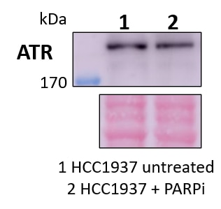
All three reviews are 5-star ratings for Western Blot. Users report clear target bands across diverse samples including human keratinocytes, cardiomyocytes, mouse skin, liver, heart tissues, and human breast cancer cells (HCC1937). One user confirmed a single band >170 kDa in PARP inhibitor-treated cells at 1:500 dilution. Others used 1:1000 dilution with good results, though one noted some background. Overall, the antibody performs reliably across species and tissue types for WB.
The reviews below have been submitted by verified Proteintech customers who received an incentive for providing their feedback.
Priya (Verified Customer) (01-17-2023) | I have used this antibody for human keratinocytes, cardiomyocytes, mouse skin and liver tissues
|
Marina (Verified Customer) (06-26-2022) | HCC1937 human breast cancer cells untreated (1) and treated with a PARP inhibitor (2). Primary antibody was incubated at 4ºC overnight in rotation. A single band >170 kDa could be observed in both conditions. Ponceau staining was used as total protein loading control.
 |
WEI (Verified Customer) (03-08-2022) | Good target band with backgrouds
|



