
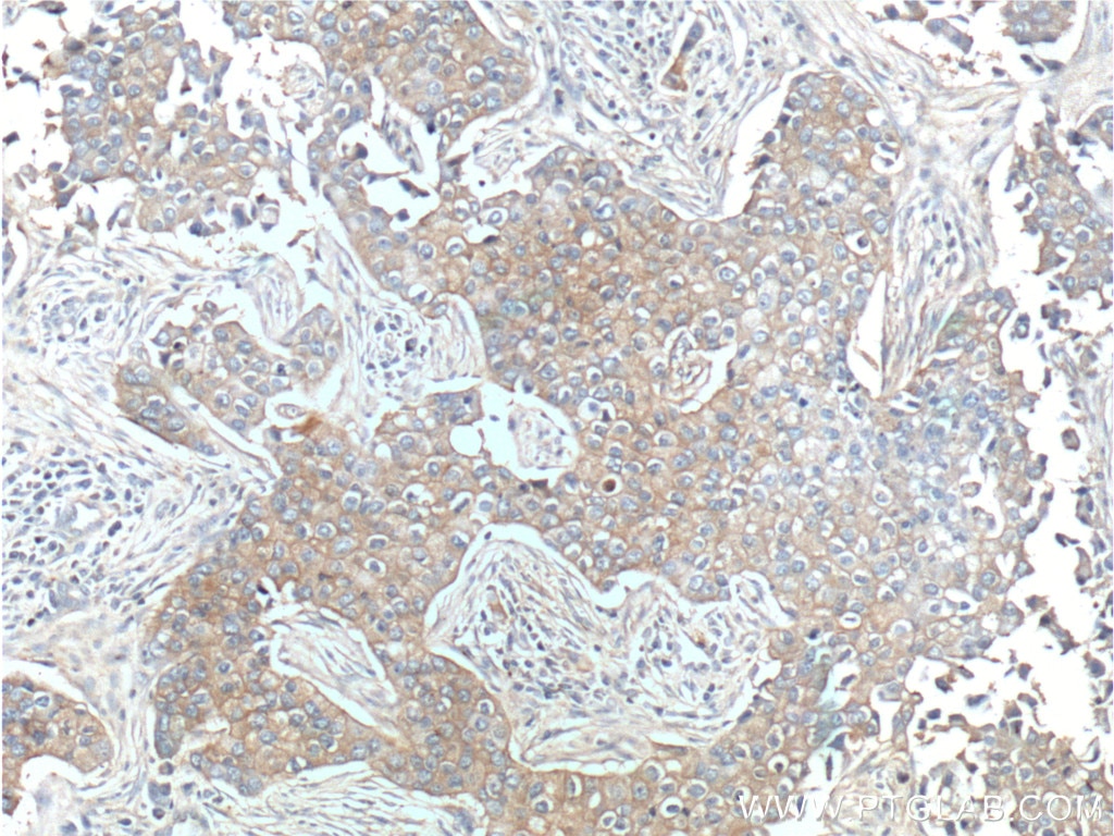
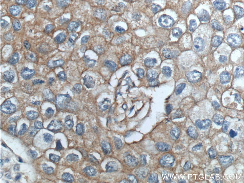
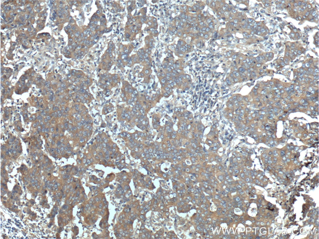
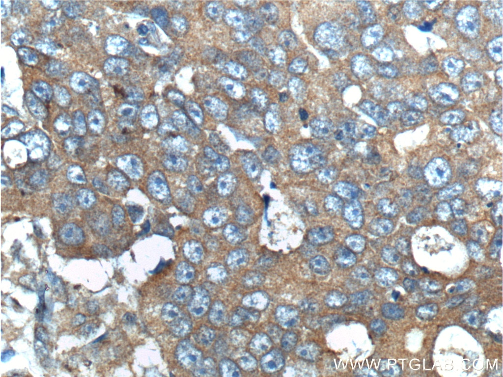

Tested Applications
| Positive IHC detected in | human breast cancer tissue, human prostate cancer tissue Note: suggested antigen retrieval with TE buffer pH 9.0; (*) Alternatively, antigen retrieval may be performed with citrate buffer pH 6.0 |
Recommended dilution
| Application | Dilution |
|---|---|
| Immunohistochemistry (IHC) | IHC : 1:50-1:500 |
| It is recommended that this reagent should be titrated in each testing system to obtain optimal results. | |
| Sample-dependent, Check data in validation data gallery. | |
Product Information
10369-1-AP targets ERBB3 in WB, IHC, ELISA, Cell treatment applications and shows reactivity with human samples.
| Tested Reactivity | human |
| Cited Reactivity | human, mouse |
| Host / Isotype | Rabbit / IgG |
| Class | Polyclonal |
| Type | Antibody |
| Immunogen |
CatNo: Ag0215 Product name: Recombinant human ERBB3 protein Source: e coli.-derived, PGEX-4T Tag: GST Domain: 88-331 aa of BC002706 Sequence: LVAMNEFSTLPLPNLRVVRGTQVYDGKFAIFVMLNYNTNSSHALRQLRLTQLTEILSGGVYIEKNDKLCHMDTIDWRDIVRDRDAEIVVKDNGRSCPPCHEVCKGRCWGPGSEDCQTLTKTICAPQCNGHCFGPNPNQCCHDECAGGCSGPQDTDCFACRHFNDSGACVPRCPQPLVYNKLTFQLEPNPHTKYQYGGVCVASCPHNFVVDQTSCVRACPPDKMEVDKNGLKMCEPCGGLCPKAF Predict reactive species |
| Full Name | v-erb-b2 erythroblastic leukemia viral oncogene homolog 3 (avian) |
| Calculated Molecular Weight | 148 kDa |
| GenBank Accession Number | BC002706 |
| Gene Symbol | ERBB3 |
| Gene ID (NCBI) | 2065 |
| RRID | AB_2099548 |
| Conjugate | Unconjugated |
| Form | Liquid |
| Purification Method | Antigen affinity purification |
| UNIPROT ID | P21860 |
| Storage Buffer | PBS with 0.02% sodium azide and 50% glycerol, pH 7.3. |
| Storage Conditions | Store at -20°C. Stable for one year after shipment. Aliquoting is unnecessary for -20oC storage. 20ul sizes contain 0.1% BSA. |
Background Information
V-erb-b2 erythroblastic leukemia viral oncogene homolog 3 (ErbB-3, HER3) is a member of the EGF receptor tyrosine kinase family including EGFR (HER1), Neu (ErbB-2, HER2), ErbB-3 (HER3), and ErbB-4 (HER4) that are frequently overexpressed in a variety of carcinomas. These family members form either homodimers or heterodimers upon ligand binding to mediate cell growth. ErbB-3 binds and is activated by neuregulins and NTAK. It forms a heterodimer with each of the other ErbB receptors. ErbB-3 is predominantly expressed in epithelial tissues and brain, and is overexpressed in a subset of human mammary tumors.
Protocols
| Product Specific Protocols | |
|---|---|
| IHC protocol for ERBB3 antibody 10369-1-AP | Download protocol |
| Standard Protocols | |
|---|---|
| Click here to view our Standard Protocols |
Publications
What published studies show
The anti-HER3 (ERBB3) antibody (Cat# 10369-1-AP) has 14 total citations. Citations appear in journals including Cell Reports, International Journal of Biological Macromolecules, Acta Pharmacologica Sinica, Liver International, Cells, Oncotarget, Journal of Molecular and Cellular Cardiology, Cancers, Frontiers in Oncology, and others. Research fields span oncology (breast, ovarian, colorectal, hepatocellular, prostate cancers), liver fibrosis, heart regeneration, pulmonary fibrosis, and sinonasal pathology, with applications primarily in Western blot and IHC.
| Species | Application | Title |
|---|---|---|
Acta Pharmacol Sin Quantitative systems pharmacology modeling of HER2-positive metastatic breast cancer for translational efficacy evaluation and combination assessment across therapeutic modalities | ||
Int J Biol Macromol Hepatitis B virus X protein increases LASP1 SUMOylation to stabilize HER2 and facilitate hepatocarcinogenesis | ||
Int J Biol Macromol CircSETD3 interrupts the bidirectional positive feedback of ErbB3 and Akt by sponging miR-4667-5p to inhibit colorectal cancer progression and cetuximab resistance | ||
Cell Rep A MYC-STAMBPL1-TOE1 positive feedback loop mediates EGFR stability in hepatocellular carcinoma | ||
Liver Int PPL(+) Cholangiocytes Define a Pro-Regenerative Subset Associated With NRG1-ERBB3-PI3K/AKT Signalling During Liver Fibrosis. | ||
Cells NOX4 Signaling Mediates Cancer Development and Therapeutic Resistance through HER3 in Ovarian Cancer Cells. |
Reviews
What customers say
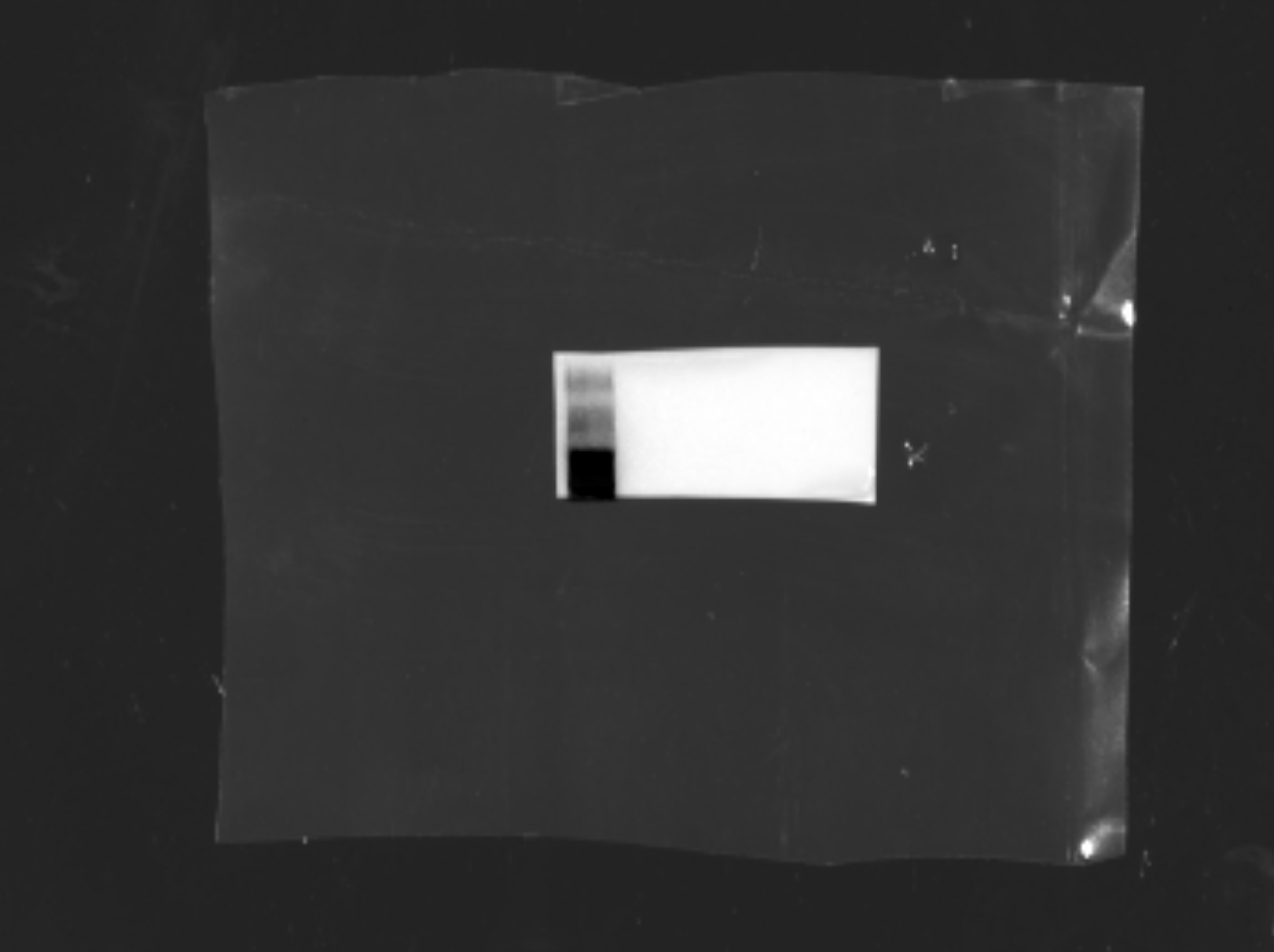
The single review for 10369-1-AP is from Morgan Woodman (October 2021), who used the antibody for Western Blot on OV90 Ovarian Cancer Cells at a 1:500 dilution. Despite giving a 4-star rating, the reviewer reported being unable to obtain any specific detection signal. They noted that the antibody band appeared at an unexpected position on the molecular weight ladder, making troubleshooting difficult. This issue was consistent across two separate Western Blot attempts. No other reviews are available for this product.
The reviews below have been submitted by verified Proteintech customers who received an incentive for providing their feedback.
Morgan (Verified Customer) (10-26-2021) | I could not get any detection for this antibody at all. It strangely appeared on the ladder and I did not know how to troubleshoot. It occurred in both western blot attempts that I tried (Please see images)
 |





