Tested Applications
| Positive WB detected in | HeLa cells, HEK-293 cells, mouse testis tissue, mouse brain tissue, K-562 cells |
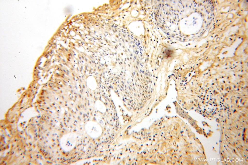
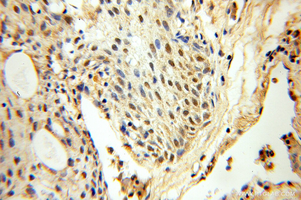
| Positive IHC detected in | human prostate cancer tissue Note: suggested antigen retrieval with TE buffer pH 9.0; (*) Alternatively, antigen retrieval may be performed with citrate buffer pH 6.0 |
Recommended dilution
| Application | Dilution |
|---|---|
| Western Blot (WB) | WB : 1:500-1:1000 |
| Immunohistochemistry (IHC) | IHC : 1:20-1:200 |
| It is recommended that this reagent should be titrated in each testing system to obtain optimal results. | |
| Sample-dependent, Check data in validation data gallery. | |
Published Applications
| KD/KO | See 2 publications below |
| WB | See 5 publications below |
| IHC | See 2 publications below |
| IF | See 5 publications below |
Product Information
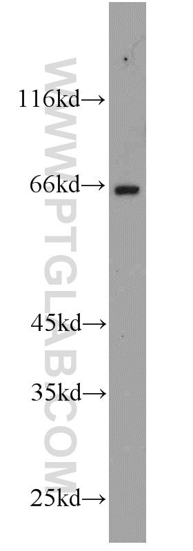
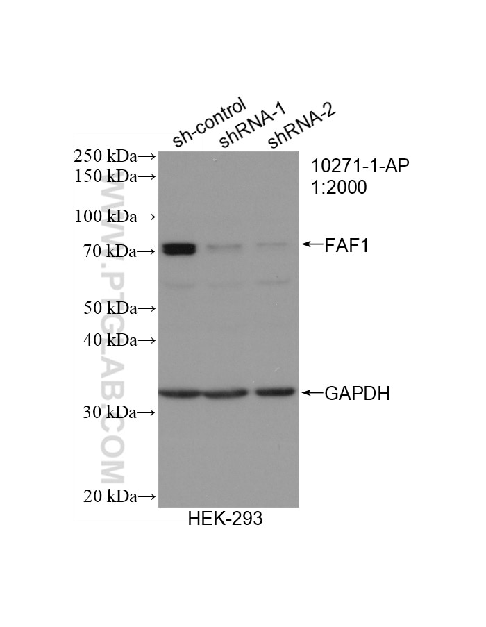
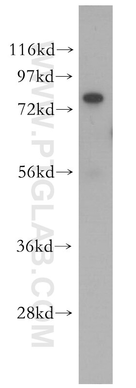
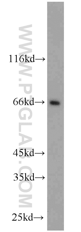
10271-1-AP targets FAF1 in WB, IF, IHC, ELISA applications and shows reactivity with human, mouse, rat samples.
| Tested Reactivity | human, mouse, rat |
| Cited Reactivity | human, mouse |
| Host / Isotype | Rabbit / IgG |
| Class | Polyclonal |
| Type | Antibody |
| Immunogen |
CatNo: Ag0407 Product name: Recombinant human FAF1 protein Source: e coli.-derived, PGEX-4T Tag: GST Domain: 268-489 aa of BC004970 Sequence: RSSPAQTREQSEEQITDVHMVSDSDGDDFEDATEFGVDDGEVFGMASSALRKSPMMPENAENEGDALLQFTAEFSSRYGDCHPVFFIGSLEAAFQEAFYVKARDRKLLAIYLHHDESVLTNVFCSQMLCAESIVSYLSQNFITWAWDLTKDSNRARFLTMCNRHFGSVVAQTIRTQKTDQFPLFLIIMGKRSSNEVLNVIQGNTTVDELMMRLMAAMEIFTA Predict reactive species |
| Full Name | Fas (TNFRSF6) associated factor 1 |
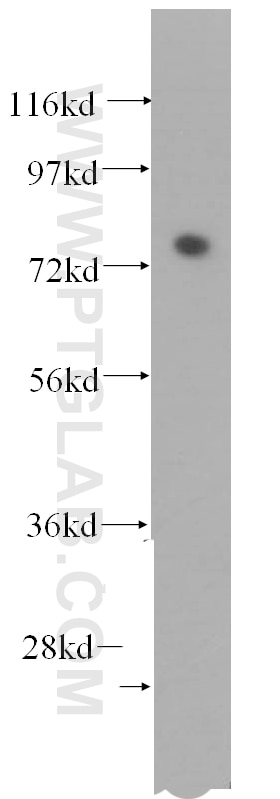
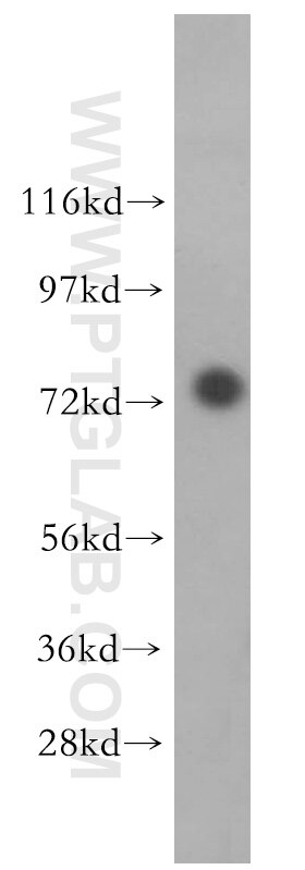
| Calculated Molecular Weight | 650aa, 74 kDa |
| Observed Molecular Weight | 74 kDa |
| GenBank Accession Number | BC004970 |
| Gene Symbol | FAF1 |
| Gene ID (NCBI) | 11124 |
| RRID | AB_2100774 |
| Conjugate | Unconjugated |
| Form | Liquid |
| Purification Method | Antigen affinity purification |
| UNIPROT ID | Q9UNN5 |
| Storage Buffer | PBS with 0.02% sodium azide and 50% glycerol, pH 7.3. |
| Storage Conditions | Store at -20°C. Stable for one year after shipment. Aliquoting is unnecessary for -20oC storage. 20ul sizes contain 0.1% BSA. |
Background Information
FAF1 was detected most abundant in testis, slightly less abundant in skeletal muscle and heart, followed by prostate, thymus, ovary, small intestine, and colon, but not in the peripheral blood leukocytes.The N-terminal region (amino acid 1∼201) including the upstream ubiquitin homology domain of hFAF1 could bind with the death domain of Fas, which mediates programmed cell death, also called apoptosis, in a number of organ systems, notably the immune and nervous systems.
Protocols
| Product Specific Protocols | |
|---|---|
| IHC protocol for FAF1 antibody 10271-1-AP | Download protocol |
| WB protocol for FAF1 antibody 10271-1-AP | Download protocol |
| Standard Protocols | |
|---|---|
| Click here to view our Standard Protocols |
Publications
| Species | Application | Title |
|---|---|---|
Cell Death Differ FAF1 mediates regulated necrosis through PARP1 activation upon oxidative stress leading to dopaminergic neurodegeneration. | ||
Hum Mol Genet Accumulation of the parkin substrate, FAF1, plays a key role in the dopaminergic neurodegeneration. | ||
Cancer Manag Res MiR-26a-5p Serves as an Oncogenic MicroRNA in Non-Small Cell Lung Cancer by Targeting FAF1.
| ||
Neuroreport Fas-associated factor 1 promotes in neurofibrillary tangle-mediated cell death of basal forebrain cholinergic neurons in P301L transgenic mice. |