
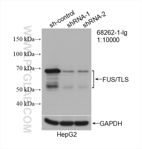
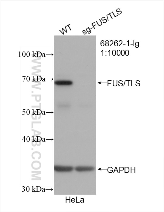
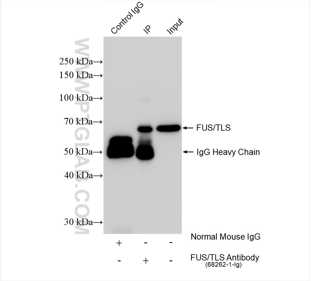
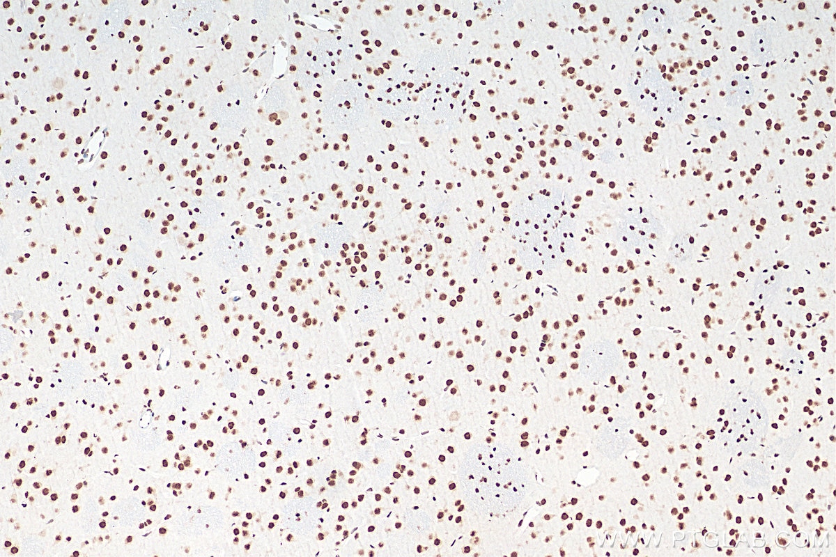

Tested Applications
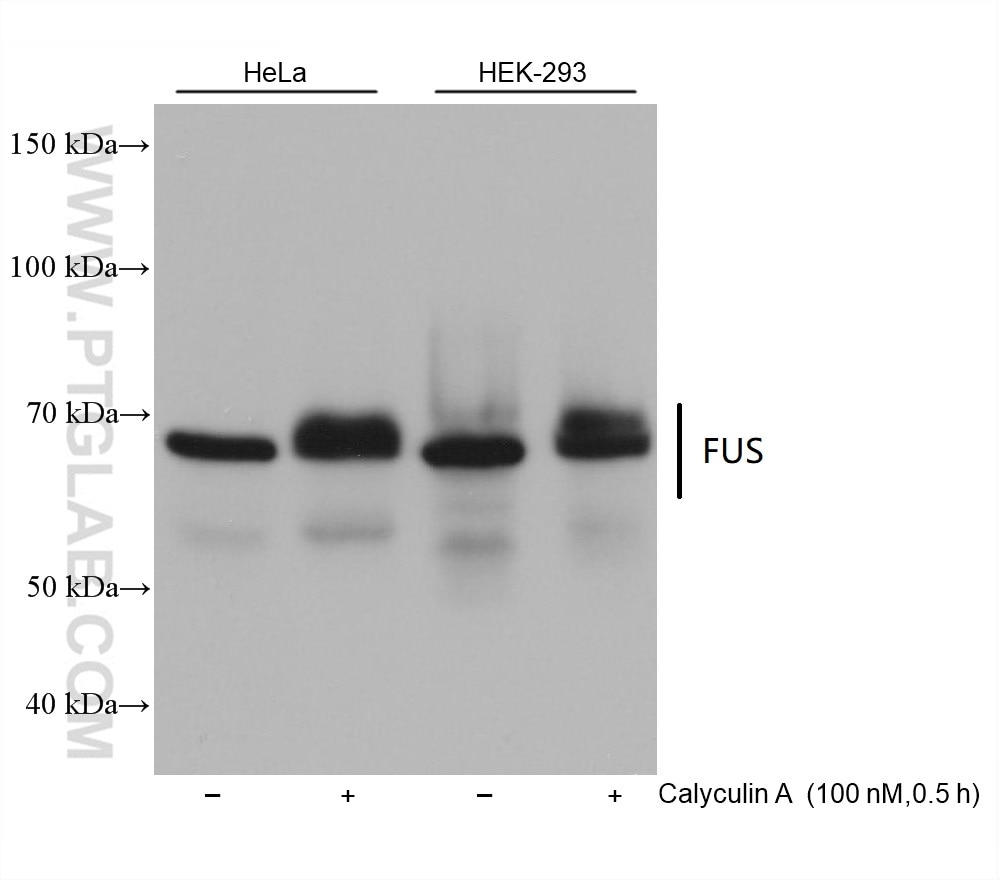
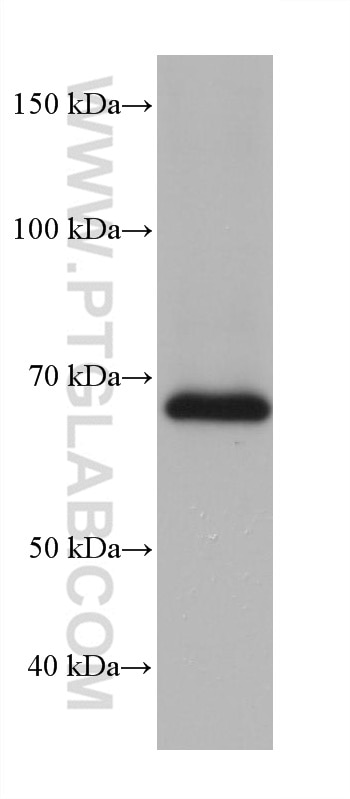
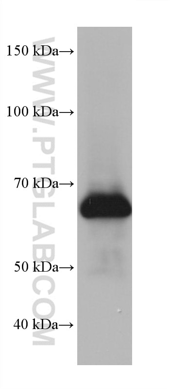
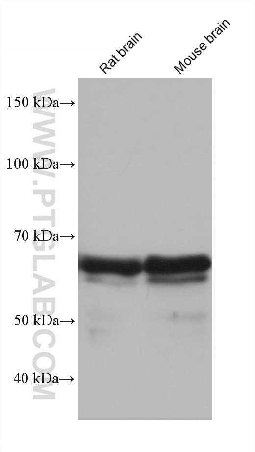
| Positive WB detected in | HeLa cells, Jurkat cells, Neuro-2a cells, rat brain tissue, HepG2 cells, HEK-293 cells, mouse brain tissue |
| Positive IP detected in | K-562 cells |
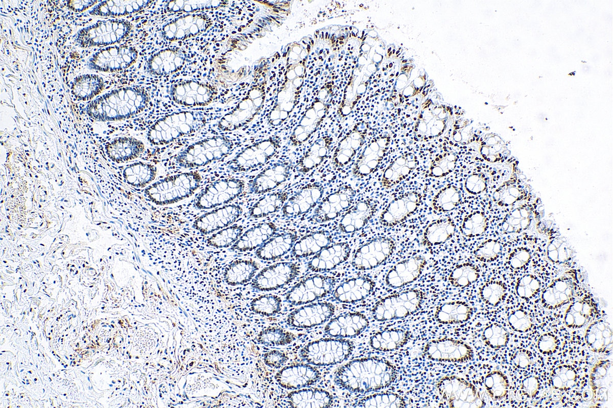
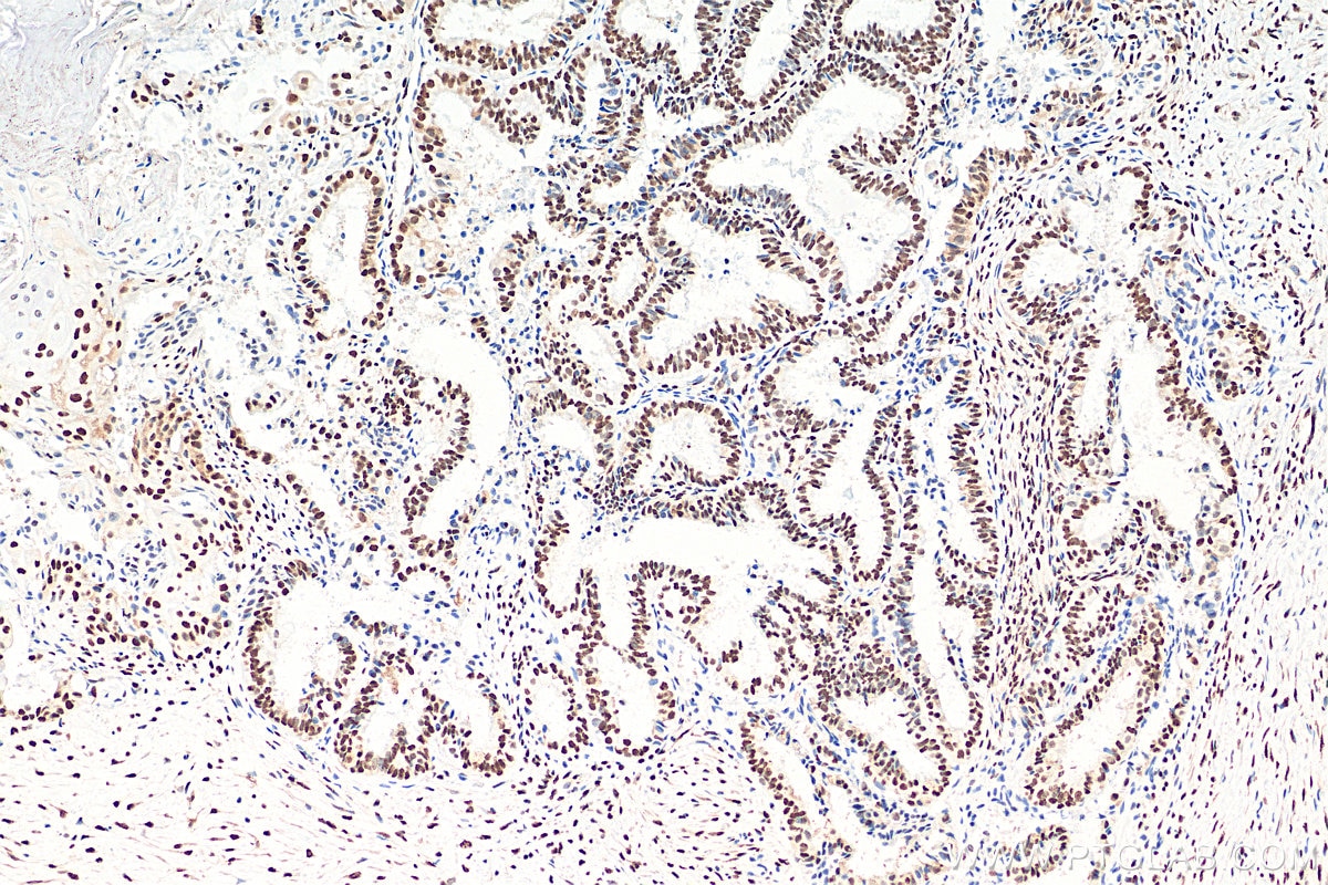
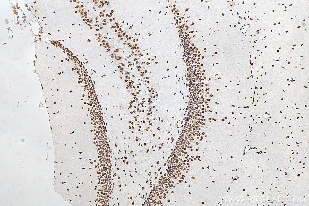
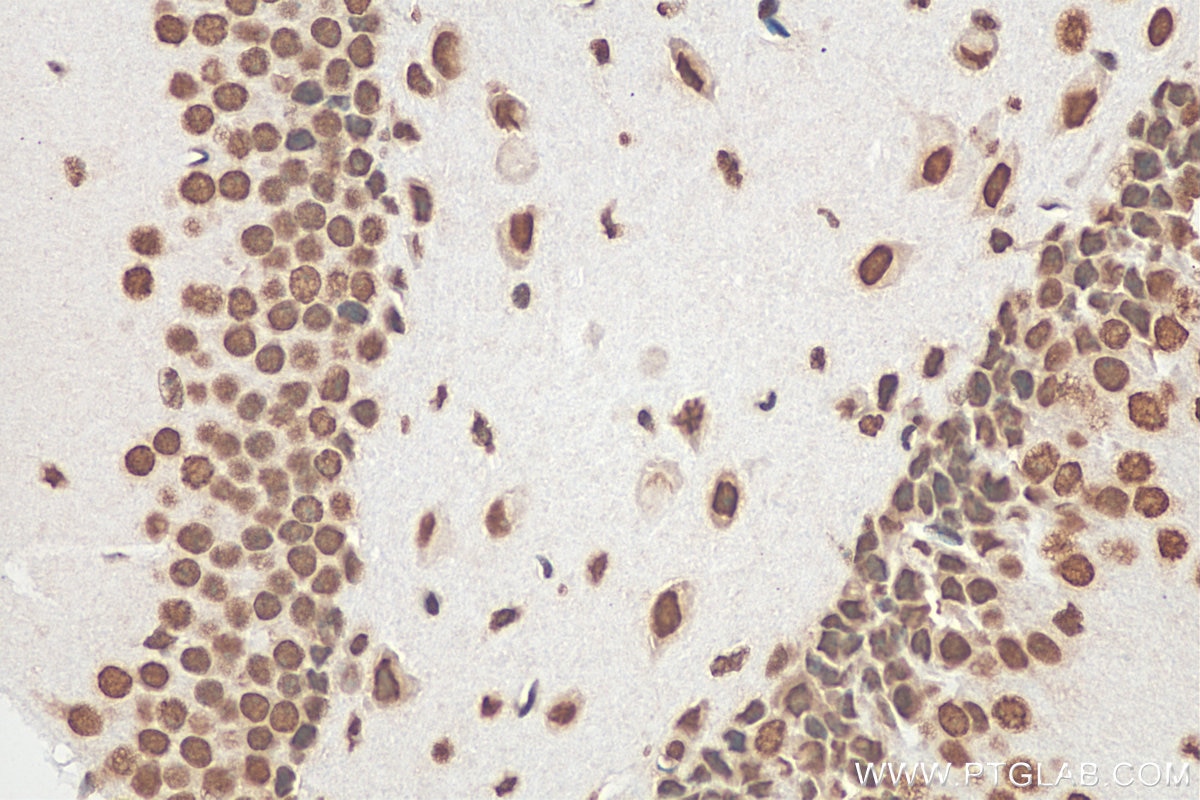
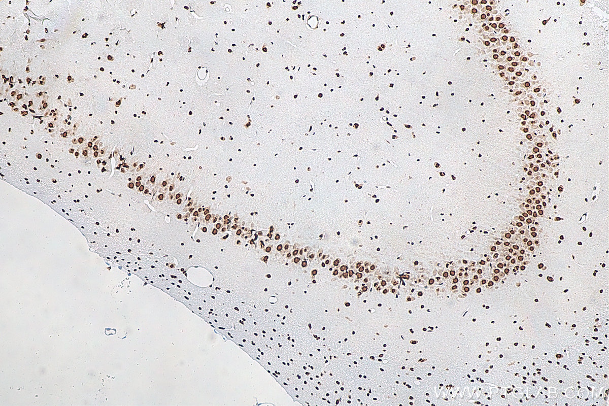
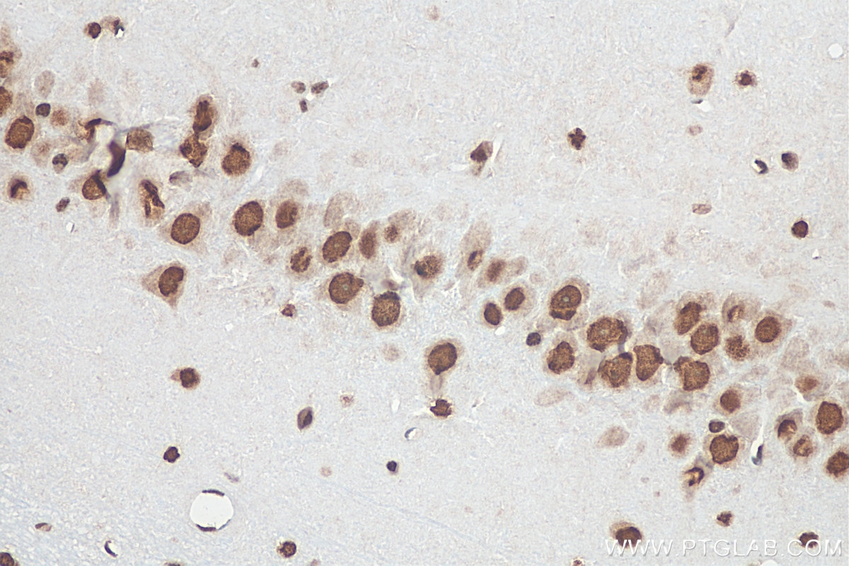
| Positive IHC detected in | rat brain tissue, human ovary tumor tissue, human colon tissue, mouse brain tissue, mouse cerebellum tissue Note: suggested antigen retrieval with TE buffer pH 9.0; (*) Alternatively, antigen retrieval may be performed with citrate buffer pH 6.0 |
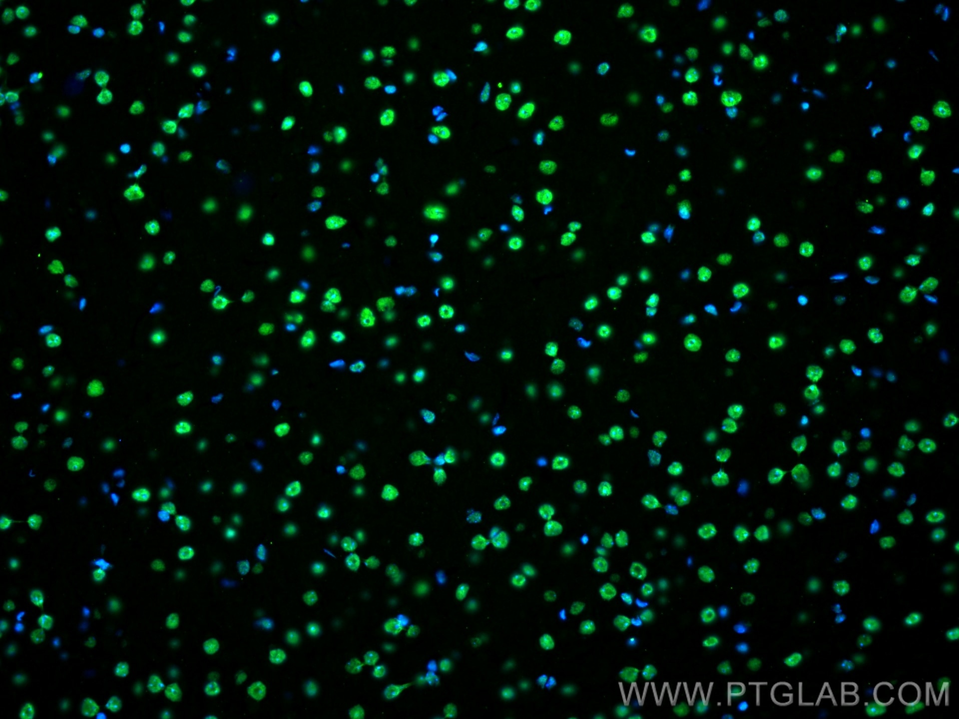
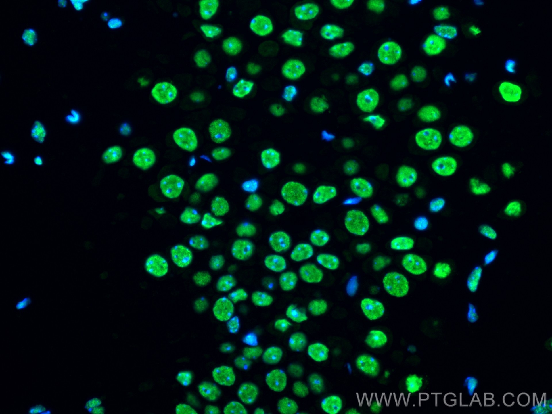
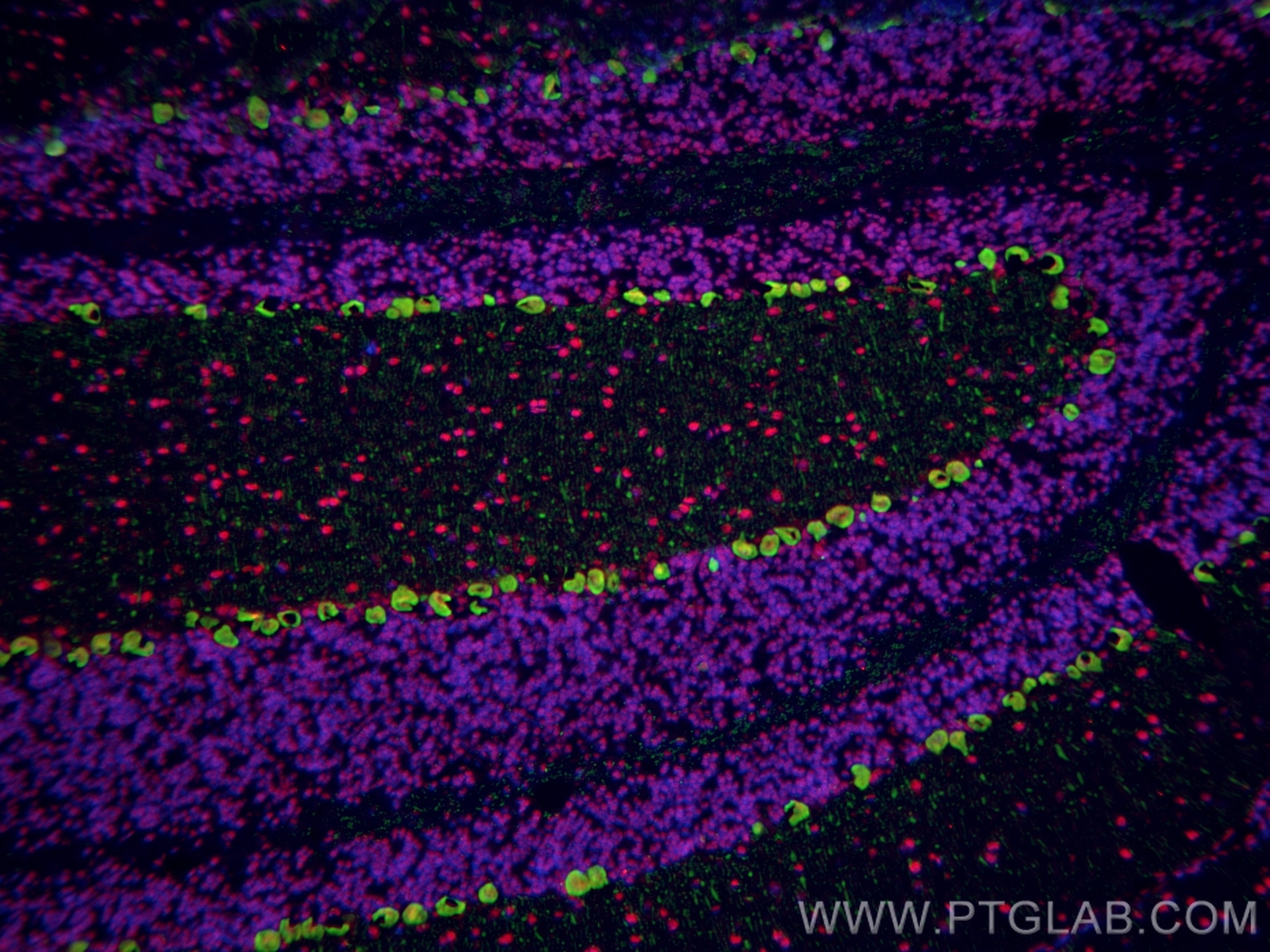
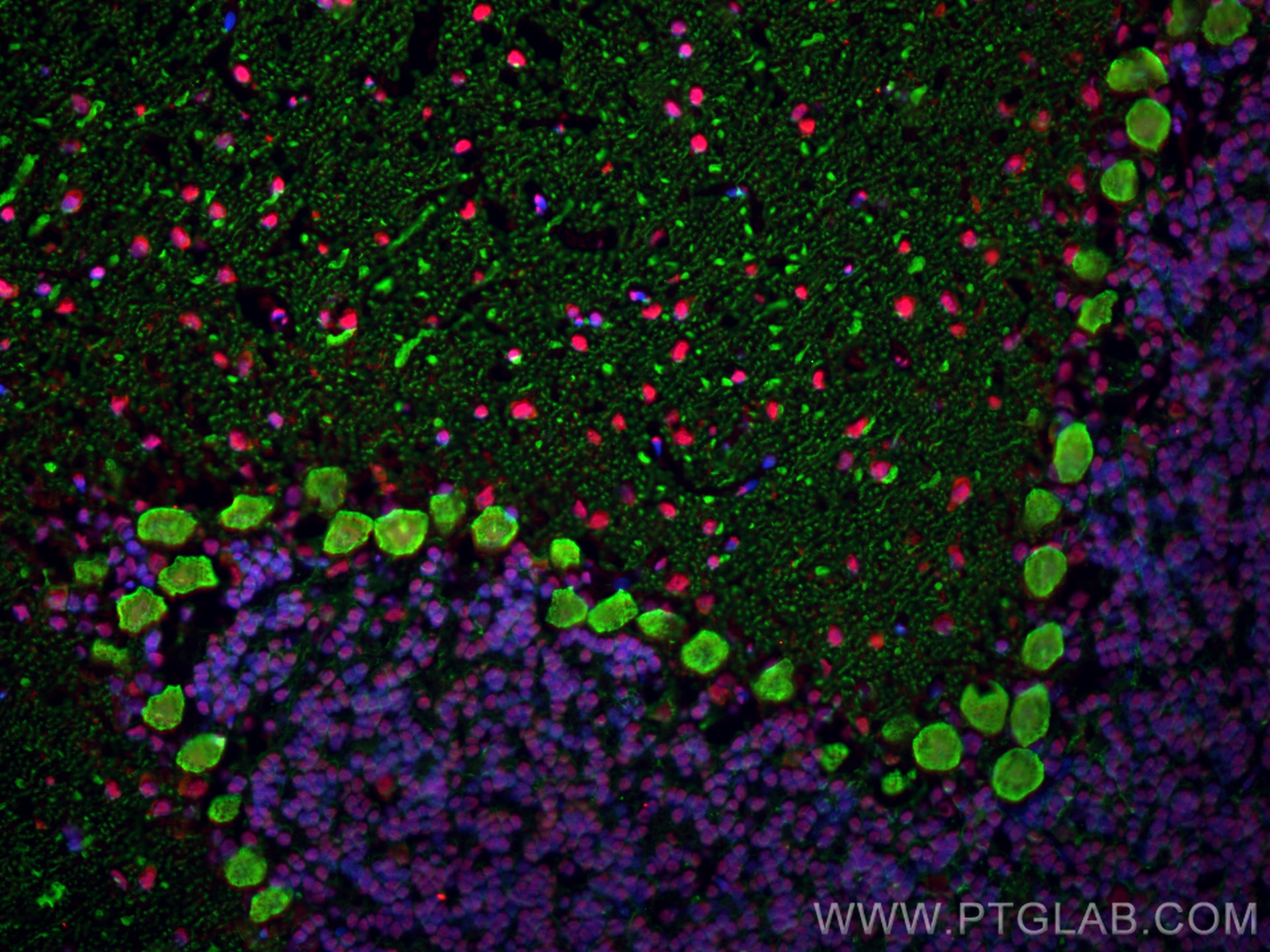
| Positive IF-P detected in | mouse brain tissue |
| Positive IF-Fro detected in | rat cerebellum tissue, mouse cerebellum tissue |
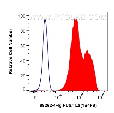
| Positive FC (Intra) detected in | HEK-293T cells |
Recommended dilution
| Application | Dilution |
|---|---|
| Western Blot (WB) | WB : 1:5000-1:50000 |
| Immunoprecipitation (IP) | IP : 0.5-4.0 ug for 1.0-3.0 mg of total protein lysate |
| Immunohistochemistry (IHC) | IHC : 1:5000-1:20000 |
| Immunofluorescence (IF)-P | IF-P : 1:200-1:800 |
| Immunofluorescence (IF)-FRO | IF-FRO : 1:200-1:800 |
| Flow Cytometry (FC) (INTRA) | FC (INTRA) : 0.40 ug per 10^6 cells in a 100 µl suspension |
| It is recommended that this reagent should be titrated in each testing system to obtain optimal results. | |
| Sample-dependent, Check data in validation data gallery. | |
Product Information
68262-1-Ig targets FUS/TLS in WB, IHC, IF-P, IF-Fro, FC (Intra), IP, ELISA applications and shows reactivity with human, mouse, rat samples.
| Tested Reactivity | human, mouse, rat |
| Cited Reactivity | human, mouse, rat, pig |
| Host / Isotype | Mouse / IgG1 |
| Class | Monoclonal |
| Type | Antibody |
| Immunogen |
CatNo: Ag2150 Product name: Recombinant human FUS/TLS protein Source: e coli.-derived, PGEX-4T Tag: GST Domain: 53-400 aa of BC026062 Sequence: SSYSSYGQSQNTGYGTQSTPQGYGSTGGYGSSQSSQSSYGQQSSYPGYGQQPAPSSTSGSYGSSSQSSSYGQPQSGSYSQQPSYGGQQQSYGQQQSYNPPQGYGQQNQYNSSSGGGGGGGGGGNYGQDQSSMSSGGGSGGGYGNQDQSGGGGSGGYGQQDRGGRGRGGSGGGGGGGGGGYNRSSGGYEPRGRGGGRGGRGGMGGSDRGGFNKFGGPRDQGSRHDSEQDNSDNNTIFVQGLGENVTIESVADYFKQIGIIKTNKKTGQPMINLYTDRETGKLKGEATVSFDDPPSAKAAIDWFDGKEFSGNPIKVSFATRRADFNRGGGNGRGGRGRGGPMGRGGYGGG Predict reactive species |
| Full Name | fusion (involved in t(12;16) in malignant liposarcoma) |
| Calculated Molecular Weight | 75 kDa |
| Observed Molecular Weight | 53 kDa, 68-75 kDa |
| GenBank Accession Number | BC026062 |
| Gene Symbol | FUS/TLS |
| Gene ID (NCBI) | 2521 |
| RRID | AB_2935347 |
| Conjugate | Unconjugated |
| Form | Liquid |
| Purification Method | Protein G purification |
| UNIPROT ID | P35637 |
| Storage Buffer | PBS with 0.02% sodium azide and 50% glycerol, pH 7.3. |
| Storage Conditions | Store at -20°C. Stable for one year after shipment. Aliquoting is unnecessary for -20oC storage. 20ul sizes contain 0.1% BSA. |
Background Information
FUS (also named TLS and POMp75) belongs to the RRM TET family. FUS may play a role in the maintenance of genomic integrity; it binds both single-stranded and double-stranded DNA and promotes ATP-independent annealing of complementary single-stranded DNAs and D-loop formation in superhelical double-stranded DNA. FUS is also an RNA-binding protein, and its links to neurodegenerative disease proffer the intriguing possibility that altered RNA metabolism or RNA processing may underlie or contribute to neuron degeneration[PMID: 22640227]. FUS may be a cause of angiomatoid fibrous histiocytoma (AFH) and is implicated in certain forms of amyotrophic lateral sclerosis (ALS) and frontotemporal dementias (FTDs) such as frontotemporal lobar dementia with ubiquitin inclusions (FTLD-U)(PMID: 22640227). Multiple phosphorylation on the N terminus of FUS caused that FUS was detected 68-75 kDa (PMID:24899704).
Protocols
| Product Specific Protocols | |
|---|---|
| FC protocol for FUS/TLS antibody 68262-1-Ig | Download protocol |
| IF protocol for FUS/TLS antibody 68262-1-Ig | Download protocol |
| IHC protocol for FUS/TLS antibody 68262-1-Ig | Download protocol |
| IP protocol for FUS/TLS antibody 68262-1-Ig | Download protocol |
| WB protocol for FUS/TLS antibody 68262-1-Ig | Download protocol |
| Standard Protocols | |
|---|---|
| Click here to view our Standard Protocols |
Publications
What published studies show
The anti-FUS antibody (Cat# 68262-1-Ig) has 14 total citations across 13 journals, including Cell, Nature Cell Biology, Nature Neuroscience, Nature Communications, Neuron, and Molecular Cell. Research fields span neurodegeneration (ALS, frontotemporal dementia), protein phase separation and condensates, RNA biology, cardiovascular disease (atherosclerosis, aortic aneurysm), gastric cancer, and mast cell-mediated inflammation. Applications are predominantly Western blot and immunofluorescence.
| Species | Application | Title |
|---|---|---|
Cell Microglial-derived C1q integrates into neuronal ribonucleoprotein complexes and impacts protein homeostasis in the aging brain | ||
Nat Cell Biol YAP maintains the dynamics of TDP-43 condensates and antagonizes TDP-43 pathological aggregates | ||
Nat Neurosci Mitochondrial respiratory complex IV deficiency recapitulates amyotrophic lateral sclerosis | ||
Mol Cell Aberrant phase separation drives membranous organelle remodeling and tumorigenesis |
Reviews
What customers say
Based on the available reviews, 68262-1-Ig has received positive feedback. One reviewer rated it 5 stars after using it at a 1:1000 dilution on HEK-293T cells for Western Blot, describing the product as "very good" and highly recommended. Overall, the limited review data suggests satisfactory performance in WB applications.
The reviews below have been submitted by verified Proteintech customers who received an incentive for providing their feedback.
Vikas (Verified Customer) (04-08-2025) | very good. Highly recomended
|




















