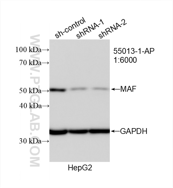
with sh-Control and sh-MAF transfected HepG2 cells.")
 at dilution of 1:4000 incubated at room temperature for 1.5 hours.")
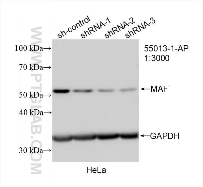
 with sh-Control and sh-MAF transfected HeLa cells.")
 at dilution of 1:300 incubated at room temperature for 1.5 hours.")
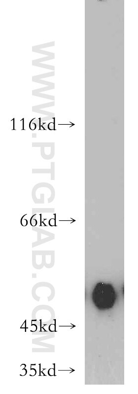
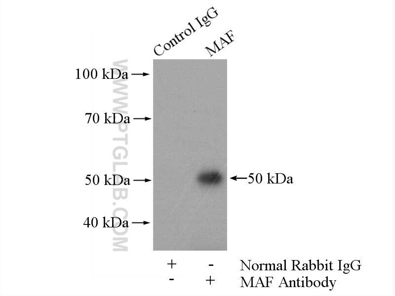
 with A431 cells lysate 2000ug.")
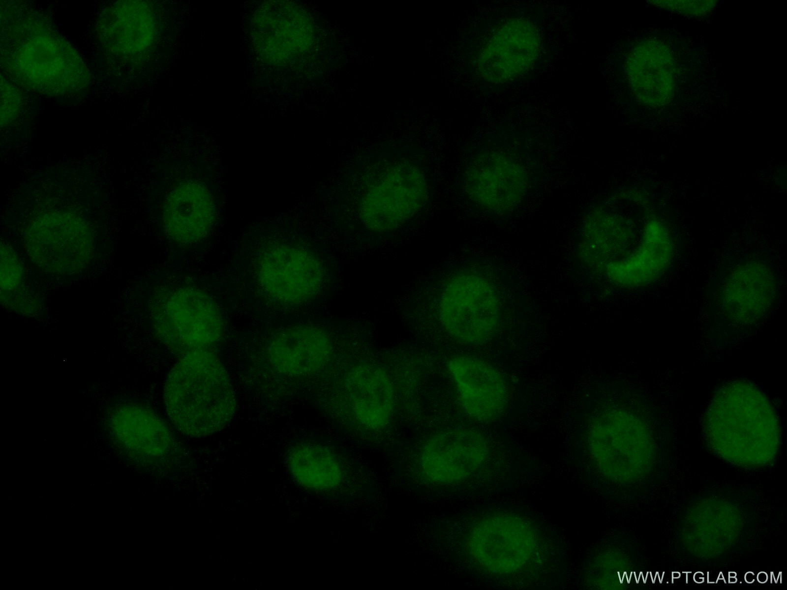
 fixed A431 cells using 55013-1-AP (c-MAF antibody) at dilution of 1:50 and Alexa Fluor 488-conjugated AffiniPure Goat Anti-Rabbit IgG(H+L).")
Tested Applications
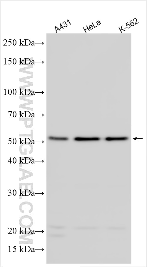
| Positive WB detected in | A431 cells, A375 cells, HeLa cells, HepG2 cells, K-562 cells |
| Positive IP detected in | A431 cells |
| Positive IF/ICC detected in | A431 cells |
Recommended dilution
| Application | Dilution |
|---|---|
| Western Blot (WB) | WB : 1:1000-1:8000 |
| Immunoprecipitation (IP) | IP : 0.5-4.0 ug for 1.0-3.0 mg of total protein lysate |
| Immunofluorescence (IF)/ICC | IF/ICC : 1:50-1:500 |
| It is recommended that this reagent should be titrated in each testing system to obtain optimal results. | |
| Sample-dependent, Check data in validation data gallery. | |
Published Applications
| KD/KO | See 1 publications below |
| WB | See 12 publications below |
| IF | See 3 publications below |
| IP | See 1 publications below |
Product Information
55013-1-AP targets c-MAF in WB, IF/ICC, IP, ELISA applications and shows reactivity with human, mouse, rat samples.
| Tested Reactivity | human, mouse, rat |
| Cited Reactivity | human, mouse |
| Host / Isotype | Rabbit / IgG |
| Class | Polyclonal |
| Type | Antibody |
| Immunogen |
Peptide Predict reactive species |
| Full Name | v-maf musculoaponeurotic fibrosarcoma oncogene homolog (avian) |
| Calculated Molecular Weight | 42 kDa |
| Observed Molecular Weight | 42-52 kDa |
| GenBank Accession Number | NM_005360 |
| Gene Symbol | MAF |
| Gene ID (NCBI) | 4094 |
| RRID | AB_10863127 |
| Conjugate | Unconjugated |
| Form | Liquid |
| Purification Method | Antigen affinity purification |
| UNIPROT ID | O75444 |
| Storage Buffer | PBS with 0.02% sodium azide and 50% glycerol, pH 7.3. |
| Storage Conditions | Store at -20°C. Stable for one year after shipment. Aliquoting is unnecessary for -20oC storage. 20ul sizes contain 0.1% BSA. |
Background Information
MAF, also named as c-Maf, belongs to the bZIP family and Maf subfamily. MAF acts as a transcriptional activator or repressor. It is involved in embryonic lens fiber cell development. MAF increases T cell susceptibility to apoptosis by interacting with MYB and decreasing BCL2 expression. Together with PAX6, it transactivates strongly the glucagon gene promoter through the G1 element. MAF activates transcription of the CD13 proximal promoter in endothelial cells. It is involved in the initial chondrocyte terminal differentiation and the disappearance of hypertrophic chondrocytes during endochondral bone development. When overexpressed, MAF represses anti-oxidant reponse element (ARE)-mediated transcription. It is involved either as an oncogene or as a tumor suppressor, depending on the cell context. A chromosomal aberration involving MAF is found in some forms of multiple myeloma (MM). Defects in MAF are the cause of cataract pulverulent juvenile-onset MAF-related (CAPJOM). Defects in MAF are the cause of cataract congenital cerulean type 4 (CCA4). The antibody is specific to MAF. And it could recognise the 50 kDa band that also be detected in the paper (PMID: 25770584 ) .
Protocols
| Product Specific Protocols | |
|---|---|
| IF protocol for c-MAF antibody 55013-1-AP | Download protocol |
| IP protocol for c-MAF antibody 55013-1-AP | Download protocol |
| WB protocol for c-MAF antibody 55013-1-AP | Download protocol |
| Standard Protocols | |
|---|---|
| Click here to view our Standard Protocols |
Publications
| Species | Application | Title |
|---|---|---|
Nat Neurosci Spinal cord Tau pathology induces tactile deficits and cognitive impairment in Alzheimer's disease via dysregulation of CCK neurons
| ||
Immunity RAS P21 Protein Activator 3 (RASA3) Specifically Promotes Pathogenic T Helper 17 Cell Generation by Repressing T-Helper-2-Cell-Biased Programs. | ||
Acta Pharmacol Sin Mebendazole elicits potent antimyeloma activity by inhibiting the USP5/c-Maf axis. | ||
Int J Mol Sci Deletion of Cdk5 in Macrophages Ameliorates Anti-Inflammatory Response during Endotoxemia through Induction of C-Maf and Il-10 | ||
Front Immunol Molecular Mechanisms Driving IL-10- Producing B Cells Functions: STAT3 and c-MAF as Underestimated Central Key Regulators? | ||
J Immunol Malat1 Suppresses Immunity to Infection through Promoting Expression of Maf and IL-10 in Th Cells. |
Reviews
The reviews below have been submitted by verified Proteintech customers who received an incentive for providing their feedback.
FH Katie (Verified Customer) (04-08-2022) | Works well for western blot. We have published with this antibody
|
FH H (Verified Customer) (04-05-2020) | This protein has a very short half life, and present in the nucleus. I could see a clear band after the nuclear-cytosolic fractionation.
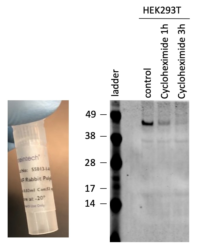 |