at dilution of 1:1500 incubated at room temperature for 1.5 hours.")
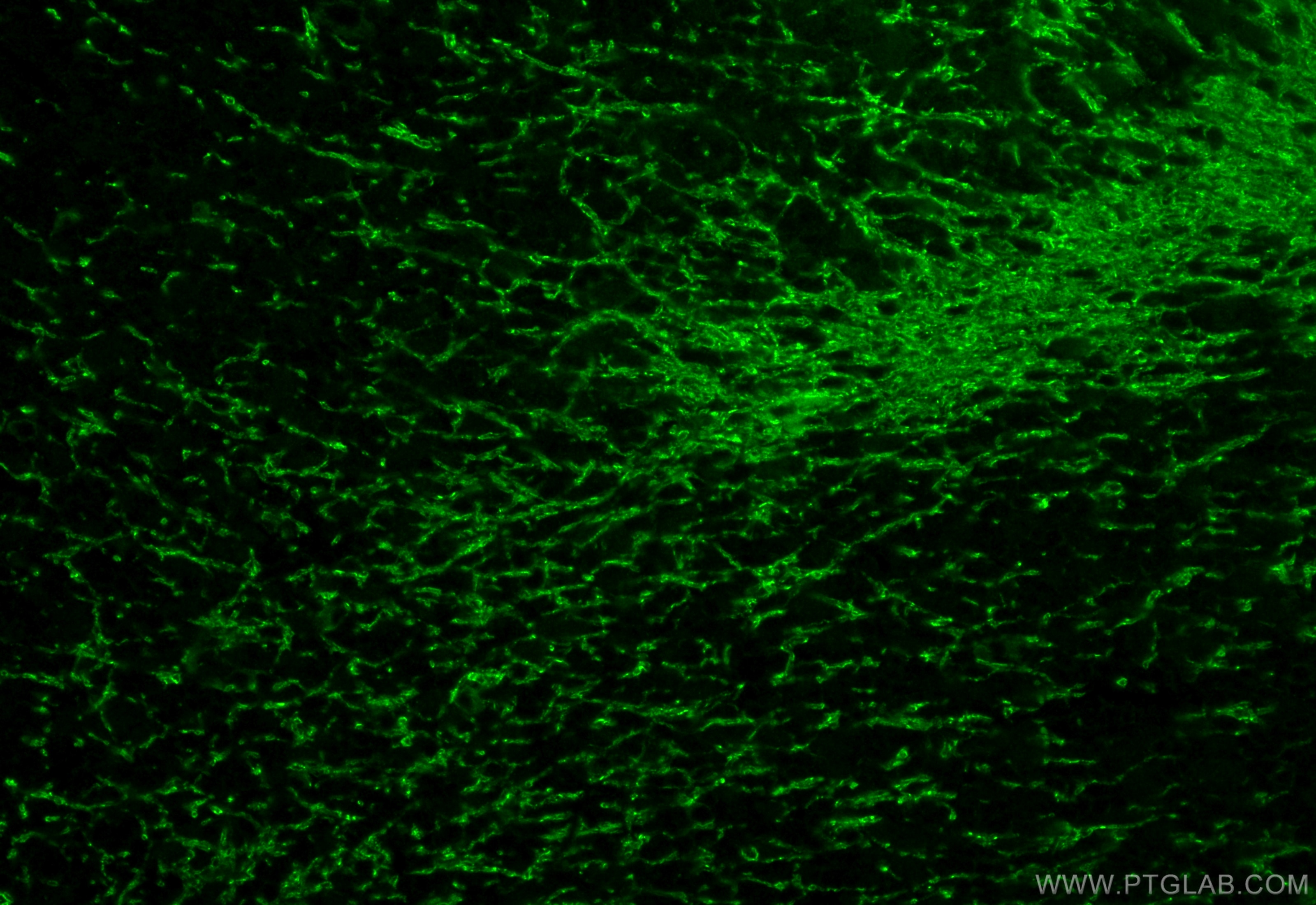
 fixed paraffin-embedded rat cerebellum tissue using MOG antibody (12690-1-AP) at dilution of 1:200 and CoraLite®488-Conjugated Goat Anti-Rabbit IgG(H+L) (SA00013-2). Heat mediated antigen retrieval with Tris-EDTA buffer (pH 9.0).")
Tested Applications
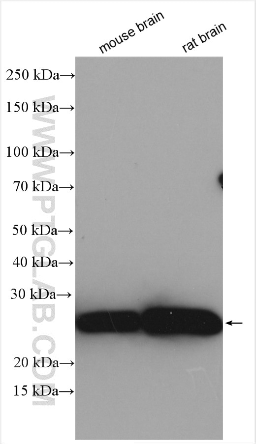
| Positive WB detected in | mouse brain tissue, rat brain tissue |
| Positive IF-P detected in | rat cerebellum tissue |
Recommended dilution
| Application | Dilution |
|---|---|
| Western Blot (WB) | WB : 1:500-1:3000 |
| Immunofluorescence (IF)-P | IF-P : 1:50-1:500 |
| It is recommended that this reagent should be titrated in each testing system to obtain optimal results. | |
| Sample-dependent, Check data in validation data gallery. | |
Published Applications
| WB | See 17 publications below |
| IHC | See 4 publications below |
| IF | See 6 publications below |
Product Information
12690-1-AP targets MOG in WB, IHC, IF-P, ELISA applications and shows reactivity with human, mouse, rat samples.
| Tested Reactivity | human, mouse, rat |
| Cited Reactivity | human, mouse, rat |
| Host / Isotype | Rabbit / IgG |
| Class | Polyclonal |
| Type | Antibody |
| Immunogen |
CatNo: Ag3273 Product name: Recombinant human MOG protein Source: e coli.-derived, PGEX-4T Tag: GST Domain: 1-295 aa of BC035938 Sequence: MASLSRPSLPSCLCSFLLLLLLQVSSSYAGQFRVIGPRHPIRALVGDEVELPCRISPGKNATGMEVGWYRPPFSRVVHLYRNGKDQDGDQAPEYRGRTELLKDAIGEGKVTLRIRNVRFSDEGGFTCFFRDHSYQEEAAMELKVEDPFYWVSPGVLVLLAVLPVLLLQITVGLVFLCLQYRLRGKLRAEIENLHRTFDPHFLRVPCWKITLFVIVPVLGPLVALIICYNWLHRRLAGQFLEELRKFSSLCYKQRIKSQERETEATRGRGGLLRDHIPRGKEELESLGGGKTPPGR Predict reactive species |
| Full Name | myelin oligodendrocyte glycoprotein |
| Calculated Molecular Weight | 295 aa, 34 kDa |
| Observed Molecular Weight | 25-28 kDa |
| GenBank Accession Number | BC035938 |
| Gene Symbol | MOG |
| Gene ID (NCBI) | 4340 |
| RRID | AB_2145527 |
| Conjugate | Unconjugated |
| Form | Liquid |
| Purification Method | Antigen affinity purification |
| UNIPROT ID | Q16653 |
| Storage Buffer | PBS with 0.02% sodium azide and 50% glycerol, pH 7.3. |
| Storage Conditions | Store at -20°C. Stable for one year after shipment. Aliquoting is unnecessary for -20oC storage. 20ul sizes contain 0.1% BSA. |
Background Information
Myelin/oligodendrocyte glycoprotein (MOG), a 23~28 kDa glycoprotein, a myelin antigen at the outer surface of the central nervous system (CNS) myelin sheath, which may trigger T-cell as well as B-cell responses. It therefore constitutes a pivotal target for autoimmune responses, which result in inflammation and also demyelination in the CNS. Its presence on the outer- most lamellae of mature CNS myelin and its late appearance during myelinogenesis suggest that it contributes to myelin maturation or maintenance. 10 isoforms of MOG produced by alternative splicing have been described, and heterodimers may be formed between the different isoforms. Defects in MOG are the cause of narcolepsy type 7 (NRCLP7), a neurological disabling sleep disorder characterized by excessive daytime sleepiness, sleep fragmentation, symptoms of abnormal rapid-eye-movement (REM) sleep, cataplexy, hypnagogic hallucinations, and sleep paralysis. Role of MOG in the pathogenesis of multiple sclerosis (MS) has been reported but remains to be clarified.
Protocols
| Product Specific Protocols | |
|---|---|
| IF protocol for MOG antibody 12690-1-AP | Download protocol |
| WB protocol for MOG antibody 12690-1-AP | Download protocol |
| Standard Protocols | |
|---|---|
| Click here to view our Standard Protocols |
Publications
| Species | Application | Title |
|---|---|---|
J Cereb Blood Flow Metab Agomelatine promotes differentiation of oligodendrocyte precursor cells and preserves white matter integrity after cerebral ischemic stroke | ||
Int J Mol Sci The Distribution of GPR17-Expressing Cells Correlates with White Matter Inflammation Status in Brain Tissues of Multiple Sclerosis Patients. | ||
Glia ST8SIA2 promotes oligodendrocyte differentiation and the integrity of myelin and axons. | ||
J Neuroinflammation Interleukin-9 regulates macrophage activation in the progressive multiple sclerosis brain. | ||
Brain Pathol Overexpression of the ubiquitin-editing enzyme A20 in the brain lesions of Multiple Sclerosis patients: moving from systemic to central nervous system inflammation. |
Reviews
The reviews below have been submitted by verified Proteintech customers who received an incentive for providing their feedback.
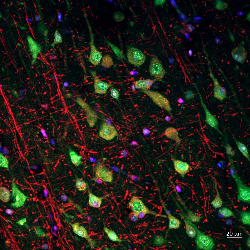
FH Reyes (Verified Customer) (03-01-2024) | MOG (in red) worked nicely showing the mielinated axons in my paraffin human brain cortex sections in IF
 |