at dilution of 1:1000 incubated at room temperature for 1.5 hours.")
 at dilution of 1:500 incubated at room temperature for 1.5 hours.")
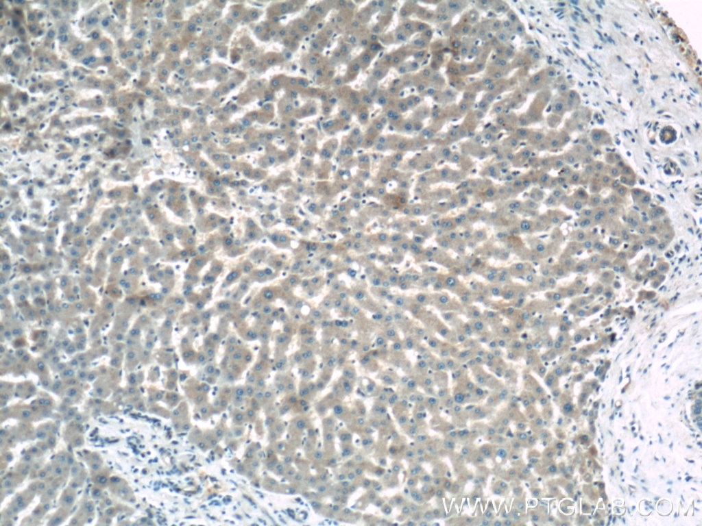
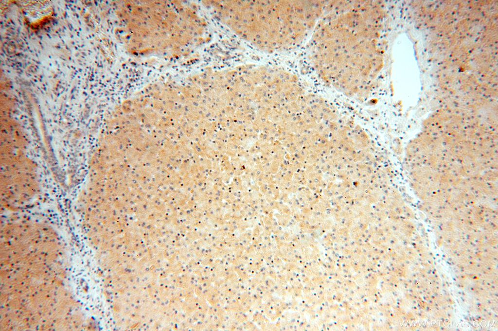
 at dilution of 1:200 (under 10x lens).")
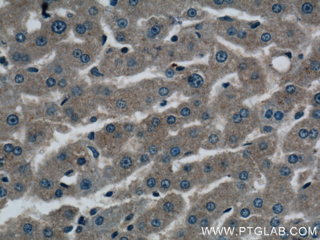
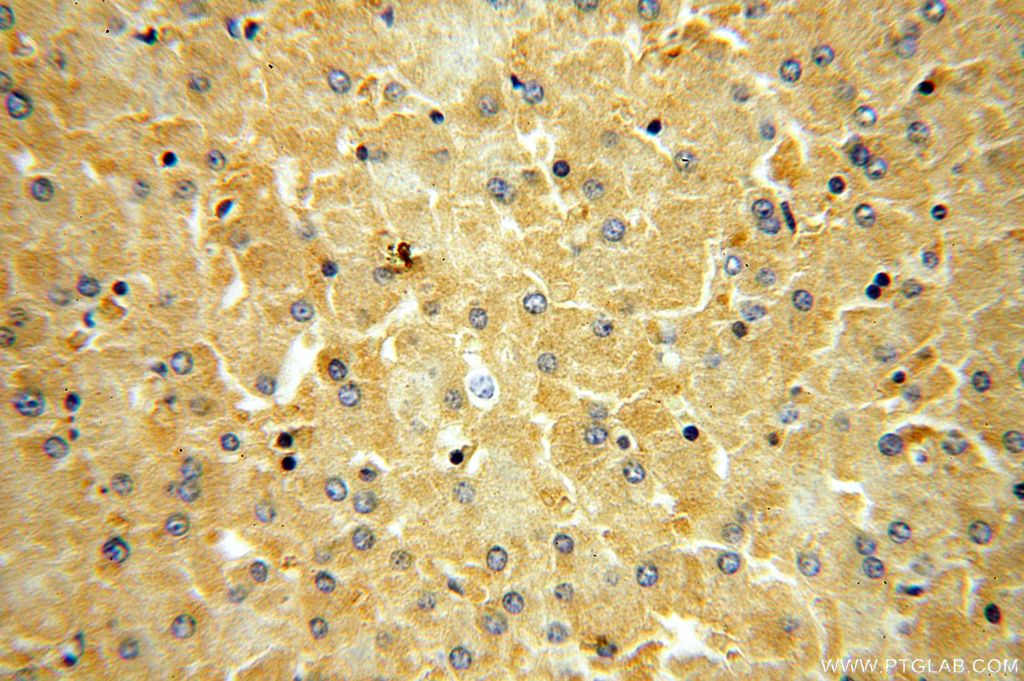
 at dilution of 1:200 (under 40x lens).")
 at dilution of 1:100 (under 10x lens).")
 at dilution of 1:100 (under 40x lens).")
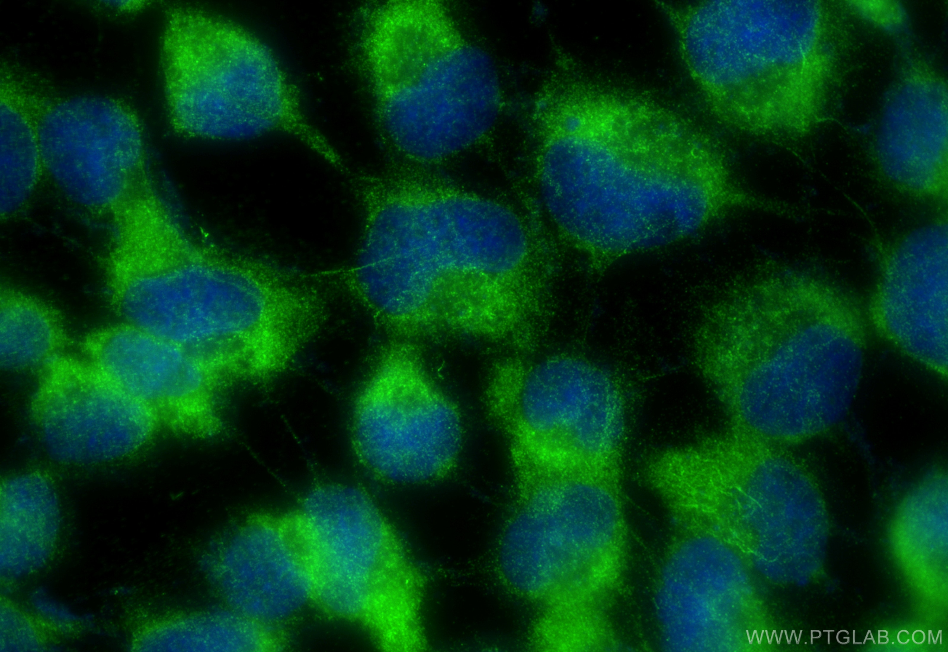
 fixed A431 cells using OAS1/3 antibody (14955-1-AP) at dilution of 1:400 and CoraLite®488-Conjugated Goat Anti-Rabbit IgG(H+L) (SA00013-2).")
Tested Applications
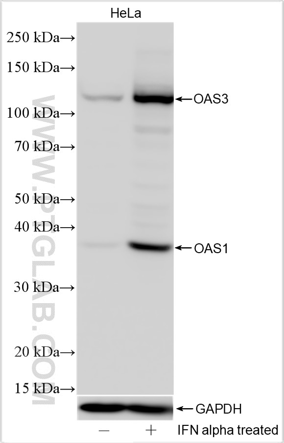
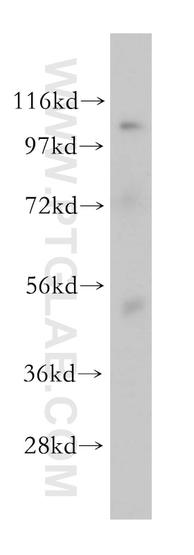
| Positive WB detected in | IFN alpha treated HeLa cells, mouse brain tissue |
| Positive IHC detected in | human liver tissue Note: suggested antigen retrieval with TE buffer pH 9.0; (*) Alternatively, antigen retrieval may be performed with citrate buffer pH 6.0 |
| Positive IF/ICC detected in | A431 cells |
Recommended dilution
| Application | Dilution |
|---|---|
| Western Blot (WB) | WB : 1:500-1:2000 |
| Immunohistochemistry (IHC) | IHC : 1:100-1:400 |
| Immunofluorescence (IF)/ICC | IF/ICC : 1:200-1:800 |
| It is recommended that this reagent should be titrated in each testing system to obtain optimal results. | |
| Sample-dependent, Check data in validation data gallery. | |
Published Applications
| KD/KO | See 3 publications below |
| WB | See 30 publications below |
| IHC | See 4 publications below |
| IF | See 2 publications below |
| CoIP | See 1 publications below |
Product Information
14955-1-AP targets OAS1/3 in WB, IHC, IF/ICC, CoIP, ELISA applications and shows reactivity with human, mouse samples.
| Tested Reactivity | human, mouse |
| Cited Reactivity | human, mouse, rat, pig |
| Host / Isotype | Rabbit / IgG |
| Class | Polyclonal |
| Type | Antibody |
| Immunogen |
CatNo: Ag6793 Product name: Recombinant human OAS1 protein Source: e coli.-derived, PGEX-4T Tag: GST Domain: 1-351 aa of BC000562 Sequence: MMDLRNTPAKSLDKFIEDYLLPDTCFRMQINHAIDIICGFLKERCFRGSSYPVCVSKVVKGGSSGKGTTLRGRSDADLVVFLSPLTTFQDQLNRRGEFIQEIRRQLEACQRERAFSVKFEVQAPRWGNPRALSFVLSSLQLGEGVEFDVLPAFDALGQLTGSYKPNPQIYVKLIEECTDLQKEGEFSTCFTELQRDFLKQRPTKLKSLIRLVKHWYQNCKKKLGKLPPQYALELLTVYAWERGSMKTHFNTAQGFRTVLELVINYQQLCIYWTKYYDFKNPIIEKYLRRQLTKPRPVILDPADPTGNLGGGDPKGWRQLAQEAEAWLNYPCFKNWDGSPVSSWILLVRPPA Predict reactive species |
| Full Name | 2',5'-oligoadenylate synthetase 1, 40/46kDa |
| Calculated Molecular Weight | 46 kDa |
| Observed Molecular Weight | ~40 kDa, 100-120 kDa |
| GenBank Accession Number | BC000562 |
| Gene Symbol | OAS1 |
| Gene ID (NCBI) | 4938 |
| RRID | AB_2158292 |
| Conjugate | Unconjugated |
| Form | Liquid |
| Purification Method | Antigen affinity purification |
| UNIPROT ID | P00973 |
| Storage Buffer | PBS with 0.02% sodium azide and 50% glycerol, pH 7.3. |
| Storage Conditions | Store at -20°C. Stable for one year after shipment. Aliquoting is unnecessary for -20oC storage. 20ul sizes contain 0.1% BSA. |
Background Information
The 2-prime,5-prime oligoadenylate synthetases (OASs), such as OAS1, are interferon-induced proteins characterized by their capacity to catalyze the synthesis of 2-prime,5-prime oligomers of adenosine (2-5As). OAS1 (type I enzymes) has some isoforms with the MW of 40, 42, 44, 46, and 48 kDa (PMID:12590567, 19383565). OAS1 is a strong candidate for determining susceptibility or resistance to viral infections(PMID:15732009). This antibody may have cross-reaction with OAS3 protein due to the high homology.
Protocols
| Product Specific Protocols | |
|---|---|
| IF protocol for OAS1/3 antibody 14955-1-AP | Download protocol |
| IHC protocol for OAS1/3 antibody 14955-1-AP | Download protocol |
| WB protocol for OAS1/3 antibody 14955-1-AP | Download protocol |
| Standard Protocols | |
|---|---|
| Click here to view our Standard Protocols |
Publications
| Species | Application | Title |
|---|---|---|
Nat Cell Biol Redox regulation of m6A methyltransferase METTL3 in β-cells controls the innate immune response in type 1 diabetes | ||
Adv Sci (Weinh) The CRISPR-Cas13a Gene-Editing System Induces Collateral Cleavage of RNA in Glioma Cells. | ||
PLoS Biol Resurrection of 2'-5'-oligoadenylate synthetase 1 (OAS1) from the ancestor of modern horseshoe bats blocks SARS-CoV-2 replication | ||
Autophagy USP19 (ubiquitin specific peptidase 19) promotes TBK1 (TANK-binding kinase 1) degradation via chaperone-mediated autophagy. |
Reviews
The reviews below have been submitted by verified Proteintech customers who received an incentive for providing their feedback.
FH Seong (Verified Customer) (11-27-2018) | OAS1 polyclonal antibody (14955-1-AP) did work by Western Blot (1:1,500) of ARPE-19 cells induced with interferon gamma but the band was very weak in uninduced cells.
|