Tested Applications
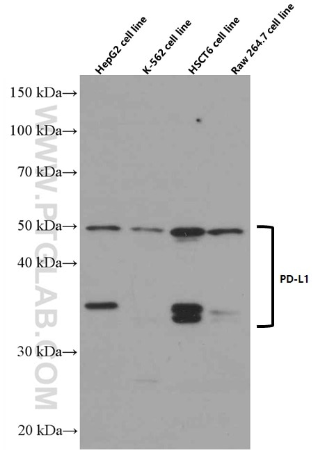
| Positive WB detected in | A375 cells, human placenta tissue, pig lung tissue, human skeletal muscle tissue, HepG2 cells, THP-1 cells, RAW 264.7 cells, A549 cells, K-562 cells, HSC-T6 cells |
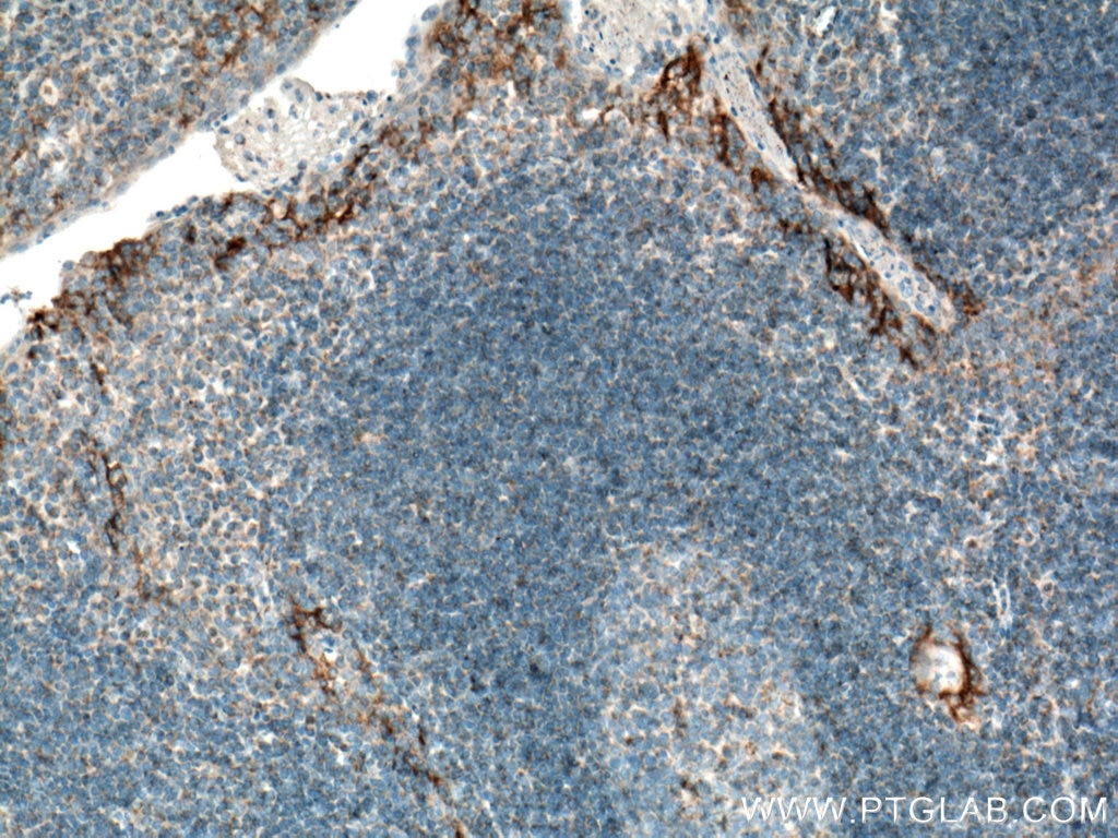
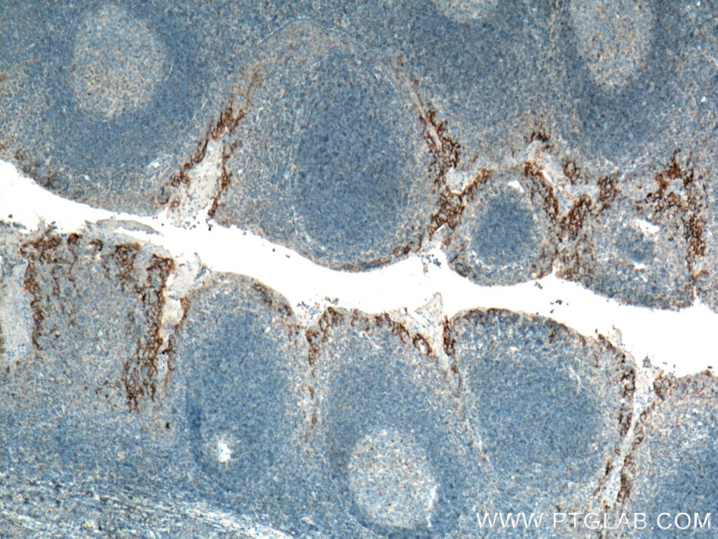
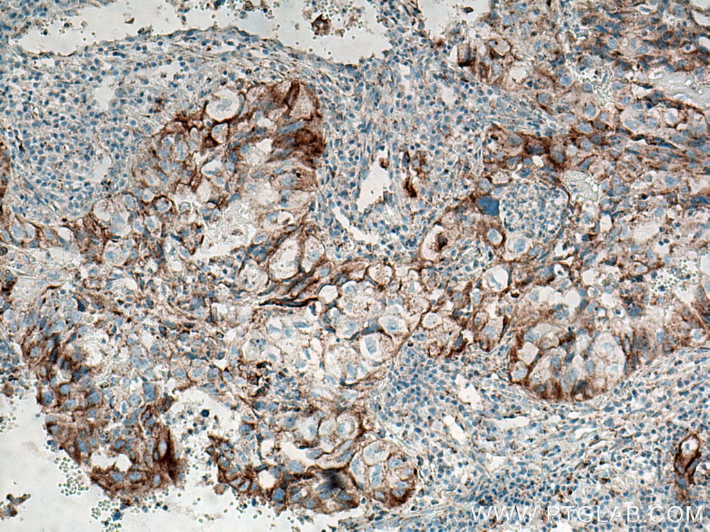
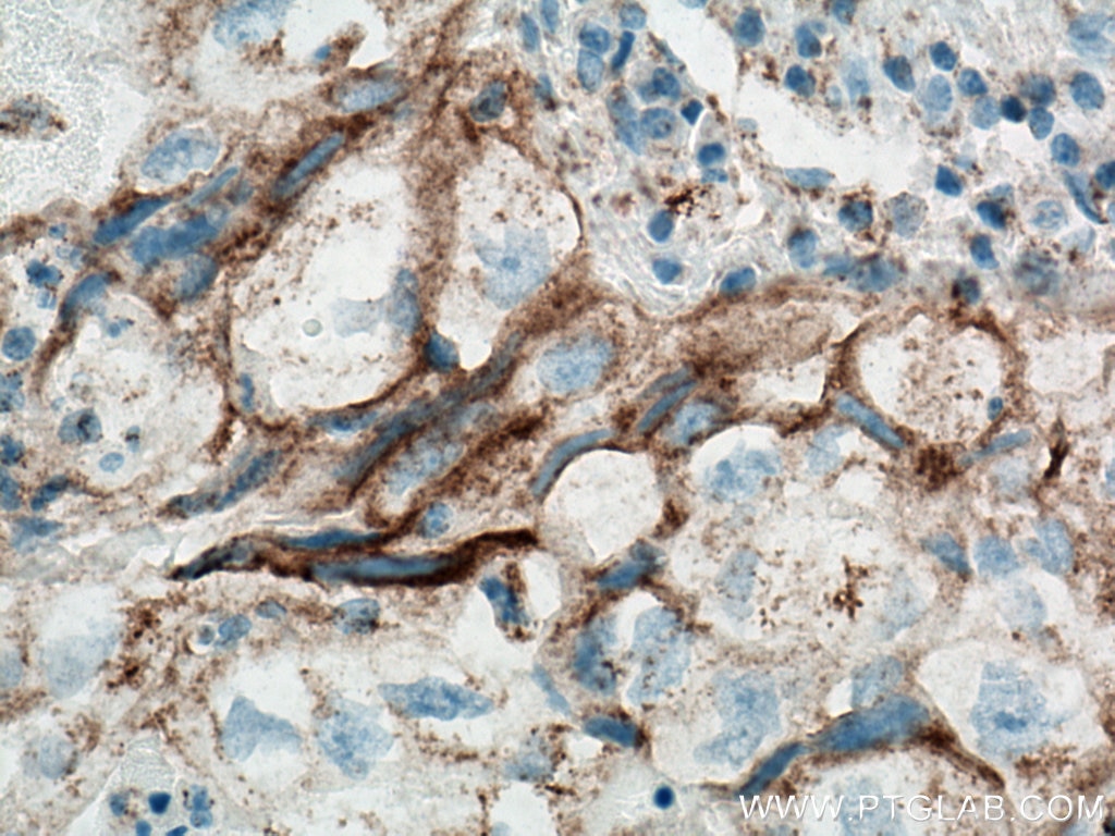
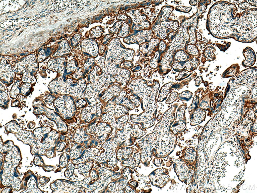
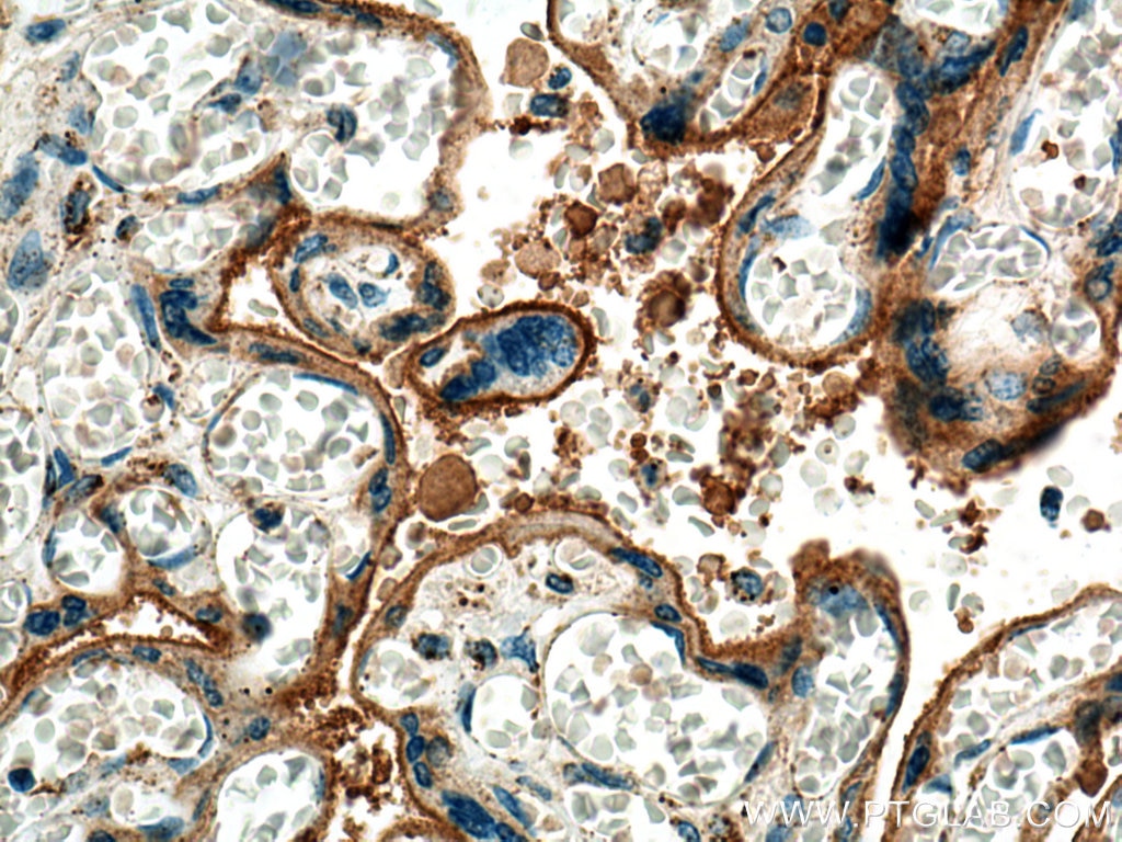
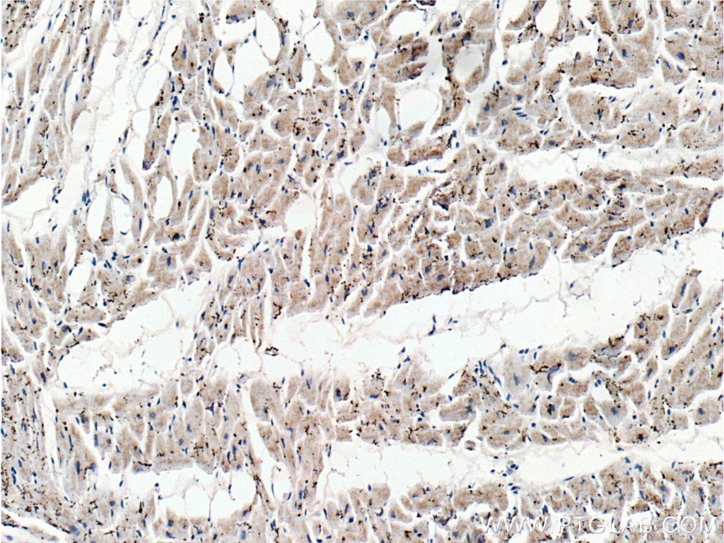
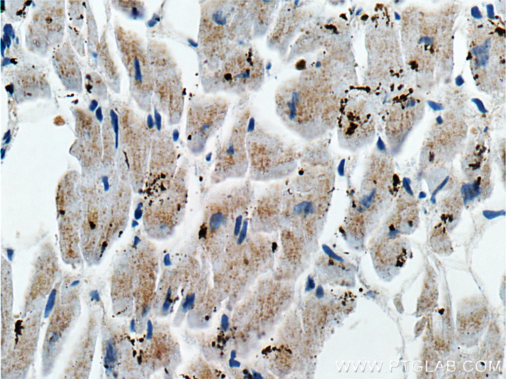
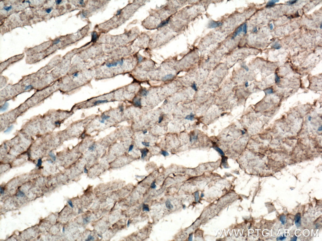
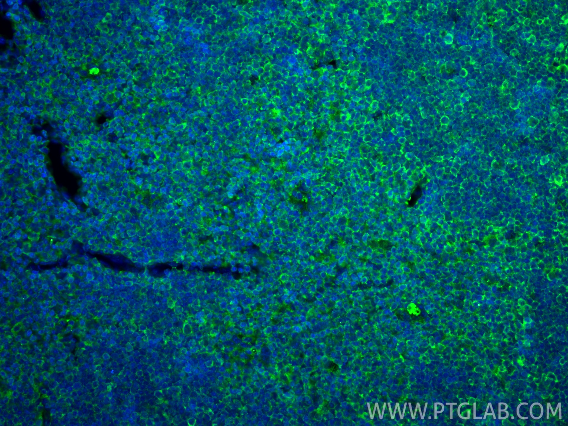
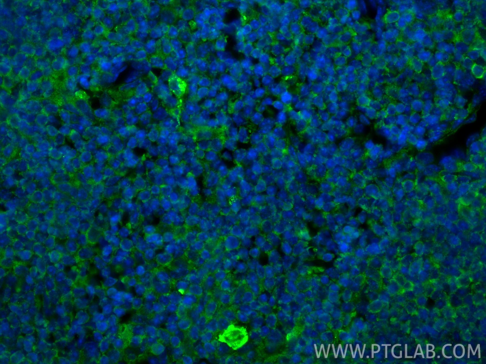
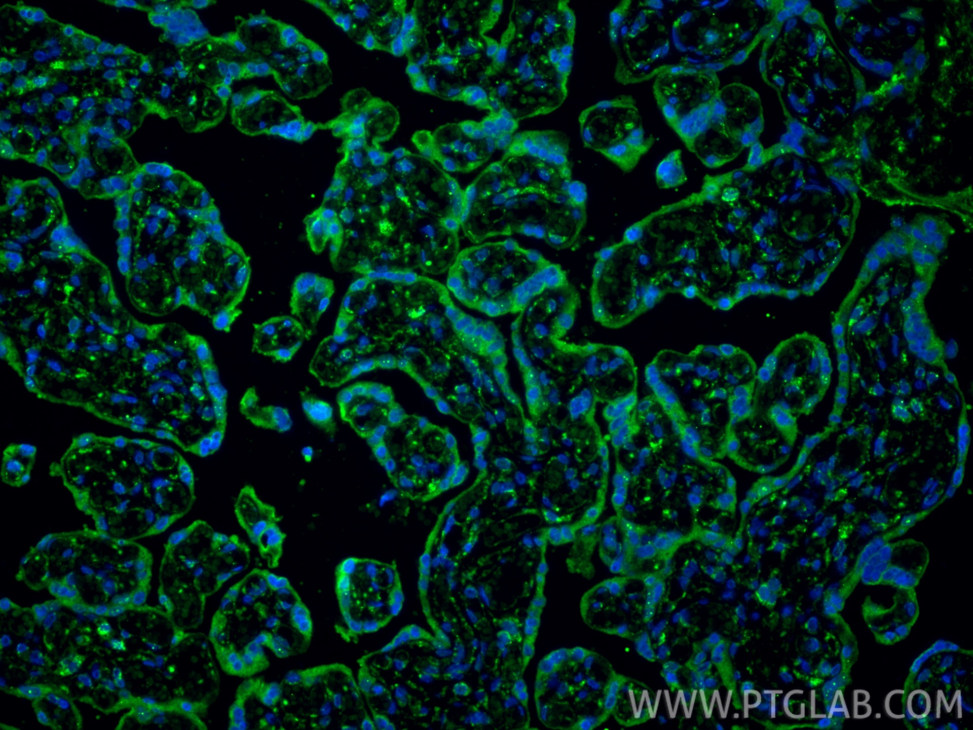
| Positive IHC detected in | human tonsillitis tissue, human heart tissue, human lung cancer tissue, human placenta tissue, mouse heart tissue Note: suggested antigen retrieval with TE buffer pH 9.0; (*) Alternatively, antigen retrieval may be performed with citrate buffer pH 6.0 |
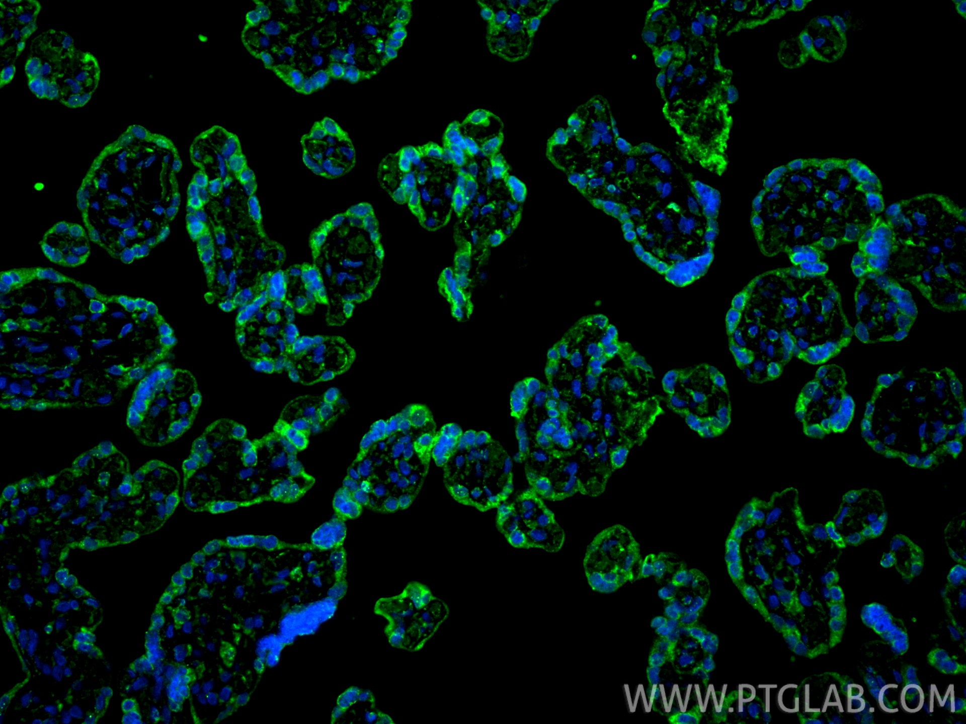
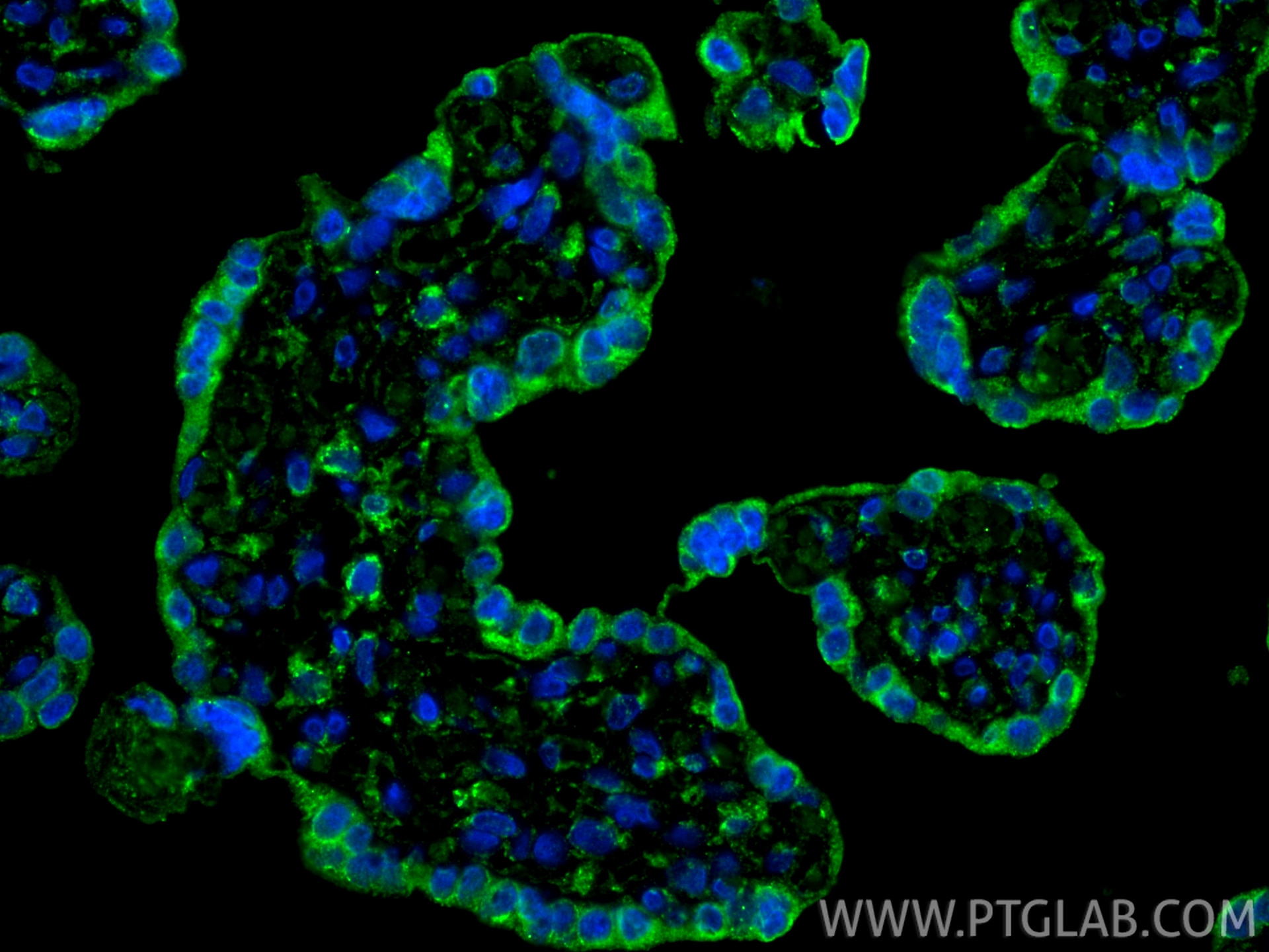
| Positive IF-P detected in | human placenta tissue, mouse thymus tissue |
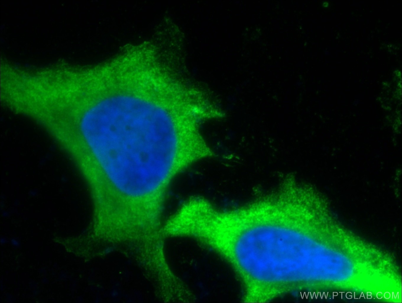
| Positive IF/ICC detected in | HeLa cells |
Recommended dilution
| Application | Dilution |
|---|---|
| Western Blot (WB) | WB : 1:2000-1:10000 |
| Immunohistochemistry (IHC) | IHC : 1:5000-1:20000 |
| Immunofluorescence (IF)-P | IF-P : 1:400-1:1600 |
| Immunofluorescence (IF)/ICC | IF/ICC : 1:50-1:500 |
| It is recommended that this reagent should be titrated in each testing system to obtain optimal results. | |
| Sample-dependent, Check data in validation data gallery. | |
Product Information
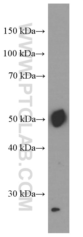
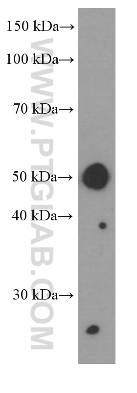
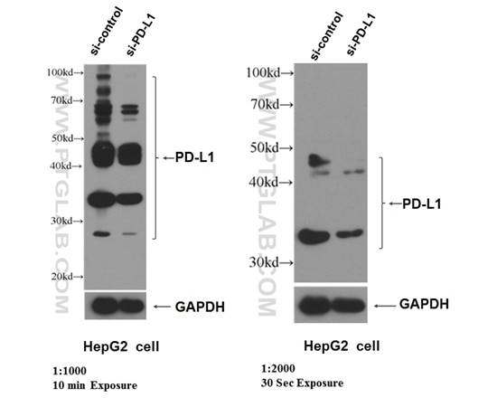
66248-1-Ig targets PD-L1/CD274 in WB, IHC, IF/ICC, IF-P, IP, CoIP, ChIP, ELISA, Blocking assay applications and shows reactivity with human, mouse, rat, pig samples.
| Tested Reactivity | human, mouse, rat, pig |
| Cited Reactivity | human, mouse, rat, pig |
| Host / Isotype | Mouse / IgG1 |
| Class | Monoclonal |
| Type | Antibody |
| Immunogen |
CatNo: Ag12443 Product name: Recombinant human PD-L1/CD274 protein Source: e coli.-derived, PET28a Tag: 6*His Domain: 25-241 aa of BC074984 Sequence: KDLYVVEYGSNMTIECKFPVEKQLDLAALIVYWEMEDKNIIQFVHGEEDLKVQHSSYRQRARLLKDQLSLGNAALQITDVKLQDAGVYRCMISYGGADYKRITVKVNAPYNKINQRILVVDPVTSEHELTCQAEGYPKAEVIWTSSDHQVLSGKTTTTNSKREEKLFNVTSTLRINTTTNEIFYCTFRRLDPEENHTAELVIPELPLAHPPNERTHL Predict reactive species |
| Full Name | CD274 molecule |
| Calculated Molecular Weight | 290 aa, 33 kDa |
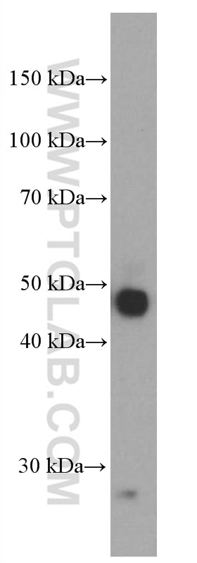
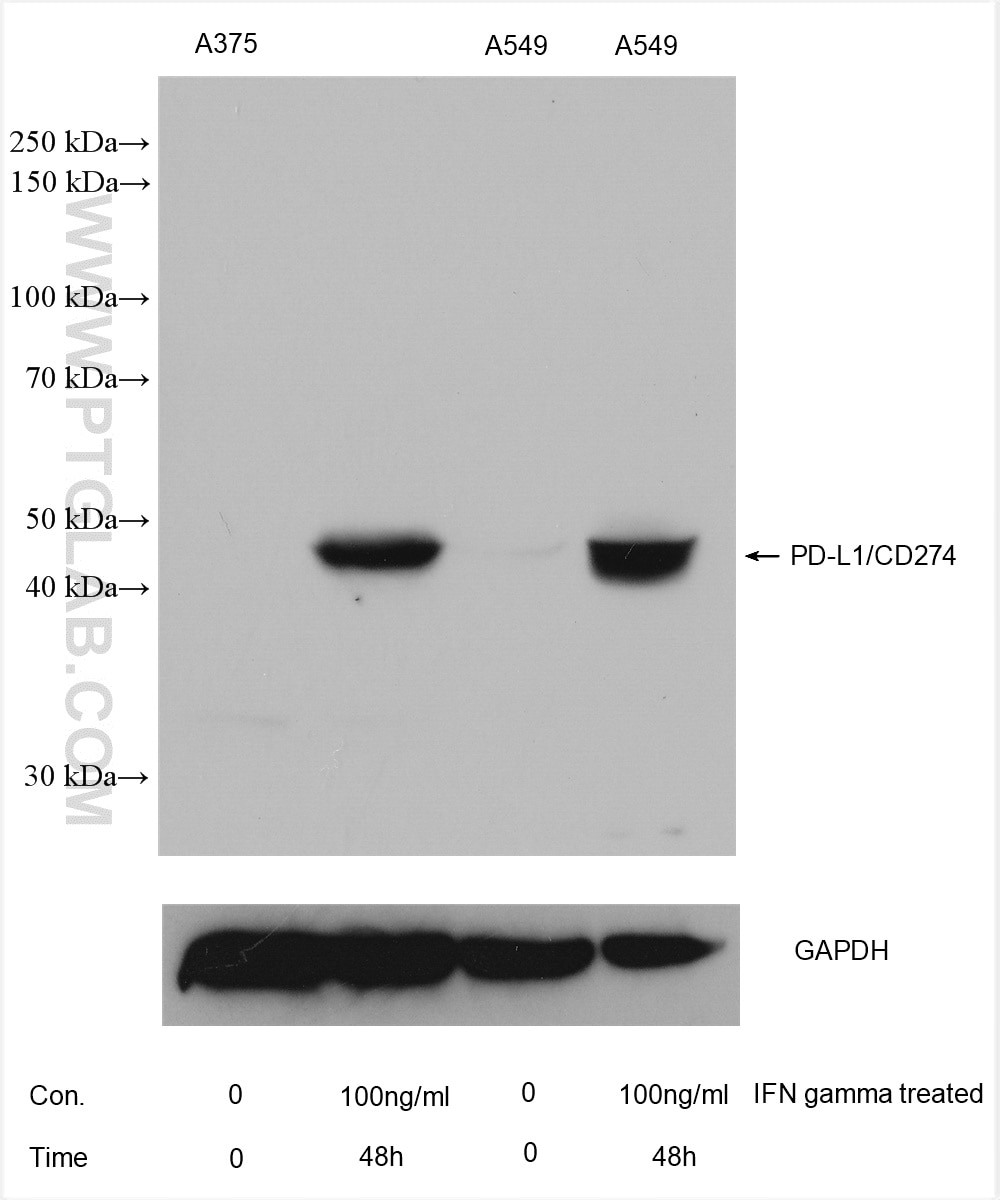
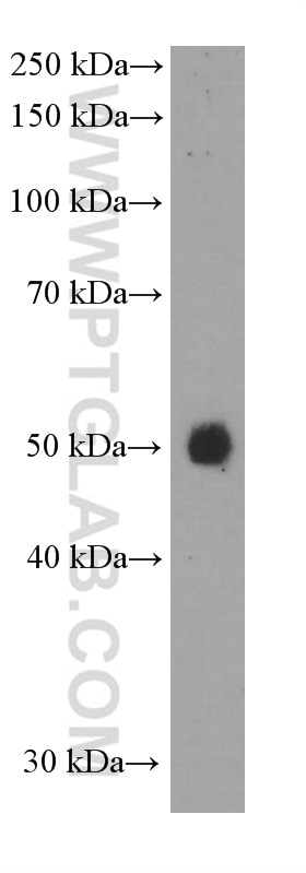
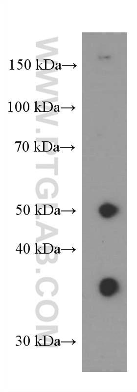
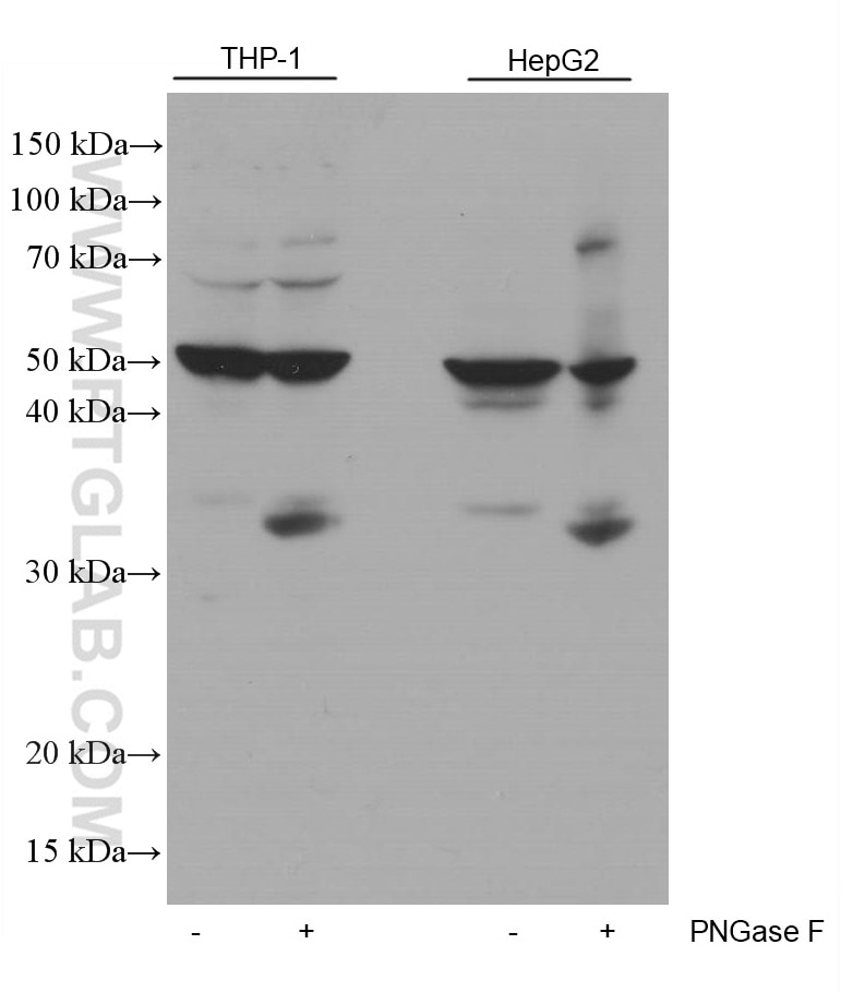
| Observed Molecular Weight | 45-50 kDa, 33 kDa |
| GenBank Accession Number | BC074984 |
| Gene Symbol | PD-L1 |
| Gene ID (NCBI) | 29126 |
| RRID | AB_2756526 |
| Conjugate | Unconjugated |
| Form | Liquid |
| Purification Method | Protein G purification |
| UNIPROT ID | Q9NZQ7 |
| Storage Buffer | PBS with 0.02% sodium azide and 50% glycerol, pH 7.3. |
| Storage Conditions | Store at -20°C. Stable for one year after shipment. Aliquoting is unnecessary for -20oC storage. 20ul sizes contain 0.1% BSA. |
Background Information
PD-L1, also known as CD274 or B7H1, stands for programmed cell death ligand 1. It is a type I transmembrane protein that is thought to repress immune responses by binding to its receptor (PD1), thus inhibiting T-cell activation, proliferation, and cytokine production. It contains V-like and C-like immunoglobulin domains. PD-L1 expression is regulated by various cytokines, such as TNF-α or LPS (ISSN: 1848-7718). Increased expression of this protein in certain types of cancers, e.g., renal cell carcinoma or colon cancer, correlates with poor prognosis.
What is the molecular weight of PD-L1?
Depending on the isoform, the calculated molecular weight of the protein varies between 20 and 33 kDa (176-290 aa).
What are the isoforms of PD-L1?
According to NCBI, three different isoforms have been identified. There are significant differences in the untranslated and protein coding regions.
What is the subcellular localization and tissue specificity of PD-L1?
It is predicted to localize in the plasma membrane of various cell types, with a particularly high expression in placental trophoblast and subsets of immune cells. High levels of PD-L1 protein have also been detected in lung and colon tissues.
What is the function of PD-L1 in immune responses?
PD-L1 is critical for the induction and maintenance of immune self-tolerance during infection or inflammation in normal tissues. The interaction of PD-L1 and its receptors is responsible for preventing auto-immune phenotypes and balancing the overall immune response in situations such as pregnancy or tissue allografts. The interaction between PD-L1 and PD-1 or B7.1 starts an inhibitory signaling cascade, which results in the decreased proliferation of antigen-specific T-cells and increased survival of regulatory T-cells (PMID: 15240681).
How can PD-L1's implication in cancer be used as a drug target?
In certain tumors, high expression of PD-L1 serves as a stop-sign to inhibit the recognition of cancer cells by T-cells (PMID: 23087408). The interaction between PD-L1 and its receptors (PD1 and B7.1) is a mechanism for the tumor to evade the host immune response (PMID: 29357948). Several mAbs have been developed to target that interaction and thus prevent the inactivation of cytotoxic T-cells by the tumor (PMIDs: 23890059, 18173375).
Protocols
| Product Specific Protocols | |
|---|---|
| IF protocol for PD-L1/CD274 antibody 66248-1-Ig | Download protocol |
| IHC protocol for PD-L1/CD274 antibody 66248-1-Ig | Download protocol |
| WB protocol for PD-L1/CD274 antibody 66248-1-Ig | Download protocol |
| Standard Protocols | |
|---|---|
| Click here to view our Standard Protocols |
Publications
| Species | Application | Title |
|---|---|---|
Cell Res Targeting ATAD3A-PINK1-mitophagy axis overcomes chemoimmunotherapy resistance by redirecting PD-L1 to mitochondria | ||
Adv Mater A Bifunctional Lysosome-Targeting Chimera Nanoplatform for Tumor-Selective Protein Degradation and Enhanced Cancer Immunotherapy | ||
Cell Metab Dual impacts of serine/glycine-free diet in enhancing antitumor immunity and promoting evasion via PD-L1 lactylation | ||
Cancer Cell ADORA1 Inhibition Promotes Tumor Immune Evasion by Regulating the ATF3-PD-L1 Axis. | ||
Cell Metab NAD+ Metabolism Maintains Inducible PD-L1 Expression to Drive Tumor Immune Evasion. |
Reviews
The reviews below have been submitted by verified Proteintech customers who received an incentive for providing their feedback.
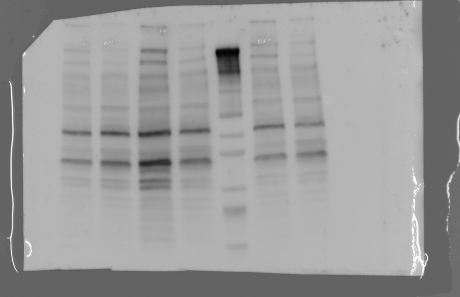
FH JIN-FENG (Verified Customer) (10-08-2025) | It works well in the PD-L1 overexpression cells. In terms of endogenous PD-L1 detection. There are a couple of non-specific bands around the target site.
 |
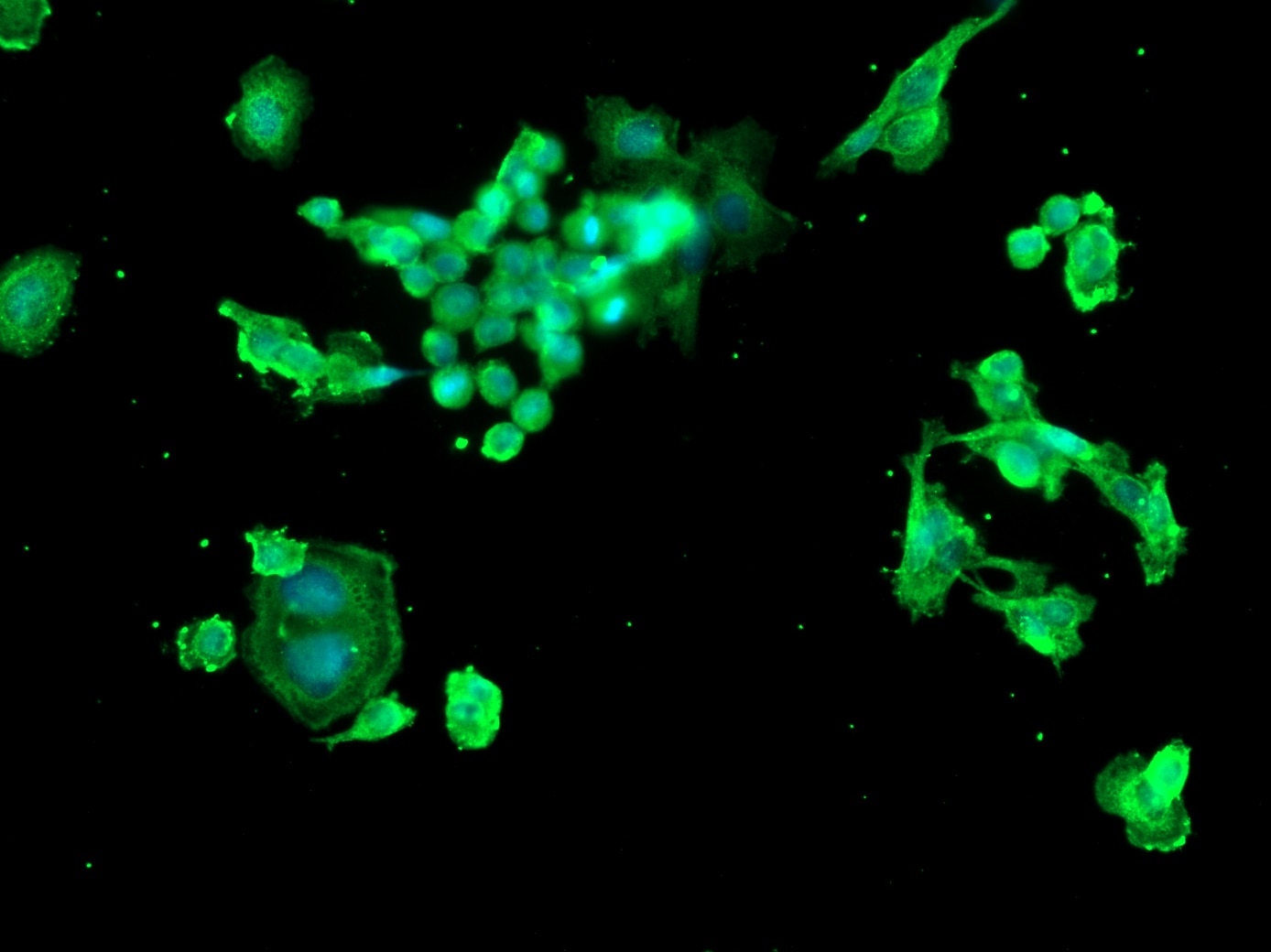
FH Margarita (Verified Customer) (11-20-2024) | Secondary antibody: AF 488
 |
FH hala (Verified Customer) (01-27-2023) | there is 2 bands one at the specific size and the other one is higher
 |
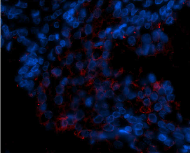
FH Emma (Verified Customer) (11-29-2021) | Used for IF on FFPE prostate tissue at 1:50 with Tris-EDTA antigen retrieval (pH 9.0).
 |
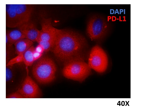
FH Marina (Verified Customer) (06-14-2021) | HCC1937 breast cancer cells were fixed with 4% PFA, permeabilized and stained overnight at 4ºC with anti-PD-L1 Ig diluted 1:300. Nuclei were stained with DAPI.
 |