


Tested Applications
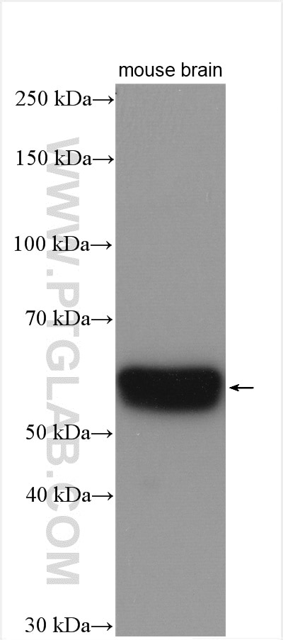
| Positive WB detected in | unboiled mouse brain tissue |
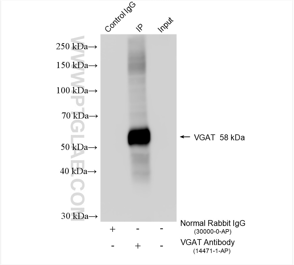
| Positive IP detected in | mouse brain tissue |
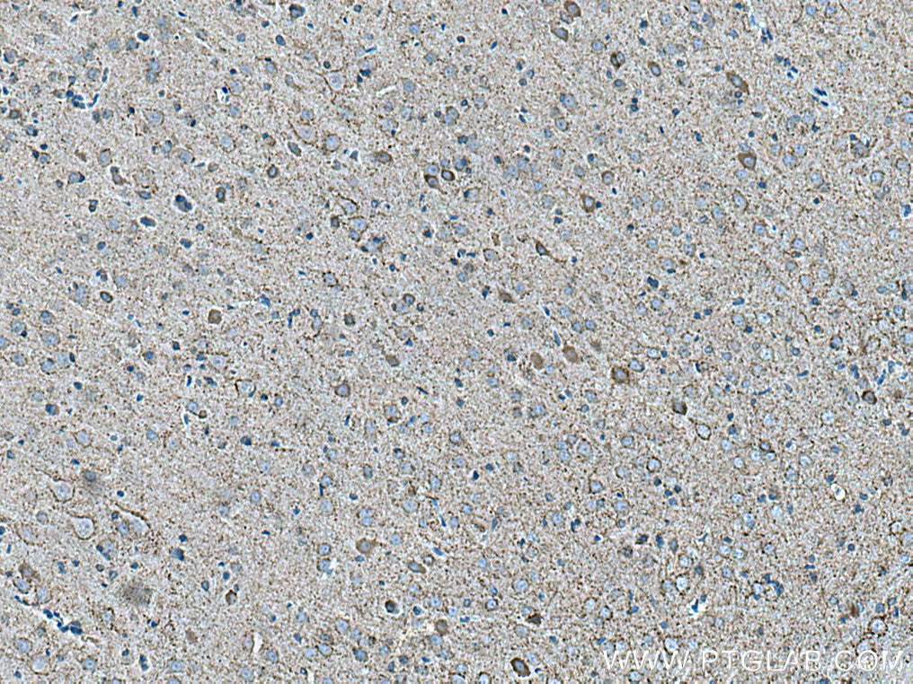
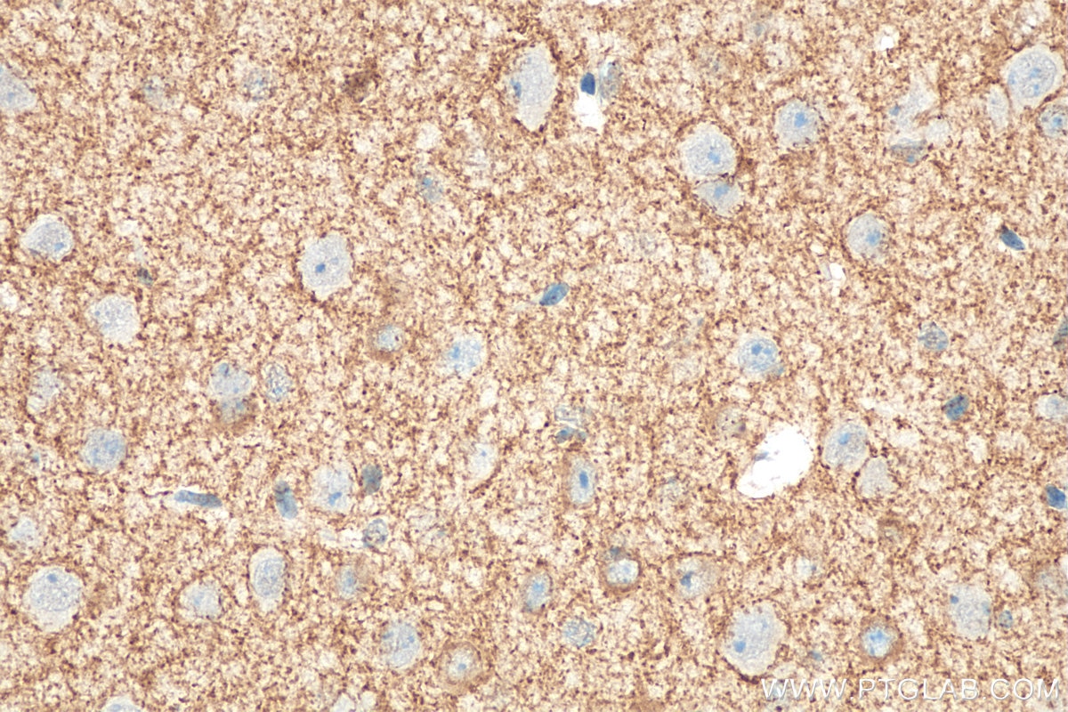
| Positive IHC detected in | rat brain tissue, mouse brain tissue Note: suggested antigen retrieval with TE buffer pH 9.0; (*) Alternatively, antigen retrieval may be performed with citrate buffer pH 6.0 |
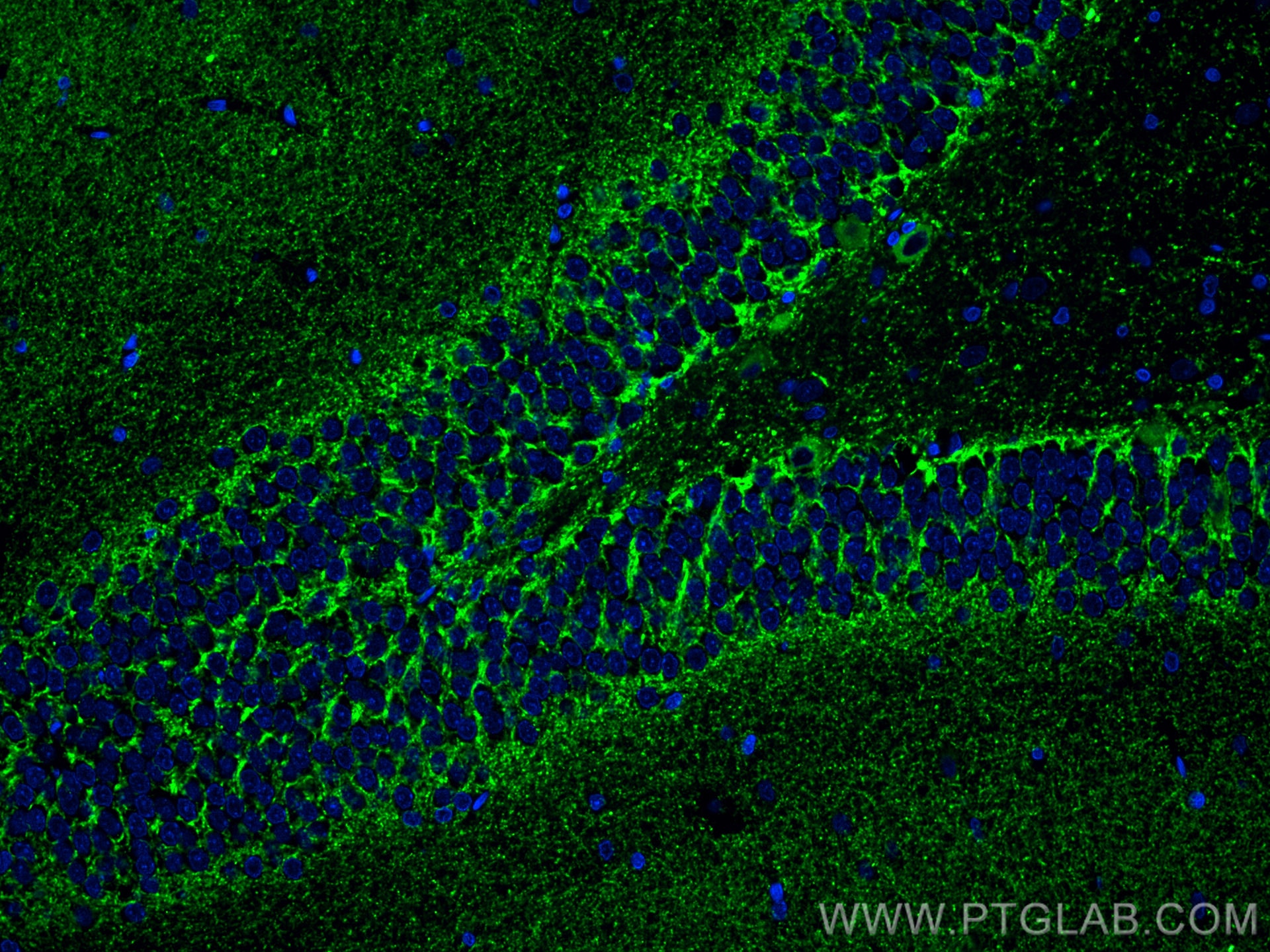
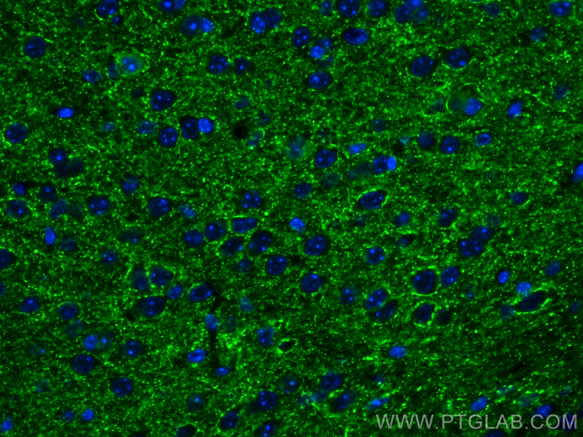
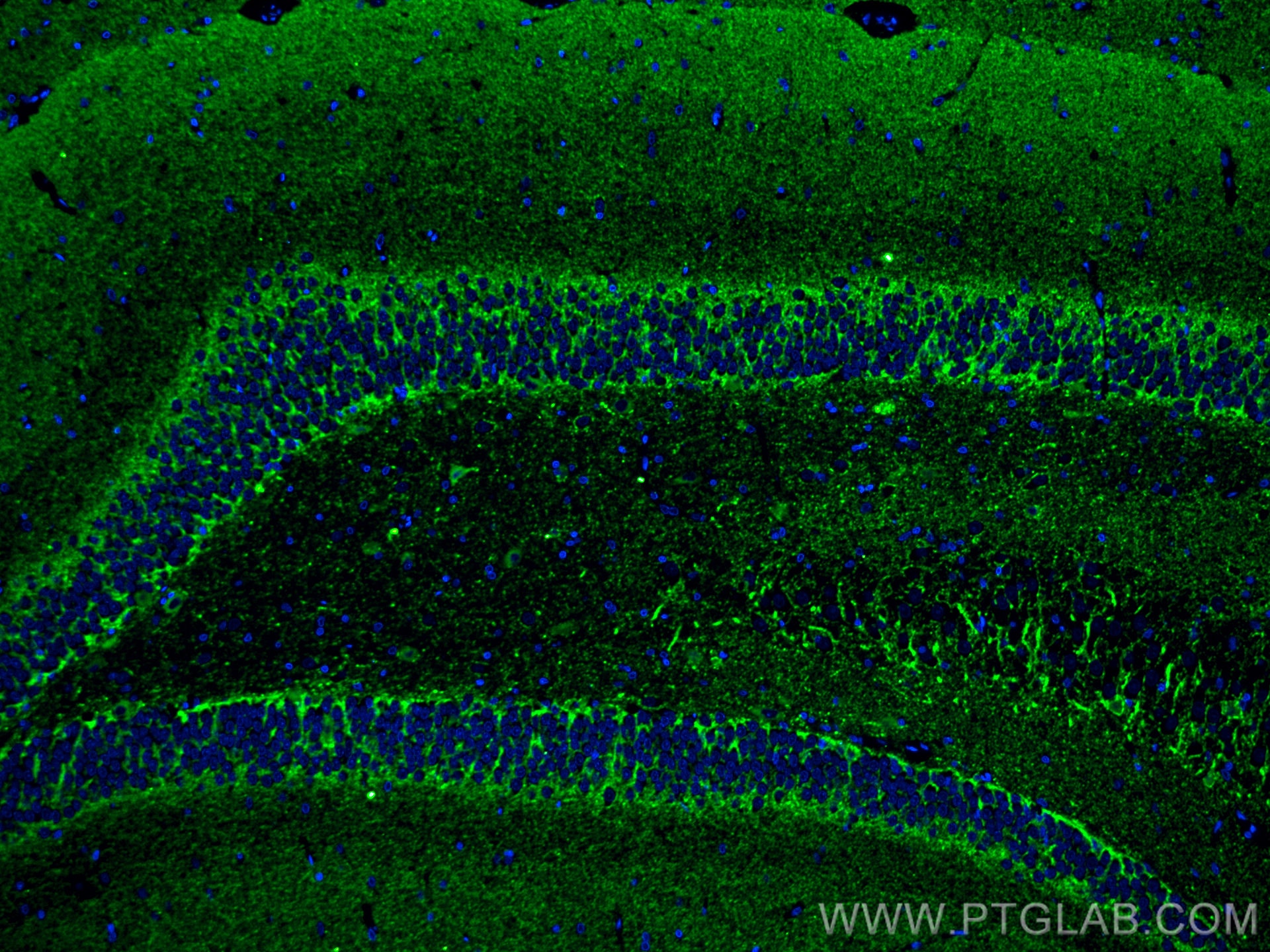
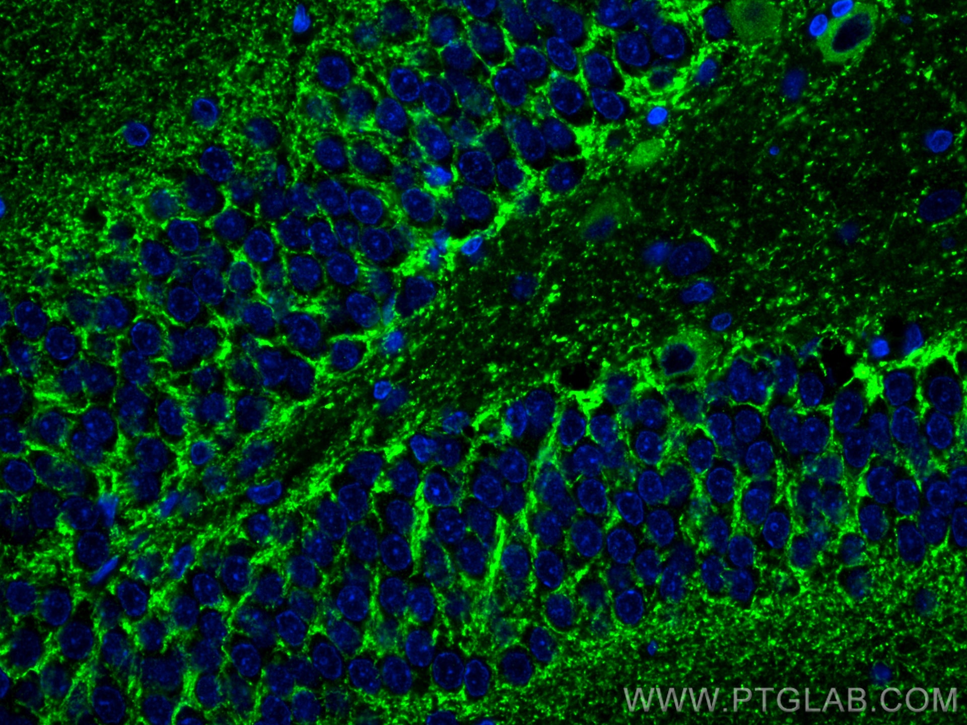
| Positive IF-P detected in | rat brain tissue, mouse brain tissue |
| Positive IF-Fro detected in | rat brain tissue |
Recommended dilution
| Application | Dilution |
|---|---|
| Western Blot (WB) | WB : 1:2000-1:10000 |
| Immunoprecipitation (IP) | IP : 0.5-4.0 ug for 1.0-3.0 mg of total protein lysate |
| Immunohistochemistry (IHC) | IHC : 1:50-1:500 |
| Immunofluorescence (IF)-P | IF-P : 1:50-1:500 |
| Immunofluorescence (IF)-FRO | IF-FRO : 1:50-1:500 |
| It is recommended that this reagent should be titrated in each testing system to obtain optimal results. | |
| Sample-dependent, Check data in validation data gallery. | |
Product Information
14471-1-AP targets SLC32A1/VGAT in WB, IHC, IF-P, IF-Fro, IP, ELISA applications and shows reactivity with human, mouse, rat samples.
| Tested Reactivity | human, mouse, rat |
| Cited Reactivity | human, mouse, rat, caenorhabditis elegans |
| Host / Isotype | Rabbit / IgG |
| Class | Polyclonal |
| Type | Antibody |
| Immunogen |
CatNo: Ag5843 Product name: Recombinant human SLC32A1 protein Source: e coli.-derived, PGEX-4T Tag: GST Domain: 1-124 aa of BC053582 Sequence: MATLLRSKLSNVATSVSNKSQAKMSGMFARMGFQAATDEEAVGFAHCDDLDFEHRQGLQMDILKAEGEPCGDEGAEAPVEGDIHYQRGSGAPLPPSGSKDQVGGGGEFGGHDKPKITAWEAGWN Predict reactive species |
| Full Name | solute carrier family 32 (GABA vesicular transporter), member 1 |
| Calculated Molecular Weight | 57 kDa |
| Observed Molecular Weight | 57 kDa |
| GenBank Accession Number | BC053582 |
| Gene Symbol | VGAT |
| Gene ID (NCBI) | 140679 |
| RRID | AB_10644324 |
| Conjugate | Unconjugated |
| Form | Liquid |
| Purification Method | Antigen affinity purification |
| UNIPROT ID | Q9H598 |
| Storage Buffer | PBS with 0.02% sodium azide and 50% glycerol, pH 7.3. |
| Storage Conditions | Store at -20°C. Stable for one year after shipment. Aliquoting is unnecessary for -20oC storage. 20ul sizes contain 0.1% BSA. |
Background Information
SLC32A1, also known as VGAT (vesicular GABA transporter), functions in the uptake of GABA and glycine into synaptic vesicles. GABA (gamma-aminobutyric acid), is the major inhibitory neurotransmitter in the CNS. VGAT transports GABA and glycine into acidic vesicles and localizes to the synaptic vesicle in glycinergic and GABAergic neurons. And VGAT antibodies are useful markers for presynaptic GABAergic and glycinergic neurons.
Protocols
| Product Specific Protocols | |
|---|---|
| IF protocol for SLC32A1/VGAT antibody 14471-1-AP | Download protocol |
| IHC protocol for SLC32A1/VGAT antibody 14471-1-AP | Download protocol |
| IP protocol for SLC32A1/VGAT antibody 14471-1-AP | Download protocol |
| WB protocol for SLC32A1/VGAT antibody 14471-1-AP | Download protocol |
| Standard Protocols | |
|---|---|
| Click here to view our Standard Protocols |
Publications
What published studies show
SLC32A1/VGAT Polyclonal antibody (Cat# 14471-1-AP) has accumulated 28 citations published in journals including Nature Neuroscience, Nature Communications, Cell Stem Cell, Theranostics, Journal of Experimental & Clinical Cancer Research, Advanced Science, EMBO Reports, ACS Omega, Scientific Reports, Movement Disorders, and others. Associated research fields encompass neuroscience (epilepsy, GABAergic signaling, neurodegeneration, chronic pain, stroke recovery, autism), oncology, and ecotoxicology.
| Species | Application | Title |
|---|---|---|
Cell Stem Cell Auditory activity sustains adult neurogenesis and cognition through the locus coeruleus-norepinephrine system. | ||
Nat Neurosci GABA-dependent microglial elimination of inhibitory synapses underlies neuronal hyperexcitability in epilepsy | ||
Nat Commun Defective ventral neurogenesis due to midfetal Chd8 mutation drives autistic-like behavior in mice. | ||
Theranostics Targeted sonogenetic modulation of GABAergic interneurons in the hippocampal CA1 region in status epilepticus | ||
J Exp Clin Cancer Res GABA induced by sleep deprivation promotes the proliferation and migration of colon tumors through miR-223-3p endogenous pathway and exosome pathway | ||
Adv Sci (Weinh) Microglia-Derived Vitamin D Binding Protein Mediates Synaptic Damage and Induces Depression by Binding to the Neuronal Receptor Megalin |
Reviews
What customers say
Users report mixed results with this vGAT antibody. For immunofluorescence, one reviewer found it functional on human brain FFPE sections at 1:100 but noted high autofluorescence complicating quantification. Another user at 1:200 on human cortical neurons was less satisfied, observing signal localized mainly to the soma rather than nerve terminals. For Western blot, a reviewer achieved good results at 1:2000 on mouse hippocampal lysate using 5% skimmed milk in 1x TBS-T as blocking solution. Overall, the antibody shows promise for WB and IF applications, though terminal staining and background may require optimization.
The reviews below have been submitted by verified Proteintech customers who received an incentive for providing their feedback.
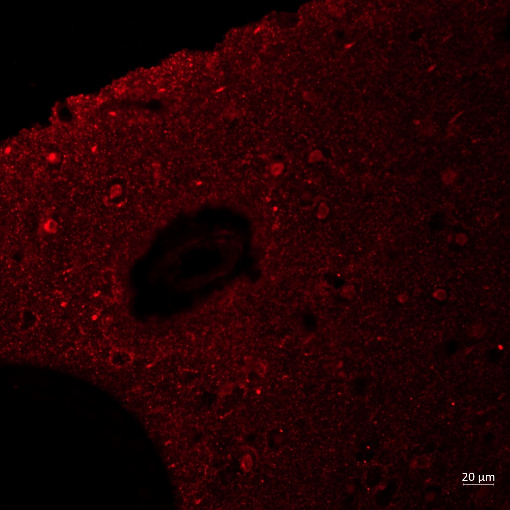
Reyes (Verified Customer) (03-04-2024) | vGAT worked on my human brain FFPE sections although the quantification would be difficult due to the high autofluorescence
 |
Mandi (Verified Customer) (03-10-2020) | Did not stain the nerve terminals well. Signal was localized to the soma mainly.
|
Ute (Verified Customer) (03-03-2020) | blocking solution 5% skimmed milk in 1x TBS-T
|










