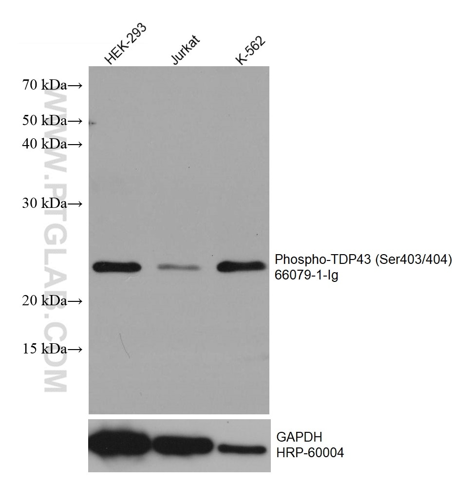
antibody) at dilution of 1:20000 incubated at room temperature for 1.5 hours. The membrane was stripped and reblotted with HRP-conjugated GAPDH Monoclonal antibody (HRP-60004) as loading control.")
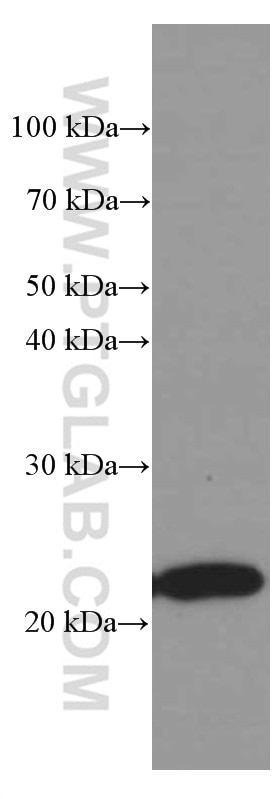
 Antibody) at dilution of 1:3000 incubated at room temperature for 1.5 hours.")
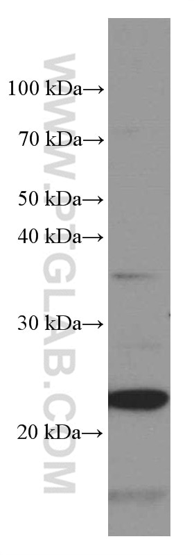
 Antibody) at dilution of 1:3000 incubated at room temperature for 1.5 hours.")
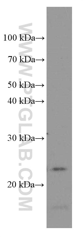
 Antibody) at dilution of 1:3000 incubated at room temperature for 1.5 hours.")
 Antibody) at dilution of 1:3000 incubated at room temperature for 1.5 hours.")
Tested Applications
| Positive WB detected in | HEK-293 cells, Jurkat cells, K-562 cells, RAW 264.7 cells, fetal human brain tissue |
Recommended dilution
| Application | Dilution |
|---|---|
| Western Blot (WB) | WB : 1:5000-1:50000 |
| It is recommended that this reagent should be titrated in each testing system to obtain optimal results. | |
| Sample-dependent, Check data in validation data gallery. | |
Published Applications
| WB | See 4 publications below |
| IHC | See 3 publications below |
| IF | See 3 publications below |
Product Information
66079-1-Ig targets Phospho-TDP43 (Ser403/404) in WB, IHC, IF, ELISA applications and shows reactivity with human, mouse samples.
| Tested Reactivity | human, mouse |
| Cited Reactivity | human, mouse, zebrafish |
| Host / Isotype | Mouse / IgG2a |
| Class | Monoclonal |
| Type | Antibody |
| Immunogen |
Peptide Predict reactive species |
| Full Name | TAR DNA binding protein |
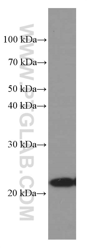
| Calculated Molecular Weight | 43 kDa |
| Observed Molecular Weight | 25 kDa |
| GenBank Accession Number | NM_007375 |
| Gene Symbol | TDP-43 |
| Gene ID (NCBI) | 23435 |
| RRID | AB_11183767 |
| Conjugate | Unconjugated |
| Form | Liquid |
| Purification Method | Protein A purification |
| UNIPROT ID | Q13148 |
| Storage Buffer | PBS with 0.02% sodium azide and 50% glycerol, pH 7.3. |
| Storage Conditions | Store at -20°C. Stable for one year after shipment. Aliquoting is unnecessary for -20oC storage. 20ul sizes contain 0.1% BSA. |
Background Information
Transactivation response (TAR) DNA-binding protein of 43 kDa (also known as TARDBP or TDP-43) was first isolated as a transcriptional inactivator binding to the TAR DNA element of the HIV-1 virus. Neumann et al. (2006) found that a hyperphosphorylated, ubiquitinated, and cleaved form of TARDBP, known as pathologic TDP-43, is the major component of the tau-negative and ubiquitin-positive inclusions that characterize amyotrophic lateral sclerosis (ALS) and the most common pathological subtype of frontotemporal lobar degeneration (FTLD-U). Various forms of TDP-43 exist, including 18-35 kDa of cleaved C-terminal fragments, 45-50 kDa phospho-protein, 55 kDa glycosylated form, 75 kDa hyperphosphorylated form, and 90-300 kDa cross-linked form. (PMID: 17023659,19823856, 21666678, 22193176).66079-1-Ig is a mouse monoclonal antibody recognizing TDP-43 only when phosphorylated at 403/404. Immunohistochemical analyses using this antibody only stain the insoluble inclusions in pathologic tissues without normal diffuse nuclear staining.
Protocols
| Product Specific Protocols | |
|---|---|
| WB protocol for Phospho-TDP43 (Ser403/404) antibody 66079-1-Ig | Download protocol |
| Standard Protocols | |
|---|---|
| Click here to view our Standard Protocols |
Publications
| Species | Application | Title |
|---|---|---|
Nat Neurosci TDP-43 condensates and lipid droplets regulate the reactivity of microglia and regeneration after traumatic brain injury | ||
EMBO J Disease-linked TDP-43 hyperphosphorylation suppresses TDP-43 condensation and aggregation. | ||
Eur J Med Chem Targeting nuclear protein TDP-43 by cell division cycle kinase 7 inhibitors: A new therapeutic approach for amyotrophic lateral sclerosis. | ||
Biochemistry Using Tetracysteine-Tagged TDP-43 with a Biarsenical Dye To Monitor Real-Time Trafficking in a Cell Model of Amyotrophic Lateral Sclerosis. | ||
PLoS One Drosophila lines with mutant and wild type human TDP-43 replacing the endogenous gene reveals phosphorylation and ubiquitination in mutant lines in the absence of viability or lifespan defects. |
Reviews
The reviews below have been submitted by verified Proteintech customers who received an incentive for providing their feedback.
FH Azita (Verified Customer) (06-17-2021) | Immunocytochemistry labelling of (4% PFA) fixed NSC-34 cells by Phospho-TDP43 (Ser403/404) Monoclonal antibody at dilution of 1:100 showed strong labelling.
|