Beta Actin Monoklonaler Antikörper
Beta Actin Monoklonal Antikörper für WB, IHC, IF/ICC, IF-P, ELISA
Wirt / Isotyp
Maus / IgM
Getestete Reaktivität
human, Maus, Pflanze, Ratte, Zebrafisch und mehr (4)
Anwendung
WB, IHC, IF/ICC, IF-P, IP, CoIP, ELISA
Konjugation
Unkonjugiert
CloneNo.
7D2C10
Kat-Nr. : 60008-1-Ig
Synonyme

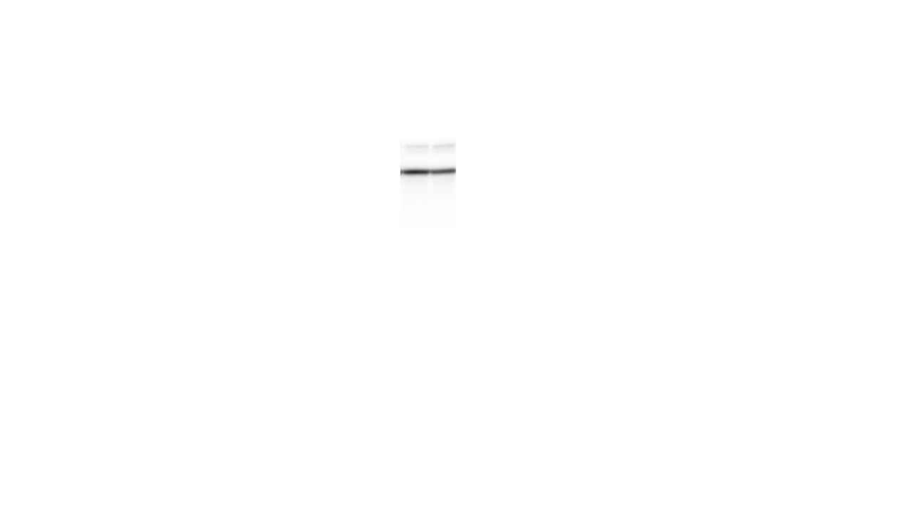
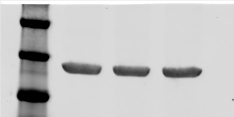
 at various dilutions.")
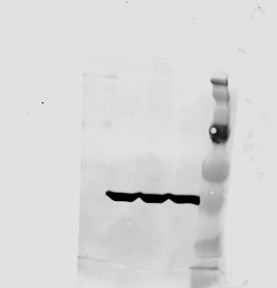
 at dilution of 1:2000 incubated at room temperature for 1.5 hours.")
 at dilution of 1:2000 incubated at room temperature for 1.5 hours.")
 at dilution of 1:8000 incubated at room temperature for 1.5 hours.")
 at dilution of 1:2000 incubated at room temperature for 1.5 hours.")
 at dilution of 1:2000 incubated at room temperature for 1.5 hours.")
 at dilution of 1:10000 incubated at room temperature for 1.5 hours.")
 at dilution of 1:200 (under 10x lens). Heat mediated antigen retrieval with Tris-EDTA buffer (pH 9.0).")
 at dilution of 1:500 (under 10x lens. Heat mediated antigen retrieval with Tris-EDTA buffer (pH 9.0).")
 at dilution of 1:500 (under 40x lens. Heat mediated antigen retrieval with Tris-EDTA buffer (pH 9.0).")
 at dilution of 1:100 (under 10x lens).")
 at dilution of 1:100 (under 40x lens).")
 at dilution of 1:100 (under 40x lens).")
 at dilution of 1:100 (under 10x lens).")
 at dilution of 1:100 (under 10x lens).")
 at dilution of 1:100 (under 40x lens).")
 at dilution of 1:100 (under 10x lens).")
 at dilution of 1:100 (under 40x lens).")
 at dilution of 1:100 (under 10x lens).")
 at dilution of 1:100 (under 40x lens).")
 at dilution of 1:100 (under 10x lens).")
 at dilution of 1:100 (under 40x lens).")
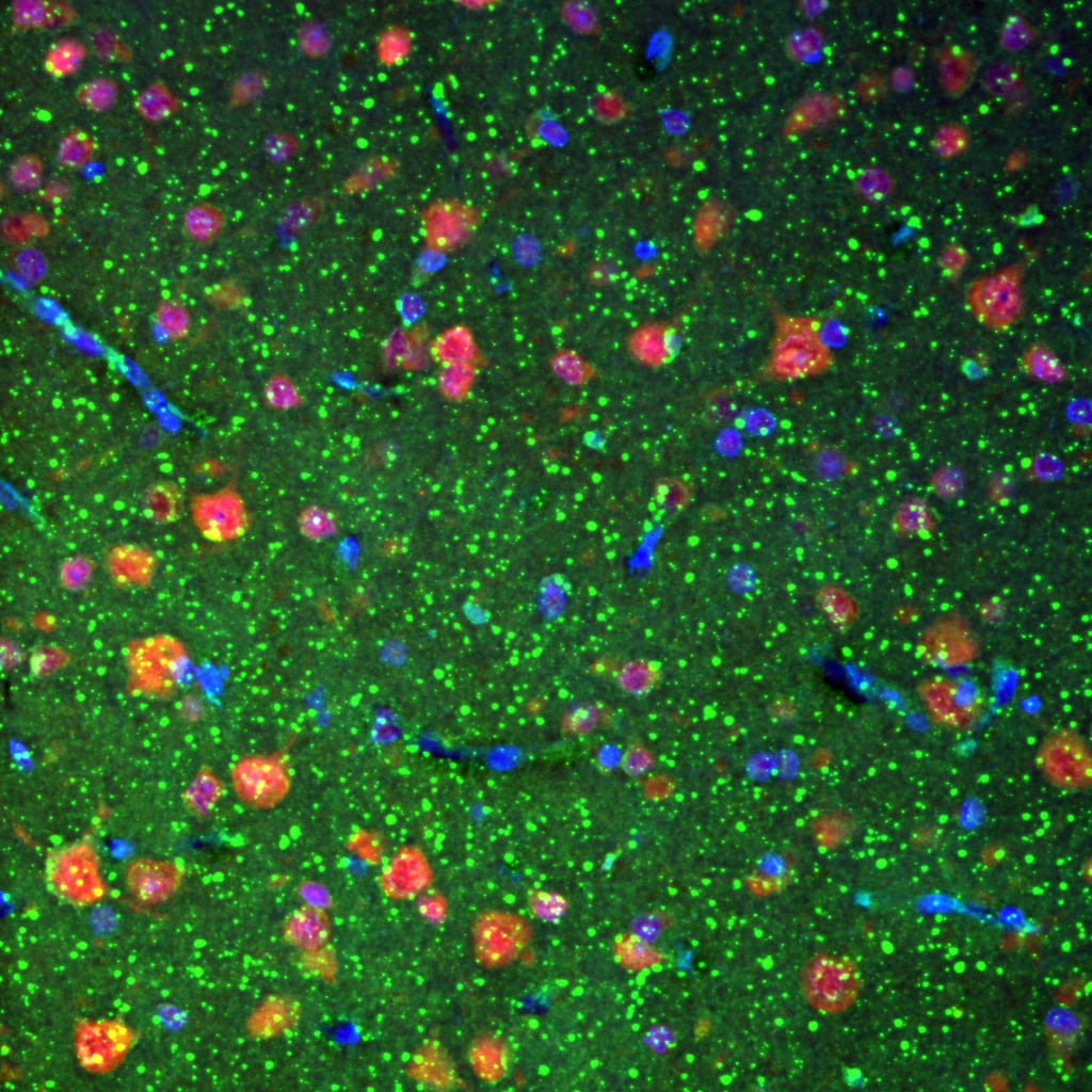
 fixed paraffin-embedded rat brain tissue using Beta Actin antibody (60008-1-Ig, Clone: 7D2C10 ) at dilution of 1:1000 and CoraLite®488-Conjugated Goat Anti-Mouse IgG(H+L) (SA00013-1). Heat mediated antigen retrieval with Tris-EDTA buffer (pH 9.0).")
 fixed MDCK cells using Beta Actin antibody (60008-1-Ig, Clone: 7D2C10 ) at dilution of 1:800 and Multi-rAb CoraLite ® Plus 488-Goat Anti-Mouse Recombinant Secondary Antibody (H+L) (RGAM002).")
Geprüfte Anwendungen
| Erfolgreiche Detektion in WB | Multizellen/-gewebe, A549-Zellen, Arabidopsis-Gewebe (aus der gesamten Pflanze), HEK-293-Zellen, HeLa-Zellen, MCF-7-Zellen, Reisgewebe (aus der gesamten Pflanze) |
| Erfolgreiche Detektion in IHC | Mausherzgewebe, humanes Hirngewebe, humanes Nierengewebe, humanes Lungengewebe, humanes Eierstockgewebe, humanes Plazenta-Gewebe, humanes Milzgewebe, humanes Hodengewebe Hinweis: Antigendemaskierung mit TE-Puffer pH 9,0 empfohlen. (*) Wahlweise kann die Antigendemaskierung auch mit Citratpuffer pH 6,0 erfolgen. |
| Erfolgreiche Detektion in IF-P | Rattenhirngewebe |
| Erfolgreiche Detektion in IF/ICC | MDCK-Zellen |
Empfohlene Verdünnung
| Anwendung | Verdünnung |
|---|---|
| Western Blot (WB) | WB : 1:5000-1:50000 |
| Immunhistochemie (IHC) | IHC : 1:50-1:500 |
| Immunfluoreszenz (IF)-P | IF-P : 1:500-1:2000 |
| Immunfluoreszenz (IF)/ICC | IF/ICC : 1:400-1:1600 |
| It is recommended that this reagent should be titrated in each testing system to obtain optimal results. | |
| Sample-dependent, check data in validation data gallery | |
Veröffentlichte Anwendungen
| WB | See 2976 publications below |
| IHC | See 4 publications below |
| IF | See 11 publications below |
| IP | See 9 publications below |
| CoIP | See 3 publications below |
Produktinformation
60008-1-Ig bindet in WB, IHC, IF/ICC, IF-P, IP, CoIP, ELISA Beta Actin und zeigt Reaktivität mit human, Maus, Pflanze, Ratten, Zebrafisch
| Getestete Reaktivität | human, Maus, Pflanze, Ratte, Zebrafisch |
| In Publikationen genannte Reaktivität | human, arabidopsis, Hefe, Huhn, Ratte, Ziege |
| Wirt / Isotyp | Maus / IgM |
| Klonalität | Monoklonal |
| Typ | Antikörper |
| Immunogen | Beta Actin fusion protein Ag0297 |
| Vollständiger Name | actin, beta |
| Berechnetes Molekulargewicht | 375 aa, 42 kDa |
| Beobachtetes Molekulargewicht | 42 kDa |
| GenBank-Zugangsnummer | BC002409 |
| Gene symbol | Beta Actin |
| Gene ID (NCBI) | 60 |
| Konjugation | Unkonjugiert |
| Form | Liquid |
| Reinigungsmethode | Caprylsäure/Ammoniumsulfat-Präzipitation |
| Lagerungspuffer | PBS with 0.02% sodium azide and 50% glycerol |
| Lagerungsbedingungen | Bei -20°C lagern. Nach dem Versand ein Jahr lang stabil Aliquotieren ist bei -20oC Lagerung nicht notwendig. 20ul Größen enthalten 0,1% BSA. |
Hintergrundinformationen
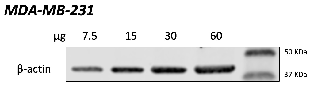
Beta actin, also named as ACTB and F-Actin, belongs to the actin family. Actins are highly conserved globular proteins that are involved in various types of cell motility and are ubiquitously expressed in all eukaryotic cells. At least six isoforms of actins are known in mammals and other vertebrates: alpha (ACTC1, cardiac muscle 1), alpha 1 (ACTA1, skeletal muscle) and 2 (ACTA2, aortic smooth muscle), beta (ACTB), gamma 1 (ACTG1) and 2 (ACTG2, enteric smooth muscle). Beta and gamma 1 are two non-muscle actin proteins. Most actins consist of 376aa, while ACTG2 (rich in muscles) has 375aa and ACTG1(found in non-muscle cells) has only 374aa. Beta actin has been widely used as the internal control in RT-PCR and Western Blotting as a 42-kDa protein. However, the 37-40 kDa cleaved fragment of beta actin can be generated during apoptosis process. This antibody can recognize all the actins.
Protokolle
| PRODUKTSPEZIFISCHE PROTOKOLLE | |
|---|---|
| WB protocol for Beta Actin antibody 60008-1-Ig | Protokoll herunterladen |
| IHC protocol for Beta Actin antibody 60008-1-Ig | Protokoll herunterladenl |
| IF protocol for Beta Actin antibody 60008-1-Ig | Protokoll herunterladen |
| STANDARD-PROTOKOLLE | |
|---|---|
| Klicken Sie hier, um unsere Standardprotokolle anzuzeigen |
Publikationen
| Species | Application | Title |
|---|---|---|
Cell Res Mitochondria-localized cGAS suppresses ferroptosis to promote cancer progression | ||
Gastroenterology PTEN deficiency facilitates exosome secretion and metastasis in cholangiocarcinoma by impairing TFEB-mediated lysosome biogenesis | ||
Immunity Microglial lipid phosphatase SHIP1 limits complement-mediated synaptic pruning in the healthy developing hippocampus | ||
Nat Methods Visualizing the native cellular organization by coupling cryofixation with expansion microscopy (Cryo-ExM). | ||
Rezensionen
The reviews below have been submitted by verified Proteintech customers who received an incentive for providing their feedback.
FH Reyes (Verified Customer) (02-10-2025) | Beta actin (in green) did not seem to work on my human FFPE brain tissue. Appart from being really autofluorescent on the tissue, it seems to have a puncta-like staining on some cells, not the usual/expected clear cytoeskeleton staining.
 |
FH P (Verified Customer) (09-23-2024) | Excellent
 |
FH Udesh (Verified Customer) (08-16-2023) | The Ab worked well for both WB and IF at mentioned dilutions
|
FH Chun (Verified Customer) (09-07-2020) | This is an excellent antibody for immunoblotting.
|
FH Nikhil (Verified Customer) (09-17-2019) | Very good and specific. I can use 1:2500 5 ml antibody solution two times.
 |
FH Leonardo (Verified Customer) (09-12-2019) | Excellent antibody. Works well since the first time.
 |
FH mark (Verified Customer) (02-05-2019) | Antibody works beautifully to detect a single Beta-actin band as a control
 |

