CEP83 Polyklonaler Antikörper
CEP83 Polyklonal Antikörper für WB, IHC, IF/ICC, ELISA
Wirt / Isotyp
Kaninchen / IgG
Getestete Reaktivität
human und mehr (1)
Anwendung
WB, IHC, IF/ICC, ELISA
Konjugation
Unkonjugiert
Kat-Nr. : 26013-1-AP
Synonyme
 at dilution of 1:600 incubated at room temperature for 1.5 hours.")
 at dilution of 1:200 (under 10x lens).")
 at dilution of 1:200 (under 40x lens).")
 fixed hTERT-RPE1 cells using CEP83 antibody (26013-1-AP) at dilution of 1:400 and CoraLite®488-Conjugated Goat Anti-Rabbit IgG(H+L), CoraLite®594 ARL13B antibody (CL594-17711, red).")
 fixed hTERT-RPE1 cells using CEP83 antibody (26013-1-AP) at dilution of 1:800 and CoraLite®594-Conjugated Goat Anti-Rabbit IgG(H+L).")
Geprüfte Anwendungen
| Erfolgreiche Detektion in WB | HEK-293-Zellen |
| Erfolgreiche Detektion in IHC | humanes Nierengewebe Hinweis: Antigendemaskierung mit TE-Puffer pH 9,0 empfohlen. (*) Wahlweise kann die Antigendemaskierung auch mit Citratpuffer pH 6,0 erfolgen. |
| Erfolgreiche Detektion in IF/ICC | hTERT-RPE1-Zellen |
Empfohlene Verdünnung
| Anwendung | Verdünnung |
|---|---|
| Western Blot (WB) | WB : 1:500-1:1000 |
| Immunhistochemie (IHC) | IHC : 1:50-1:500 |
| Immunfluoreszenz (IF)/ICC | IF/ICC : 1:200-1:800 |
| It is recommended that this reagent should be titrated in each testing system to obtain optimal results. | |
| Sample-dependent, check data in validation data gallery | |
Veröffentlichte Anwendungen
| WB | See 7 publications below |
| IF | See 6 publications below |
Produktinformation
26013-1-AP bindet in WB, IHC, IF/ICC, ELISA CEP83 und zeigt Reaktivität mit human
| Getestete Reaktivität | human |
| In Publikationen genannte Reaktivität | human, Maus |
| Wirt / Isotyp | Kaninchen / IgG |
| Klonalität | Polyklonal |
| Typ | Antikörper |
| Immunogen | CEP83 fusion protein Ag22826 |
| Vollständiger Name | coiled-coil domain containing 41 |
| Berechnetes Molekulargewicht | 693 aa, 82 kDa |
| Beobachtetes Molekulargewicht | 83 kDa, 68 kDa |
| GenBank-Zugangsnummer | BC125087 |
| Gene symbol | CEP83 |
| Gene ID (NCBI) | 51134 |
| Konjugation | Unkonjugiert |
| Form | Liquid |
| Reinigungsmethode | Antigen-Affinitätsreinigung |
| Lagerungspuffer | PBS with 0.02% sodium azide and 50% glycerol |
| Lagerungsbedingungen | Bei -20°C lagern. Nach dem Versand ein Jahr lang stabil Aliquotieren ist bei -20oC Lagerung nicht notwendig. 20ul Größen enthalten 0,1% BSA. |
Hintergrundinformationen
CEP83 (centrosomal protein, 83 kDa), also known as CCDC41 (coiled-coil domain containing 41), is required for ciliary vesicle docking to the mother centriole. It is a key component of the centriolar distal appendages. CCDC41 may collaborate with IFT20 in the trafficking of ciliary membrane proteins from the Golgi complex to the cilium during the initiation of primary cilium assembly (PMID: 23530209). Alternative splicing results in transcript variants encoding two isoforms with calculated molecular weights of 83 kDa and 68 kDa, respectively.
Protokolle
| PRODUKTSPEZIFISCHE PROTOKOLLE | |
|---|---|
| WB protocol for CEP83 antibody 26013-1-AP | Protokoll herunterladen |
| IHC protocol for CEP83 antibody 26013-1-AP | Protokoll herunterladenl |
| IF protocol for CEP83 antibody 26013-1-AP | Protokoll herunterladen |
| STANDARD-PROTOKOLLE | |
|---|---|
| Klicken Sie hier, um unsere Standardprotokolle anzuzeigen |
Publikationen
| Species | Application | Title |
|---|---|---|
Cell Res NudCL2 is an autophagy receptor that mediates selective autophagic degradation of CP110 at mother centrioles to promote ciliogenesis. | ||
J Cell Biol The Cby3/ciBAR1 complex positions the annulus along the sperm flagellum during spermiogenesis | ||
PLoS Biol The evolutionary conserved proteins CEP90, FOPNL, and OFD1 recruit centriolar distal appendage proteins to initiate their assembly | ||
Cell Mol Life Sci Requirement of NPHP5 in the hierarchical assembly of basal feet associated with basal bodies of primary cilia. | ||
J Biol Chem The C7orf43/TRAPPC14 component links the TRAPPII complex to RABIN8 for preciliary vesicle tethering at the mother centriole during ciliogenesis. |
Rezensionen
The reviews below have been submitted by verified Proteintech customers who received an incentive for providing their feedback.
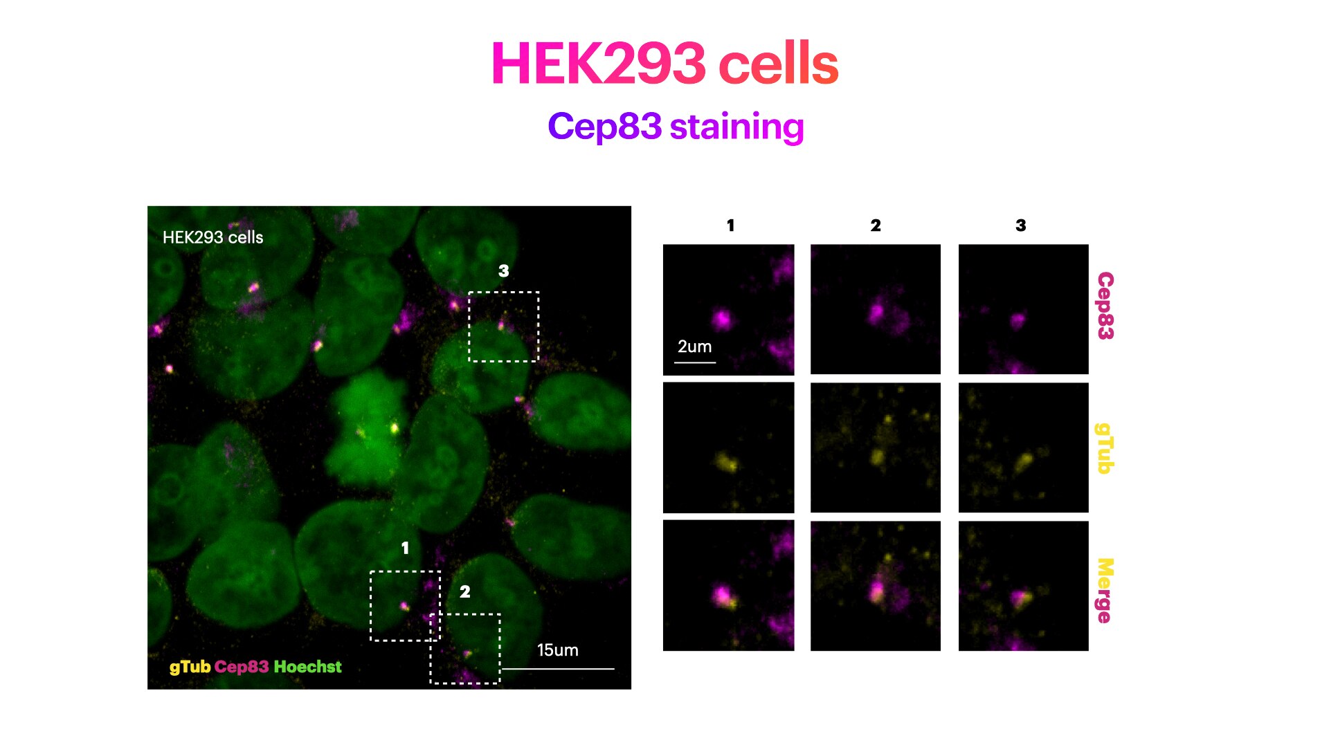
FH Elisa (Verified Customer) (03-01-2022) | HEK293 cells stained for Hoechst (DNA marker, in green), Cep83 (mother centriole distal appendage marker, in magenta) and g-Tubulin (pericentriolar matrix marker, in green). HEK293 cells were plated on Poly-lysine coated coverslips and fixed in cold methanol for 2' at -20C. Cells were then rehydrated with PBS for 5'. Membrane permeabilization was then performed with 0.1% Triton + 0.1% Tween +0.01%SDS in PBS for 5'. Cells were finally incubated with blocking buffer (5% BSA+ 0.1% Tween in PBS) for 30' at RT. Primary antibody was diluted in blocking buffer 1:200 and incubated for 1h at room temperature. Alexa-555-Anti-rabbit was used as secondary antibody (1:600 dilution) (1h at room temperature).
 |
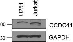
FH Sarah (Verified Customer) (07-03-2019) | Total cell lysate (15 ug) was resolved on a 4-12% Bis-Tris gel and transferred to nitrocellulose membrane. Membrane was incubated in blocking buffer (5% milk/0.1% Tween-20) for 1h. Membrane was incubated with anti-CCDC41 in blocking buffer (1:1000) at 4C overnight. After washing, membrane was incubated in anti-rabbit-HRP in blocking bufffer (1:3000) for 1h at room temperature. Protein was detected using ECL reagent and imaged on a chemiluminescence detection system.
 |

