CD107a / LAMP1 Monoklonaler Antikörper
CD107a / LAMP1 Monoklonal Antikörper für WB, IF/ICC, FC (Intra)
Wirt / Isotyp
Maus / IgG1, kappa
Getestete Reaktivität
human und mehr (2)
Anwendung
WB, IF/ICC, FC (Intra)
Konjugation
Unkonjugiert
CloneNo.
H4A3
Kat-Nr. : 65051-1-Ig
Synonyme
Geprüfte Anwendungen
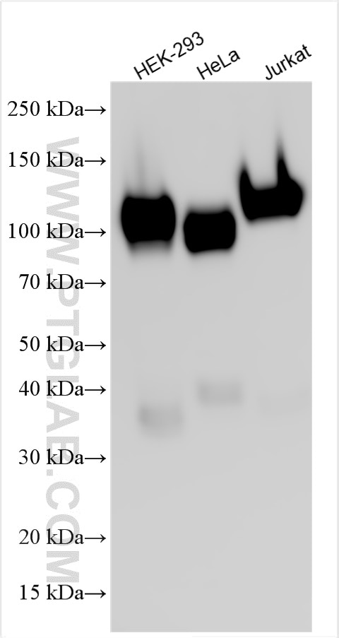
| Erfolgreiche Detektion in WB | HEK-293-Zellen, HeLa-Zellen, Jurkat-Zellen |
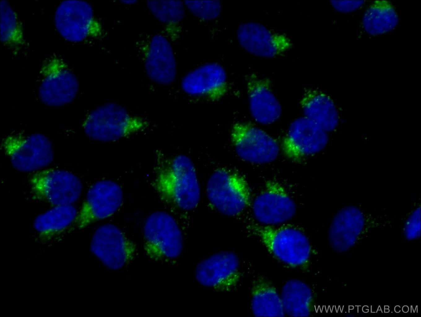
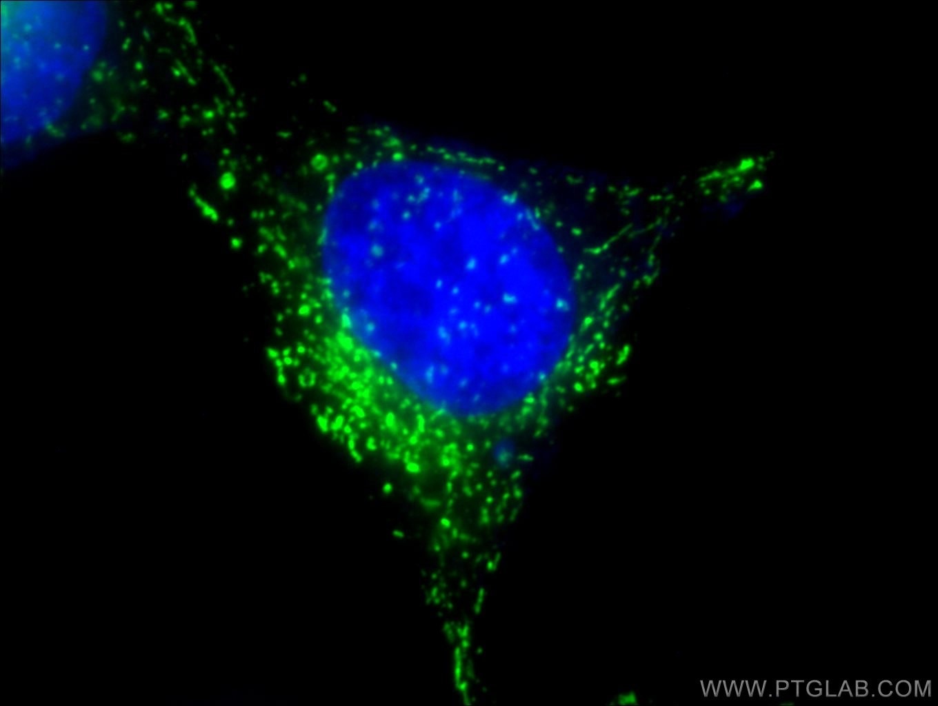
| Erfolgreiche Detektion in IF/ICC | HeLa-Zellen |
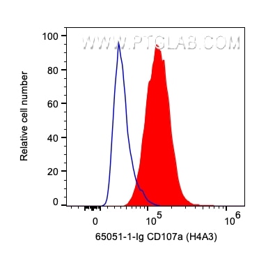
| Erfolgreiche Detektion in FC (Intra) | HeLa-Zellen |
Empfohlene Verdünnung
| Anwendung | Verdünnung |
|---|---|
| Western Blot (WB) | WB : 1:1000-1:8000 |
| Immunfluoreszenz (IF)/ICC | IF/ICC : 1:50-1:500 |
| This reagent has been tested for flow cytometric analysis. It is recommended that this reagent should be titrated in each testing system to obtain optimal results. | |
| Sample-dependent, check data in validation data gallery | |
Veröffentlichte Anwendungen
| WB | See 3 publications below |
| IF | See 8 publications below |
Produktinformation
65051-1-Ig bindet in WB, IF/ICC, FC (Intra) CD107a / LAMP1 und zeigt Reaktivität mit human
| Getestete Reaktivität | human |
| In Publikationen genannte Reaktivität | human, Affe, Maus |
| Wirt / Isotyp | Maus / IgG1, kappa |
| Klonalität | Monoklonal |
| Typ | Antikörper |
| Immunogen | Humane adhärente periphere Blutzellen |
| Vollständiger Name | lysosomal-associated membrane protein 1 |
| Berechnetes Molekulargewicht | 45 kDa |
| Beobachtetes Molekulargewicht | 100-120 kDa |
| GenBank-Zugangsnummer | BC006345 |
| Gene symbol | LAMP1 |
| Gene ID (NCBI) | 3916 |
| Konjugation | Unkonjugiert |
| Form | Liquid |
| Reinigungsmethode | Protein-G-Reinigung |
| Lagerungspuffer | PBS with 0.09% sodium azide |
| Lagerungsbedingungen | Store at 2-8°C. Stable for one year after shipment. |
Hintergrundinformationen
LAMP1 (CD107a) is a heavily glycosylated membrane protein enriched in the lysosomal membrane. LAMP1 is extensively glycosylated with asparagine-linked oligosaccharides which protect it from intracellular proteolysis (PMID: 10521503). Although LAMP1 is expressed largely in the endosome-lysosomal membrane of cells, it is also found on the plasma membrane (PMID: 16168398). Elevated LAMP1 expression at the cell surface has also been detected during platelet and granulocytic cell activation, as well as in some tumor cells (PMID: 29085473). LAMP1 functions to provide selectins with carbohydrate ligands. This protein has also been shown to be a marker of degranulation on lymphocytes such as CD8+ and NK cells and may also play a role in tumor cell differentiation and metastasis (PMID: 18835598; 29085473; 9426697).
Protokolle
| PRODUKTSPEZIFISCHE PROTOKOLLE | |
|---|---|
| WB protocol for CD107a / LAMP1 antibody 65051-1-Ig | Protokoll herunterladen |
| IF protocol for CD107a / LAMP1 antibody 65051-1-Ig | Protokoll herunterladen |
| STANDARD-PROTOKOLLE | |
|---|---|
| Klicken Sie hier, um unsere Standardprotokolle anzuzeigen |
Publikationen
| Species | Application | Title |
|---|---|---|
Eur J Pharmacol Cycloheptylprodigiosin from marine bacterium Spartinivicinus ruber MCCC 1K03745T induces a novel form of cell death characterized by Golgi disruption and enhanced secretion of cathepsin D in Non-small cell lung cancer cell lines | ||
Autophagy ATP6V1D drives hepatocellular carcinoma stemness and progression via both lysosome acidification-dependent and -independent mechanisms | ||
Cell Rep A MYC-STAMBPL1-TOE1 positive feedback loop mediates EGFR stability in hepatocellular carcinoma | ||
MAbs Targeted protein degradation through site-specific antibody conjugation with mannose 6-phosphate glycan | ||
CNS Neurosci Ther Inhibition of Salt-Inducible Kinase 2 Protects Motor Neurons From Degeneration in ALS by Activating Autophagic Flux and Enhancing mTORC1 Activity |
Rezensionen
The reviews below have been submitted by verified Proteintech customers who received an incentive for providing their feedback.
FH Parijat (Verified Customer) (09-23-2025) | Works well for IF. Typical lysosomal pattern was observed.
|
FH Christine (Verified Customer) (10-02-2024) | I randomly tried this antibody for western blotting (because this clone has been shown to work in WB in publications) and it worked beautifully at 1:500 on total lysates of HEK293T cells. Could be diluted much further.
|


