CEP250/CNAP1 Polyklonaler Antikörper
CEP250/CNAP1 Polyklonal Antikörper für WB, IHC, IF/ICC, IP, ELISA
Wirt / Isotyp
Kaninchen / IgG
Getestete Reaktivität
human und mehr (1)
Anwendung
WB, IHC, IF/ICC, IP, ELISA
Konjugation
Unkonjugiert
Kat-Nr. : 14498-1-AP
Synonyme
 at dilution of 1:1500 incubated at room temperature for 1.5 hours.")
 with HEK-293 cells lysate 1800 ug.")
.")
.")
 fixed hTERT-RPE1 cells using CEP250/CNAP1 antibody (14498-1-AP) at dilution of 1:100 and Cy3-conjugated Affinipure Goat Anti-Rabbit IgG(H+L), SCLT1 antibody (14875-1-AP, Magenta), CoraLite®488 ARL13B antibody (CL488-17711, green).")
Geprüfte Anwendungen
| Erfolgreiche Detektion in WB | A431-Zellen, HEK-293T-Zellen, HeLa-Zellen |
| Erfolgreiche IP | HEK-293-Zellen |
| Erfolgreiche Detektion in IHC | humanes Plazenta-Gewebe Hinweis: Antigendemaskierung mit TE-Puffer pH 9,0 empfohlen. (*) Wahlweise kann die Antigendemaskierung auch mit Citratpuffer pH 6,0 erfolgen. |
| Erfolgreiche Detektion in IF/ICC | hTERT-RPE1-Zellen |
Empfohlene Verdünnung
| Anwendung | Verdünnung |
|---|---|
| Western Blot (WB) | WB : 1:500-1:3000 |
| Immunpräzipitation (IP) | IP : 0.5-4.0 ug for 1.0-3.0 mg of total protein lysate |
| Immunhistochemie (IHC) | IHC : 1:50-1:500 |
| Immunfluoreszenz (IF)/ICC | IF/ICC : 1:50-1:500 |
| It is recommended that this reagent should be titrated in each testing system to obtain optimal results. | |
| Sample-dependent, check data in validation data gallery | |
Veröffentlichte Anwendungen
| WB | See 5 publications below |
| IHC | See 1 publications below |
| IF | See 31 publications below |
Produktinformation
14498-1-AP bindet in WB, IHC, IF/ICC, IP, ELISA CEP250/CNAP1 und zeigt Reaktivität mit human
| Getestete Reaktivität | human |
| In Publikationen genannte Reaktivität | human, Maus |
| Wirt / Isotyp | Kaninchen / IgG |
| Klonalität | Polyklonal |
| Typ | Antikörper |
| Immunogen | CEP250/CNAP1 fusion protein Ag5925 |
| Vollständiger Name | centrosomal protein 250kDa |
| Berechnetes Molekulargewicht | 281 kDa |
| Beobachtetes Molekulargewicht | 250 kDa |
| GenBank-Zugangsnummer | BC001433 |
| Gene symbol | CEP250/CNAP1 |
| Gene ID (NCBI) | 11190 |
| Konjugation | Unkonjugiert |
| Form | Liquid |
| Reinigungsmethode | Antigen-Affinitätsreinigung |
| Lagerungspuffer | PBS with 0.02% sodium azide and 50% glycerol |
| Lagerungsbedingungen | Bei -20°C lagern. Nach dem Versand ein Jahr lang stabil Aliquotieren ist bei -20oC Lagerung nicht notwendig. 20ul Größen enthalten 0,1% BSA. |
Hintergrundinformationen
CEP250, also known as C-Nap1, is a 250 kDa coiled-coil protein that localizes to the proximal ends of mother and daughter centrioles. It is required for centriole-centriole cohesion during interphase of the cell cycle. It dissociates from the centrosomes when parental centrioles separate at the beginning of mitosis. The protein associates with and is phosphorylated by NIMA-related kinase 2, which is also associated with the centrosome.
Protokolle
| PRODUKTSPEZIFISCHE PROTOKOLLE | |
|---|---|
| WB protocol for CEP250/CNAP1 antibody 14498-1-AP | Protokoll herunterladen |
| IHC protocol for CEP250/CNAP1 antibody 14498-1-AP | Protokoll herunterladenl |
| IF protocol for CEP250/CNAP1 antibody 14498-1-AP | Protokoll herunterladen |
| IP protocol for CEP250/CNAP1 antibody 14498-1-AP | Protokoll herunterladen |
| STANDARD-PROTOKOLLE | |
|---|---|
| Klicken Sie hier, um unsere Standardprotokolle anzuzeigen |
Publikationen
| Species | Application | Title |
|---|---|---|
Science A liquid-like spindle domain promotes acentrosomal spindle assembly in mammalian oocytes. | ||
Nat Commun Microtubule asters anchored by FSD1 control axoneme assembly and ciliogenesis. | ||
Nat Commun Cytoplasmic E2f4 forms organizing centres for initiation of centriole amplification during multiciliogenesis. | ||
PLoS Biol Stable centrosomal roots disentangle to allow interphase centriole independence. | ||
Proc Natl Acad Sci U S A Dishevelled is a NEK2 kinase substrate controlling dynamics of centrosomal linker proteins. | ||
J Cell Biol Uncoordinated centrosome cycle underlies the instability of non-diploid somatic cells in mammals. |
Rezensionen
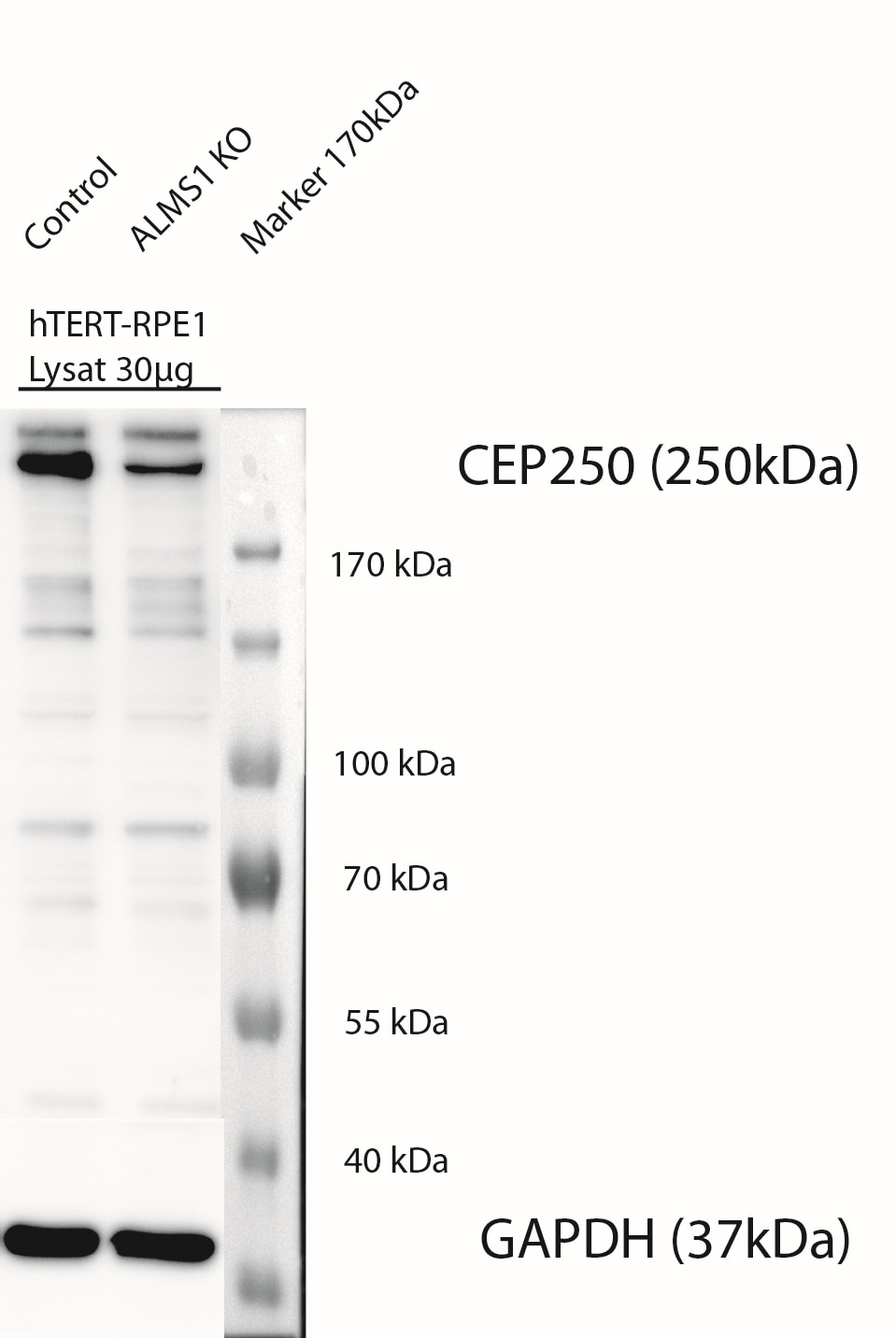
The reviews below have been submitted by verified Proteintech customers who received an incentive for providing their feedback.
FH Karsten (Verified Customer) (12-22-2022) | WB: 1:600 dilution in 5% Milch in 1xTBST over nigth at 4°C, 30µg Protein loaded: result show prominent band at a hight of 250 kDa. unspecific bands also visible, but not prominent. worked fine IFS: 1:100, MetOH (-20°) fixation for 5 min at RT , Permeabilisation with 0.3% PBST (Triton) for 5 min at RT, Blocking with 1% BSA in PBS for 1h at RT. Antibody dilution 1:100 in 1%BSA in PBS 1h RT, sek Ak 1h at RT- nice basal body stainings at cilia in hTERT-RPE1 cells, that were starved for 3 days.
 |
FH Hairuo (Verified Customer) (12-16-2019) | Used in IF in neonatal mouse testis cry-sections.Works well.
|

