- Featured Product
- KD/KO Validated
Cytochrome c Polyklonaler Antikörper
Cytochrome c Polyklonal Antikörper für WB, IHC, IF-P, FC (Intra), ELISA
Wirt / Isotyp
Kaninchen / IgG
Getestete Reaktivität
human, Maus, Ratte und mehr (6)
Anwendung
WB, IHC, IF-P, FC (Intra), IP, ELISA
Konjugation
Unkonjugiert
Kat-Nr. : 10993-1-AP
Synonyme
 at dilution of 1:4000 incubated at room temperature for 1.5 hours.")
 with sh-Control and sh-Cytochrome c transfected HepG2 cells.")
 with sh-Control and sh-Cytochrome c transfected HEK-293 cells.")
 at dilution of 1:8000 incubated at room temperature for 1.5 hours.")
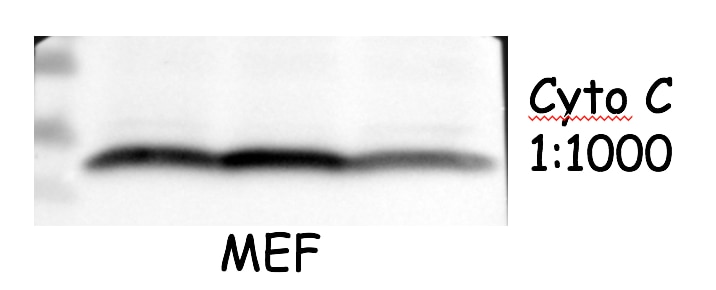
 at dilution of 1:1000 incubated at room temperature for 1.5 hours.")
 at dilution of 1:800 incubated at room temperature for 1.5 hours.")
 at dilution of 1:800 incubated at room temperature for 1.5 hours.")
 at dilution of 1:800 incubated at room temperature for 1.5 hours.")
 at dilution of 1:1000 (under 40x lens). Heat mediated antigen retrieval with Tris-EDTA buffer (pH 9.0).")
 at dilution of 1:1000 (under 10x lens). Heat mediated antigen retrieval with Tris-EDTA buffer (pH 9.0).")
 at dilution of 1:1000 (under 40x lens). Heat mediated antigen retrieval with Tris-EDTA buffer (pH 9.0).")
 at dilution of 1:1000 (under 10x lens). Heat mediated antigen retrieval with Tris-EDTA buffer (pH 9.0).")
 at dilution of 1:3000 (under 20x lens). Heat mediated antigen retrieval with Tris-EDTA buffer (pH 9.0).")
 at dilution of 1:1000 (under 10x lens). Heat mediated antigen retrieval with Tris-EDTA buffer (pH 9.0).")
 at dilution of 1:50 (under 10x lens).")
 at dilution of 1:50 (under 40x lens).")
 fixed paraffin-embedded mouse eye tissue using Cytochrome c antibody (10993-1-AP) at dilution of 1:200 and CoraLite®488-Conjugated AffiniPure Goat Anti-Rabbit IgG(H+L) (SA00013-2). Heat mediated antigen retrieval with Tris-EDTA buffer (pH 9.0).")
 fixed paraffin-embedded mouse eye tissue using Cytochrome c antibody (10993-1-AP) at dilution of 1:200 and CoraLite®488-Conjugated AffiniPure Goat Anti-Rabbit IgG(H+L) (SA00013-2). Heat mediated antigen retrieval with Tris-EDTA buffer (pH 9.0).")
 and CoraLite®488-Conjugated AffiniPure Goat Anti-Rabbit IgG(H+L) at dilution 1:1000 (red), or 0.4 ug Isotype Control. Cells were fixed with 4% PFA and permeabilized with Flow Cytometry Perm Buffer (PF00011-C).")
Geprüfte Anwendungen
| Erfolgreiche Detektion in WB | HeLa-Zellen, C2C12-Zellen, HEK-293-Zellen, HepG2-Zellen, Jurkat-Zellen, Mausnierengewebe, Maus-Skelettmuskelgewebe, NIH/3T3-Zellen, Rattennierengewebe, Rattenlebergewebe, Ratten-Skelettmuskelgewebe |
| Erfolgreiche Detektion in IHC | humanes Lebergewebe, humanes Mammakarzinomgewebe, humanes Kolonkarzinomgewebe, Maushirngewebe Hinweis: Antigendemaskierung mit TE-Puffer pH 9,0 empfohlen. (*) Wahlweise kann die Antigendemaskierung auch mit Citratpuffer pH 6,0 erfolgen. |
| Erfolgreiche Detektion in IF-P | Maus-Augengewebe |
| Erfolgreiche Detektion in FC (Intra) | HepG2-Zellen |
Empfohlene Verdünnung
| Anwendung | Verdünnung |
|---|---|
| Western Blot (WB) | WB : 1:1000-1:8000 |
| Immunhistochemie (IHC) | IHC : 1:500-1:2000 |
| Immunfluoreszenz (IF)-P | IF-P : 1:50-1:500 |
| Durchflusszytometrie (FC) (INTRA) | FC (INTRA) : 0.40 ug per 10^6 cells in a 100 µl suspension |
| It is recommended that this reagent should be titrated in each testing system to obtain optimal results. | |
| Sample-dependent, check data in validation data gallery | |
Veröffentlichte Anwendungen
| WB | See 582 publications below |
| IHC | See 33 publications below |
| IF | See 38 publications below |
| IP | See 1 publications below |
| FC | See 1 publications below |
Produktinformation
10993-1-AP bindet in WB, IHC, IF-P, FC (Intra), IP, ELISA Cytochrome c und zeigt Reaktivität mit human, Maus, Ratten
| Getestete Reaktivität | human, Maus, Ratte |
| In Publikationen genannte Reaktivität | human, Affe, hamster, Hausschwein, Huhn, Maus, Ratte, Ziege, Hippospongia |
| Wirt / Isotyp | Kaninchen / IgG |
| Klonalität | Polyklonal |
| Typ | Antikörper |
| Immunogen | Cytochrome c fusion protein Ag1455 |
| Vollständiger Name | cytochrome c, somatic |
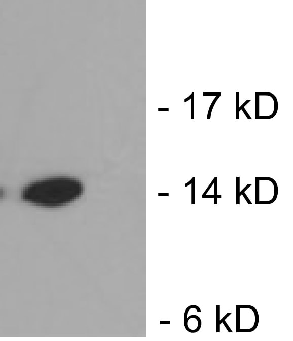
| Berechnetes Molekulargewicht | 12 kDa |
| Beobachtetes Molekulargewicht | 12-15 kDa |
| GenBank-Zugangsnummer | BC009578 |
| Gene symbol | Cytochrome c |
| Gene ID (NCBI) | 54205 |
| Konjugation | Unkonjugiert |
| Form | Liquid |
| Reinigungsmethode | Antigen-Affinitätsreinigung |
| Lagerungspuffer | PBS with 0.02% sodium azide and 50% glycerol |
| Lagerungsbedingungen | Bei -20°C lagern. Nach dem Versand ein Jahr lang stabil Aliquotieren ist bei -20oC Lagerung nicht notwendig. 20ul Größen enthalten 0,1% BSA. |
Hintergrundinformationen
Cytochrome c is a 12-15 kDa electron transporting protein located in the inner mitochondrial membrane. As a part of respiratory chain, cytochrome c plays a critical role in the process of oxidative phosphorylation and ATP producing. Besides, cytochrome c also gets implicated in apoptosis process. Upon apoptotic stimulation, cytochrome c ca99n be released from mitochondria into cytoplasm, which is required for caspase-3 activation and the occurrence of apoptosis.
Protokolle
| PRODUKTSPEZIFISCHE PROTOKOLLE | |
|---|---|
| WB protocol for Cytochrome c antibody 10993-1-AP | Protokoll herunterladen |
| IHC protocol for Cytochrome c antibody 10993-1-AP | Protokoll herunterladenl |
| IF protocol for Cytochrome c antibody 10993-1-AP | Protokoll herunterladen |
| STANDARD-PROTOKOLLE | |
|---|---|
| Klicken Sie hier, um unsere Standardprotokolle anzuzeigen |
Publikationen
| Species | Application | Title |
|---|---|---|
Cell Tau interactome maps synaptic and mitochondrial processes associated with neurodegeneration. | ||
Nat Commun MYG1 drives glycolysis and colorectal cancer development through nuclear-mitochondrial collaboration | ||
Mol Cell Filamentous GLS1 promotes ROS-induced apoptosis upon glutamine deprivation via insufficient asparagine synthesis. | ||
Adv Sci (Weinh) Hierarchical Targeting Nanodrug with Holistic DNA Protection for Effective Treatment of Acute Kidney Injury | ||
Adv Sci (Weinh) Mitochondrial tRNAGlu 14693A>G Mutation, an "Enhancer" to the Phenotypic Expression of Leber's Hereditary Optic Neuropathy | ||
Acta Pharm Sin B Honokiol alleviated neurodegeneration by reducing oxidative stress and improving mitochondrial function in mutant SOD1 cellular and mouse models of amyotrophic lateral sclerosis |
Rezensionen
The reviews below have been submitted by verified Proteintech customers who received an incentive for providing their feedback.
FH Kazu (Verified Customer) (12-14-2022) | Worked well for 4% PFA fixed mouse optic nerve using this antibody at dilution of 1:200. Little background observed.
|
FH Azita (Verified Customer) (06-02-2021) | Western blot analysis using Cytochrome c polyclonal antibody in NSC34 cell line at dilution of 1:1000.
|
FH Ying (Verified Customer) (04-22-2021) | good antibody. work well with mouse cells
 |
FH Chi (Verified Customer) (09-26-2019) | The antibody works very well with good signal and low background.
 |
FH Xiaoping (Verified Customer) (10-22-2018) | The signal is good and the size is between 10-15kDa.
 |

