- Featured Product
- KD/KO Validated
E-cadherin Monoklonaler Antikörper
E-cadherin Monoklonal Antikörper für WB, IHC, IF/ICC, IF-P, IF-Fro, ELISA
Wirt / Isotyp
Maus / IgG2b
Getestete Reaktivität
Hausschwein, human, Maus, Ratte und mehr (1)
Anwendung
WB, IHC, IF/ICC, IF-P, IF-Fro, ELISA
Konjugation
Unkonjugiert
CloneNo.
6B11F11
Kat-Nr. : 60335-1-Ig
Synonyme
 with sh-Control and sh-E-cadherin transfected A431 cells.")
 at dilution of 1:4000 incubated at room temperature for 1.5 hours.")
 at dilution of 1:1000 incubated at room temperature for 1.5 hours.")
 at dilution of 1:8000 incubated at room temperature for 1.5 hours.")
 at dilution of 1:8000 incubated at room temperature for 1.5 hours.")
 at dilution of 1:2000 (under 10x lens). Heat mediated antigen retrieval with Tris-EDTA buffer (pH 9.0).")
 at dilution of 1:2000 (under 40x lens). Heat mediated antigen retrieval with Tris-EDTA buffer (pH 9.0).")
 at dilution of 1:2000 (under 10x lens. Heat mediated antigen retrieval with Tris-EDTA buffer (pH 9.0).")
 at dilution of 1:2000 (under 40x lens. Heat mediated antigen retrieval with Tris-EDTA buffer (pH 9.0).")
 at dilution of 1:8000 (under 10x lens. Heat mediated antigen retrieval with Tris-EDTA buffer (pH 9.0).")
 at dilution of 1:10000 (under 10x lens). Heat mediated antigen retrieval with Tris-EDTA buffer (pH 9.0).")
 at dilution of 1:10000 (under 40x lens). Heat mediated antigen retrieval with Tris-EDTA buffer (pH 9.0).")
 at dilution of 1:300 (under 10x lens). Heat mediated antigen retrieval with Tris-EDTA buffer (pH 9.0).")
 at dilution of 1:300 (under 40x lens). Heat mediated antigen retrieval with Tris-EDTA buffer (pH 9.0).")
 at dilution of 1:4000 (under 10x lens. Heat mediated antigen retrieval with Tris-EDTA buffer (pH 9.0).")
 at dilution of 1:4000 (under 40x lens. Heat mediated antigen retrieval with Tris-EDTA buffer (pH 9.0).")
 at dilution of 1:2000 (under 10x lens). Heat mediated antigen retrieval with Tris-EDTA buffer (pH 9.0).")
 at dilution of 1:2000 (under 40x lens). Heat mediated antigen retrieval with Tris-EDTA buffer (pH 9.0).")
 fixed human kidney tissue using E-cadherin antibody (60335-1-Ig, Clone: 6B11F11 ) at dilution of 1:300 and CoraLite®594-Conjugated AffiniPure Goat Anti-Mouse IgG(H+L), (18150-1-AP, green). DNA was stained by DAPI (blue).")
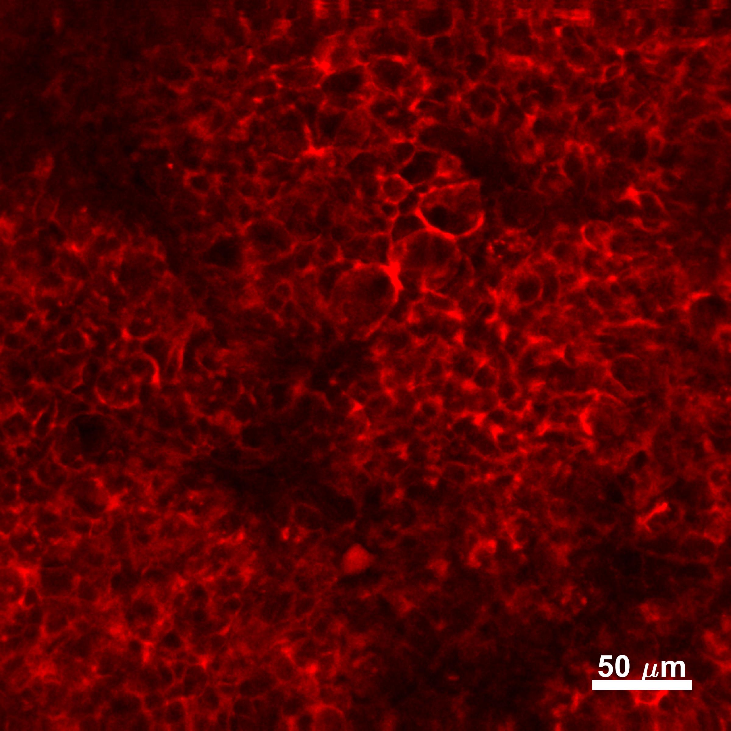
 fixed human breast cancer tissue using E-cadherin antibody (60335-1-Ig, Clone: 6B11F11 ) at dilution of 1:400 and CoraLite®488-Conjugated AffiniPure Goat Anti-Mouse IgG(H+L).")
 fixed frozen OCT-embedded mouse brain tissue using E-cadherin antibody (60335-1-Ig, Clone: 6B11F11 ) at dilution of 1:400 and CoraLite®488-Conjugated Goat Anti-Mouse IgG(H+L) (SA00013-1).")
 fixed MCF-7 cells using E-cadherin antibody (60335-1-Ig, Clone: 6B11F11 ) at dilution of 1:400 and CoraLite®488-Conjugated AffiniPure Goat Anti-Mouse IgG(H+L).")
 fixed mouse breast cancer using E-cadherin antibody (60335-1-Ig, Clone: 6B11F11 ) at dilution of 1:800 and CoraLite®488-Conjugated Goat Anti-Mouse IgG(H+L) (SA00013-1).")
Geprüfte Anwendungen
| Erfolgreiche Detektion in WB | PC-3-Zellen, A431-Zellen, MCF-7-Zellen, MKN-45-Zellen, Hausschwein-Hirngewebe, SGC-7901-Zellen |
| Erfolgreiche Detektion in IHC | humanes Mammakarzinomgewebe, humanes Kolongewebe, Ratten-Kolongewebe, Ratten-Magengewebe Hinweis: Antigendemaskierung mit TE-Puffer pH 9,0 empfohlen. (*) Wahlweise kann die Antigendemaskierung auch mit Citratpuffer pH 6,0 erfolgen. |
| Erfolgreiche Detektion in IF-P | humanes Mammakarzinomgewebe, humanes Nierengewebe, MCF-7-Zellen |
| Erfolgreiche Detektion in IF-Fro | Maushirngewebe |
| Erfolgreiche Detektion in IF/ICC | MCF-7-Zellen, mouse breast cancer |
Empfohlene Verdünnung
| Anwendung | Verdünnung |
|---|---|
| Western Blot (WB) | WB : 1:2000-1:8000 |
| Immunhistochemie (IHC) | IHC : 1:1000-1:4000 |
| Immunfluoreszenz (IF)-P | IF-P : 1:200-1:800 |
| Immunfluoreszenz (IF)-FRO | IF-FRO : 1:200-1:800 |
| Immunfluoreszenz (IF)/ICC | IF/ICC : 1:200-1:800 |
| It is recommended that this reagent should be titrated in each testing system to obtain optimal results. | |
| Sample-dependent, check data in validation data gallery | |
Veröffentlichte Anwendungen
| WB | See 190 publications below |
| IHC | See 27 publications below |
| IF | See 57 publications below |
Produktinformation
60335-1-Ig bindet in WB, IHC, IF/ICC, IF-P, IF-Fro, ELISA E-cadherin und zeigt Reaktivität mit Hausschwein, human, Maus, Ratten
| Getestete Reaktivität | Hausschwein, human, Maus, Ratte |
| In Publikationen genannte Reaktivität | human, Affe, Hausschwein, Maus, Ratte |
| Wirt / Isotyp | Maus / IgG2b |
| Klonalität | Monoklonal |
| Typ | Antikörper |
| Immunogen | E-cadherin fusion protein Ag15085 |
| Vollständiger Name | cadherin 1, type 1, E-cadherin (epithelial) |
| Berechnetes Molekulargewicht | 882 aa, 97 kDa |
| Beobachtetes Molekulargewicht | 120 kDa |
| GenBank-Zugangsnummer | BC141838 |
| Gene symbol | E-cadherin |
| Gene ID (NCBI) | 999 |
| Konjugation | Unkonjugiert |
| Form | Liquid |
| Reinigungsmethode | Protein-A-Reinigung |
| Lagerungspuffer | PBS with 0.02% sodium azide and 50% glycerol |
| Lagerungsbedingungen | Bei -20°C lagern. Nach dem Versand ein Jahr lang stabil Aliquotieren ist bei -20oC Lagerung nicht notwendig. 20ul Größen enthalten 0,1% BSA. |
Hintergrundinformationen
Cadherins are a family of transmembrane glycoproteins that mediate calcium-dependent cell-cell adhesion and play an important role in the maintenance of normal tissue architecture. E-cadherin (epithelial cadherin), also known as CDH1 (cadherin 1) or CAM 120/80, is a classical member of the cadherin superfamily which also include N-, P-, R-, and B-cadherins. E-cadherin is expressed on the cell surface in most epithelial tissues. The extracellular region of E-cadherin establishes calcium-dependent homophilic trans binding, providing specific interaction with adjacent cells, while the cytoplasmic domain is connected to the actin cytoskeleton through the interaction with p120-, α-, β-, and γ-catenin (plakoglobin). E-cadherin is important in the maintenance of the epithelial integrity, and is involved in mechanisms regulating proliferation, differentiation, and survival of epithelial cell. E-cadherin may also play a role in tumorigenesis. It is considered to be an invasion suppressor protein and its loss is an indicator of high tumor aggressiveness.
Protokolle
| PRODUKTSPEZIFISCHE PROTOKOLLE | |
|---|---|
| WB protocol for E-cadherin antibody 60335-1-Ig | Protokoll herunterladen |
| IHC protocol for E-cadherin antibody 60335-1-Ig | Protokoll herunterladenl |
| IF protocol for E-cadherin antibody 60335-1-Ig | Protokoll herunterladen |
| STANDARD-PROTOKOLLE | |
|---|---|
| Klicken Sie hier, um unsere Standardprotokolle anzuzeigen |
Publikationen
| Species | Application | Title |
|---|---|---|
Theranostics Vitamin D binding protein (VDBP) hijacks twist1 to inhibit vasculogenic mimicry in hepatocellular carcinoma | ||
Theranostics FSH induces EMT in ovarian cancer via ALKBH5-regulated Snail m6A demethylation | ||
Biomaterials Urinary exosomes-based Engineered Nanovectors for Homologously Targeted Chemo-Chemodynamic Prostate Cancer Therapy via abrogating IGFR/AKT/NF-kB/IkB signaling. | ||
Redox Biol Riboflavin deficiency leads to irreversible cellular changes in the RPE and disrupts retinal function through alterations in cellular metabolic homeostasis. |
Rezensionen
The reviews below have been submitted by verified Proteintech customers who received an incentive for providing their feedback.
FH Aqib (Verified Customer) (09-02-2024) | It works quite well. recommended
|
FH Saba (Verified Customer) (06-14-2022) | The IF staining was very good and satisfying.
|
FH Silvia (Verified Customer) (02-09-2022) | The antibody worked well on HT-29 cells at 1:800 dilution for IF.
|
FH Joshua (Verified Customer) (12-27-2019) | Caco-2 cells fixed in 4% paraformaldehyde. Stained overnight at 4C. Bright stain, minimal background
 |
FH Louisiane (Verified Customer) (02-06-2019) | Cells were fixed with 4% PFA for 10 min, permeabilized with 0.1% Triton-X100 for 5 min and blocked with 1% FBS/1% BSA in PBS for 3 h. Antibodies were diluted in 1% FBS/1% BSA in PBS. Primary antibody: 2 h. Alexa Fluor anti-mouse secondary antibody (1:250): 1 h.Cells were imaged by confocal microscopy - no labeling was observed.
|

