- Featured Product
- KD/KO Validated
E2F1 Monoklonaler Antikörper
E2F1 Monoklonal Antikörper für WB, ELISA
Wirt / Isotyp
Maus / IgG2b
Getestete Reaktivität
human, Ratte und mehr (1)
Anwendung
WB, IHC, IF, IP, CoIP, ChIP, ELISA
Konjugation
Unkonjugiert
CloneNo.
5D7G8
Kat-Nr. : 66515-1-Ig
Synonyme
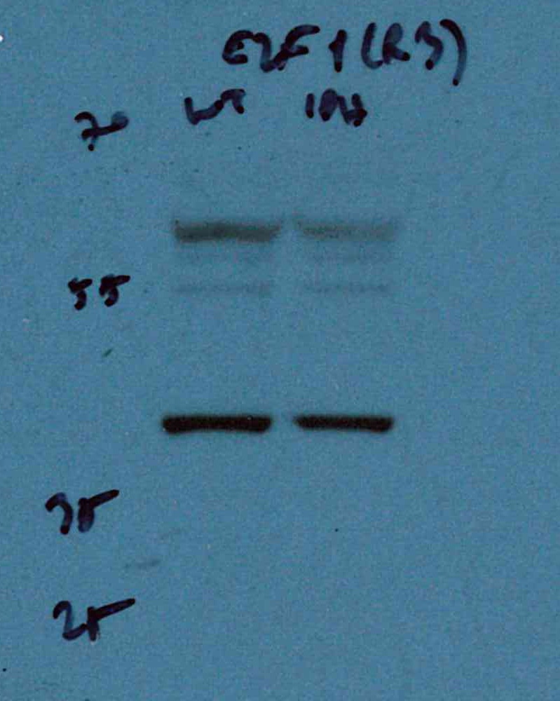
 at dilution of 1:3000 incubated at room temperature for 1.5 hours.")
 with sh-Control and sh-E2F1 transfected HepG2 cells.")
Geprüfte Anwendungen
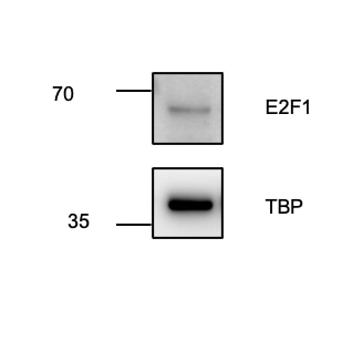
| Erfolgreiche Detektion in WB | LNCaP-Zellen, HEK-293-Zellen, HeLa-Zellen, HepG2-Zellen, Jurkat-Zellen |
Empfohlene Verdünnung
| Anwendung | Verdünnung |
|---|---|
| Western Blot (WB) | WB : 1:1000-1:6000 |
| It is recommended that this reagent should be titrated in each testing system to obtain optimal results. | |
| Sample-dependent, check data in validation data gallery | |
Veröffentlichte Anwendungen
| KD/KO | See 10 publications below |
| WB | See 59 publications below |
| IHC | See 5 publications below |
| IF | See 4 publications below |
| IP | See 4 publications below |
| CoIP | See 4 publications below |
| ChIP | See 12 publications below |
Produktinformation
66515-1-Ig bindet in WB, IHC, IF, IP, CoIP, ChIP, ELISA E2F1 und zeigt Reaktivität mit human, Ratten
| Getestete Reaktivität | human, Ratte |
| In Publikationen genannte Reaktivität | human, Maus, Ratte |
| Wirt / Isotyp | Maus / IgG2b |
| Klonalität | Monoklonal |
| Typ | Antikörper |
| Immunogen | E2F1 fusion protein Ag17363 |
| Vollständiger Name | E2F transcription factor 1 |
| Berechnetes Molekulargewicht | 437 aa, 47 kDa |
| Beobachtetes Molekulargewicht | 55-60 kDa |
| GenBank-Zugangsnummer | BC050369 |
| Gene symbol | E2F1 |
| Gene ID (NCBI) | 1869 |
| Konjugation | Unkonjugiert |
| Form | Liquid |
| Reinigungsmethode | Protein-A-Reinigung |
| Lagerungspuffer | PBS with 0.02% sodium azide and 50% glycerol |
| Lagerungsbedingungen | Bei -20°C lagern. Nach dem Versand ein Jahr lang stabil Aliquotieren ist bei -20oC Lagerung nicht notwendig. 20ul Größen enthalten 0,1% BSA. |
Hintergrundinformationen
Transcription factor E2F1 (E2F1), also known as RBBP3, is a transcription activator that binds DNA cooperatively with dp proteins through the E2 recognition site, 5'-TTTC[CG]CGC-3' found in the promoter region of a number of genes whose products are involved in cell cycle regulation or in DNA replication. The DRTF1/E2F complex functions in the control of cell-cycle progression from G1 to S phase. E2F-1 binds preferentially RB1 protein, in a cell-cycle dependent manner. It can mediate both cell proliferation and p53-dependent apoptosis. The calculated molecular weight of E2F1 is 47 kDa, but the sumoylated E2F1 is bout 55-60 kDa.
Protokolle
| PRODUKTSPEZIFISCHE PROTOKOLLE | |
|---|---|
| WB protocol for E2F1 antibody 66515-1-Ig | Protokoll herunterladen |
| STANDARD-PROTOKOLLE | |
|---|---|
| Klicken Sie hier, um unsere Standardprotokolle anzuzeigen |
Publikationen
| Species | Application | Title |
|---|---|---|
Cancer Res LncRNA AGPG confers endocrine resistance in breast cancer by promoting E2F1 activity | ||
J Adv Res Transducin-like enhancer of split 3 protects against lipopolysaccharide-induced inflammation through DDX5-ATF1-PPP2R5A signaling | ||
Cell Death Dis Identification of STAM-binding protein as a target for the treatment of gemcitabine resistance pancreatic cancer in a nutrient-poor microenvironment
| ||
Cell Death Dis ZNF652 exerts a tumor suppressor role in lung cancer by transcriptionally downregulating cyclin D3 | ||
Cell Rep FAK-mediated phosphorylation at Y464 regulates p85β nuclear translocation to promote tumorigenesis of ccRCC by repressing RB1 expression |
Rezensionen
The reviews below have been submitted by verified Proteintech customers who received an incentive for providing their feedback.
FH Umut (Verified Customer) (08-30-2023) | It works very well, even though it is diluted a lot, i.e., 1:2000. The total protein concentration of my samples were around 2 ug/ml before adding 1:4 laemmli buffer and denature, and it is able to capture the target at low exposure times.
 |
FH Juliane (Verified Customer) (05-11-2023) | The antibody worked fine, but only under using ECL sensitve reagent and high exposure when imaging.
 |

