- Featured Product
- KD/KO Validated
EGFR Monoklonaler Antikörper
EGFR Monoklonal Antikörper für WB, IHC, ELISA
Wirt / Isotyp
Maus / IgG1
Getestete Reaktivität
human
Anwendung
WB, IHC, IF, ELISA
Konjugation
Unkonjugiert
CloneNo.
2A2H10
Kat-Nr. : 66455-1-Ig
Synonyme
 at dilution of 1:20000 incubated at room temperature for 1.5 hours.")
 at dilution of 1:20000 incubated at room temperature for 1.5 hours.")
 with sh-Control and sh-EGFR transfected HepG2 cells.")
 at dilution of 1:20000 incubated at room temperature for 1.5 hours.")
 at dilution of 1:9700 incubated at room temperature for 1.5 hours.")
 at dilution of 1:9700 incubated at room temperature for 1.5 hours.")
 at dilution of 1:9700 incubated at room temperature for 1.5 hours.")
 at dilution of 1:9700 incubated at room temperature for 1.5 hours.")
 at dilution of 1:9700 incubated at room temperature for 1.5 hours.")
 at dilution of 1:9700 incubated at room temperature for 1.5 hours.")
 at dilution of 1:9700 incubated at room temperature for 1.5 hours.")
 at dilution of 1:2000 (under 10x lens). Heat mediated antigen retrieval with Tris-EDTA buffer (pH 9.0).")
 at dilution of 1:4000 (under 10x lens). Heat mediated antigen retrieval with Tris-EDTA buffer (pH 9.0).")
 at dilution of 1:4000 (under 40x lens). Heat mediated antigen retrieval with Tris-EDTA buffer (pH 9.0).")
 at dilution of 1:4000 (under 20x lens). Heat mediated antigen retrieval with Tris-EDTA buffer (pH 9.0).")
 at dilution of 1:4000 (under 20x lens). Heat mediated antigen retrieval with Tris-EDTA buffer (pH 9.0).")
 at dilution of 1:800 (under 10x lens. Heat mediated antigen retrieval with Tris-EDTA buffer (pH 9.0).")
 at dilution of 1:800 (under 40x lens. Heat mediated antigen retrieval with Tris-EDTA buffer (pH 9.0).")
 at dilution of 1:1000 (under 10x lens. Heat mediated antigen retrieval with Tris-EDTA buffer (pH 9.0).")
 at dilution of 1:1000 (under 40x lens. Heat mediated antigen retrieval with Tris-EDTA buffer (pH 9.0).")
 at dilution of 1:1000 (under 10x lens. Heat mediated antigen retrieval with Tris-EDTA buffer (pH 9.0).")
 at dilution of 1:1000 (under 40x lens. Heat mediated antigen retrieval with Tris-EDTA buffer (pH 9.0).")
 at dilution of 1:2000 (under 10x lens). Heat mediated antigen retrieval with Tris-EDTA buffer (pH 9.0).")
 at dilution of 1:2000 (under 40x lens). Heat mediated antigen retrieval with Tris-EDTA buffer (pH 9.0).")
 at dilution of 1:2000 (under 10x lens). Heat mediated antigen retrieval with Tris-EDTA buffer (pH 9.0).")
 at dilution of 1:2000 (under 40x lens). Heat mediated antigen retrieval with Tris-EDTA buffer (pH 9.0).")
 at dilution of 1:2000 (under 10x lens). Heat mediated antigen retrieval with Tris-EDTA buffer (pH 9.0).")
 at dilution of 1:2000 (under 40x lens). Heat mediated antigen retrieval with Tris-EDTA buffer (pH 9.0).")
Geprüfte Anwendungen
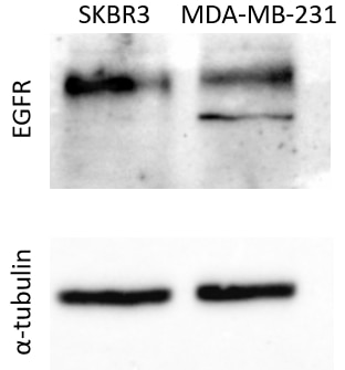
| Erfolgreiche Detektion in WB | A431-Zellen, A549-Zellen, EC109-Zellen, HeLa-Zellen, HepG2-Zellen, LNCaP-Zellen, MDA-MB-231-Zellen, MDA-MB-468-Zellen, PC-3-Zellen, SKOV-3-Zellen |
| Erfolgreiche Detektion in IHC | humanes Tonsillitisgewebe, humanes Mammakarzinomgewebe, humanes Zervixkarzinomgewebe, humanes Kolonkarzinomgewebe, humanes Gliomgewebe, humanes Lungenkarzinomgewebe, humanes Plazenta-Gewebe, humanes Hautkrebsgewebe Hinweis: Antigendemaskierung mit TE-Puffer pH 9,0 empfohlen. (*) Wahlweise kann die Antigendemaskierung auch mit Citratpuffer pH 6,0 erfolgen. |
Empfohlene Verdünnung
| Anwendung | Verdünnung |
|---|---|
| Western Blot (WB) | WB : 1:5000-1:50000 |
| Immunhistochemie (IHC) | IHC : 1:2000-1:8000 |
| It is recommended that this reagent should be titrated in each testing system to obtain optimal results. | |
| Sample-dependent, check data in validation data gallery | |
Veröffentlichte Anwendungen
| KD/KO | See 6 publications below |
| WB | See 85 publications below |
| IHC | See 14 publications below |
| IF | See 10 publications below |
Produktinformation
66455-1-Ig bindet in WB, IHC, IF, ELISA EGFR und zeigt Reaktivität mit human
| Getestete Reaktivität | human |
| In Publikationen genannte Reaktivität | human |
| Wirt / Isotyp | Maus / IgG1 |
| Klonalität | Monoklonal |
| Typ | Antikörper |
| Immunogen | EGFR fusion protein Ag24947 |
| Vollständiger Name | epidermal growth factor receptor (erythroblastic leukemia viral (v-erb-b) oncogene homolog, avian) |
| Berechnetes Molekulargewicht | 1210 aa, 134 kDa |
| Beobachtetes Molekulargewicht | 145-165 kDa |
| GenBank-Zugangsnummer | BC094761 |
| Gene symbol | EGFR |
| Gene ID (NCBI) | 1956 |
| Konjugation | Unkonjugiert |
| Form | Liquid |
| Reinigungsmethode | Protein-G-Reinigung |
| Lagerungspuffer | PBS with 0.02% sodium azide and 50% glycerol |
| Lagerungsbedingungen | Bei -20°C lagern. Nach dem Versand ein Jahr lang stabil Aliquotieren ist bei -20oC Lagerung nicht notwendig. 20ul Größen enthalten 0,1% BSA. |
Hintergrundinformationen
EGFR, also named as ERBB1, is a cell-surface receptor for members of the epidermal growth factor family (EGF-family) of extracellular protein ligands. Binding of the protein to a ligand induces receptor dimerization and tyrosine autophosphorylation and leads to cell proliferation. The gene resides on chromosome 7p12, encoding a 170 kDa membrane-associated glycoprotein. Recent studies have shown EGFR plays a critical role in cancer development and progression, including cell proliferation, apoptosis, angiogenesis, and metastatic spread. Mutations in this gene are associated with lung cancer.
Protokolle
| PRODUKTSPEZIFISCHE PROTOKOLLE | |
|---|---|
| WB protocol for EGFR antibody 66455-1-Ig | Protokoll herunterladen |
| IHC protocol for EGFR antibody 66455-1-Ig | Protokoll herunterladenl |
| STANDARD-PROTOKOLLE | |
|---|---|
| Klicken Sie hier, um unsere Standardprotokolle anzuzeigen |
Publikationen
| Species | Application | Title |
|---|---|---|
Nat Commun EGFR core fucosylation, induced by hepatitis C virus, promotes TRIM40-mediated-RIG-I ubiquitination and suppresses interferon-I antiviral defenses | ||
Mol Cell N7-Methylguanosine tRNA modification enhances oncogenic mRNA translation and promotes intrahepatic cholangiocarcinoma progression.
| ||
Cancer Res Inhibition of EGFR Overcomes Acquired Lenvatinib Resistance Driven by STAT3-ABCB1 Signaling in Hepatocellular Carcinoma | ||
Pharmacol Res Upregulation of CSNK1A1 induced by ITGB5 confers to hepatocellular carcinoma resistance to sorafenib in vivo by disrupting the EPS15/EGFR complex | ||
Cell Death Dis Neurokinin-1 receptor promotes non-small cell lung cancer progression through transactivation of EGFR. |
Rezensionen
The reviews below have been submitted by verified Proteintech customers who received an incentive for providing their feedback.
FH k. (Verified Customer) (10-26-2023) | This antibody worked well for human cells and mouse liver cell proteins at 1:500 or 1:1000 concentrations at 4 °C over a night of incubation.
|
FH Christos (Verified Customer) (02-13-2023) | -35ug protein extract were loaded per well -Transfer was performed for 2hr at 400mA at 4oC, on a Nitrocellulose Blotting Membrane -Membrane blocking was performed in 5% non-fat milk in PBS-Tween20 at room temperature, under mild shacking -Antibodies dilutions were performed in 5% non-fat milk in PBS-Tween20. -Incubations with the primary antibodies were performed as followed: 1)EGFR: 1:1000 for 1.5hr at 4oC 2)tubulin (sc32293, Santa Cruz): 1:5000 for 1.5hr at 4oC -Incubations with the secondary antibodies were performed with Rb pAb to Ms IgG (HRP) (ab 6728, Abcam) at a 1:20000 dilution, for 1hr at 4oC.
 |
FH Guorong (Verified Customer) (03-31-2022) | A band of approximately 160 kDa was detected
 |
FH Carly (Verified Customer) (11-17-2020) | Tested using EDTA plasma on an antibody microarray
|

