- Featured Product
- KD/KO Validated
ENDOG Polyklonaler Antikörper
ENDOG Polyklonal Antikörper für WB, IHC, IP, ELISA
Wirt / Isotyp
Kaninchen / IgG
Getestete Reaktivität
human, Maus, Ratte
Anwendung
WB, IHC, IF, IP, CoIP, ELISA
Konjugation
Unkonjugiert
Kat-Nr. : 22148-1-AP
Synonyme
 at dilution of 1:8000 incubated at room temperature for 1.5 hours.")
 at dilution of 1:1000 incubated at room temperature for 1.5 hours.")
 at dilution of 1:1000 incubated at 4 degree celsius over night.")
 at dilution of 1:500 incubated at room temperature for 1.5 hours.")
 at dilution of 1:1000 incubated at room temperature for 1.5 hours.")
 at dilution of 1:1000 incubated at room temperature for 1.5 hours.")
 at dilution of 1:1000 incubated at room temperature for 1.5 hours.")
 with mouse heart tissue lysate 4000ug.")
 at dilution of 1:800 (under 20x lens). Heat mediated antigen retrieval with Tris-EDTA buffer (pH 9.0).")
Geprüfte Anwendungen
| Erfolgreiche Detektion in WB | Mausherzgewebe, Mausnierengewebe, Mauslebergewebe, Maus-Skelettmuskelgewebe, Rattennierengewebe, Rattenlebergewebe |
| Erfolgreiche IP | Mausherzgewebe |
| Erfolgreiche Detektion in IHC | human ovary cancer tissue Hinweis: Antigendemaskierung mit TE-Puffer pH 9,0 empfohlen. (*) Wahlweise kann die Antigendemaskierung auch mit Citratpuffer pH 6,0 erfolgen. |
Empfohlene Verdünnung
| Anwendung | Verdünnung |
|---|---|
| Western Blot (WB) | WB : 1:2000-1:16000 |
| Immunpräzipitation (IP) | IP : 0.5-4.0 ug for 1.0-3.0 mg of total protein lysate |
| Immunhistochemie (IHC) | IHC : 1:400-1:1600 |
| It is recommended that this reagent should be titrated in each testing system to obtain optimal results. | |
| Sample-dependent, check data in validation data gallery | |
Veröffentlichte Anwendungen
| KD/KO | See 1 publications below |
| WB | See 12 publications below |
| IHC | See 1 publications below |
| IF | See 1 publications below |
| CoIP | See 1 publications below |
Produktinformation
22148-1-AP bindet in WB, IHC, IF, IP, CoIP, ELISA ENDOG und zeigt Reaktivität mit human, Maus, Ratten
| Getestete Reaktivität | human, Maus, Ratte |
| In Publikationen genannte Reaktivität | human, Maus, Ratte |
| Wirt / Isotyp | Kaninchen / IgG |
| Klonalität | Polyklonal |
| Typ | Antikörper |
| Immunogen | ENDOG fusion protein Ag17739 |
| Vollständiger Name | endonuclease G |
| Berechnetes Molekulargewicht | 297 aa, 33 kDa |
| Beobachtetes Molekulargewicht | 27-30 kDa |
| GenBank-Zugangsnummer | BC016351 |
| Gene symbol | ENDOG |
| Gene ID (NCBI) | 2021 |
| Konjugation | Unkonjugiert |
| Form | Liquid |
| Reinigungsmethode | Antigen-Affinitätsreinigung |
| Lagerungspuffer | PBS with 0.02% sodium azide and 50% glycerol |
| Lagerungsbedingungen | Bei -20°C lagern. Nach dem Versand ein Jahr lang stabil Aliquotieren ist bei -20oC Lagerung nicht notwendig. 20ul Größen enthalten 0,1% BSA. |
Hintergrundinformationen
Endonuclease G, also named as EndoG, is a mitochondrial protein. It's a nuclease which was first characterized in bovine heart mitochondrial extracts. It's involved in many cellular process, including apoptosis, paternal mitochondrial elimination and autophage (PMID:33473107). It is a nuclear encoded, sugar-non-specific (PMID:15066427) and well-conserved nuclease (PMID:17244531). It can be released from the mitochondria and translocated to the nucleus where it induces fragmentation of DNA, leading to apoptosis (PMID:11452314). EndoG is a 297-amino-acid long protein with a molecular weight of 30-35 kDa. There is a homodimer form with MW about 60-70 kDa.
Protokolle
| PRODUKTSPEZIFISCHE PROTOKOLLE | |
|---|---|
| WB protocol for ENDOG antibody 22148-1-AP | Protokoll herunterladen |
| IHC protocol for ENDOG antibody 22148-1-AP | Protokoll herunterladenl |
| IP protocol for ENDOG antibody 22148-1-AP | Protokoll herunterladen |
| STANDARD-PROTOKOLLE | |
|---|---|
| Klicken Sie hier, um unsere Standardprotokolle anzuzeigen |
Publikationen
| Species | Application | Title |
|---|---|---|
Chem Biol Interact O-Alkylated derivatives of quercetin induce apoptosis of MCF-7 cells via a caspase-independent mitochondrial pathway. | ||
Inflammation Chlorogenic Acid Alleviates Hepatic Ischemia-Reperfusion Injury by Inhibiting Oxidative Stress, Inflammation, and Mitochondria-Mediated Apoptosis In Vivo and In Vitro | ||
Front Pharmacol Quercitrin Attenuates Acetaminophen-Induced Acute Liver Injury by Maintaining Mitochondrial Complex I Activity. | ||
Front Pharmacol Emodin Induced SREBP1-Dependent and SREBP1-Independent Apoptosis in Hepatocellular Carcinoma Cells. | ||
Metallomics Induction of mitochondrial apoptosis pathway mediated through caspase-8 and c-Jun N-terminal kinase by cadmium-activated Fas in rat cortical neurons. | ||
Int J Mol Sci Proteomics Analysis of Tangeretin-Induced Apoptosis through Mitochondrial Dysfunction in Bladder Cancer Cells. |
Rezensionen
The reviews below have been submitted by verified Proteintech customers who received an incentive for providing their feedback.
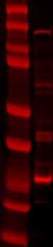
FH Lana (Verified Customer) (06-11-2021) | SDS-PAGE: 40 ug/ul RIPA protein lysate, 4-12% Bis-Tris gradient gel. Transfer: Immobilon-FL transfer membranes (Millipore) O/N at 30V, 4C. Blocking: SEA Block Blocking Buffer 1h, room T. Primary Ab: O/N incubation at 4C, 1:1000. Secondary Ab: IRDye 680LT Goat anti-Rabbit, 1:15000. Lines of WB image: 1 – protein ladder, 2 – mitochondria fraction lysate.
 |

