ERCC2 Polyklonaler Antikörper
ERCC2 Polyklonal Antikörper für WB, IP, IHC, ELISA
Wirt / Isotyp
Kaninchen / IgG
Getestete Reaktivität
human und mehr (2)
Anwendung
WB, IHC, IF, IP, ELISA
Konjugation
Unkonjugiert
Kat-Nr. : 10818-1-AP
Synonyme
Geprüfte Anwendungen
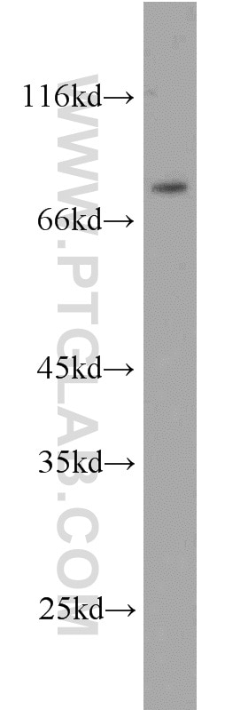
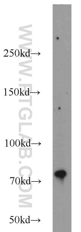
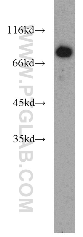
| Erfolgreiche Detektion in WB | HeLa-Zellen, HEK-293-Zellen, K-562-Zellen |
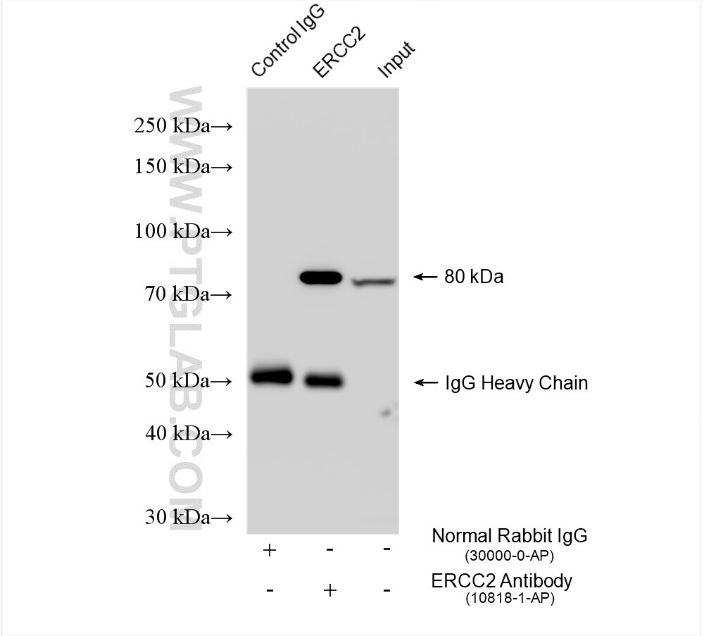
| Erfolgreiche IP | HEK-293-Zellen |
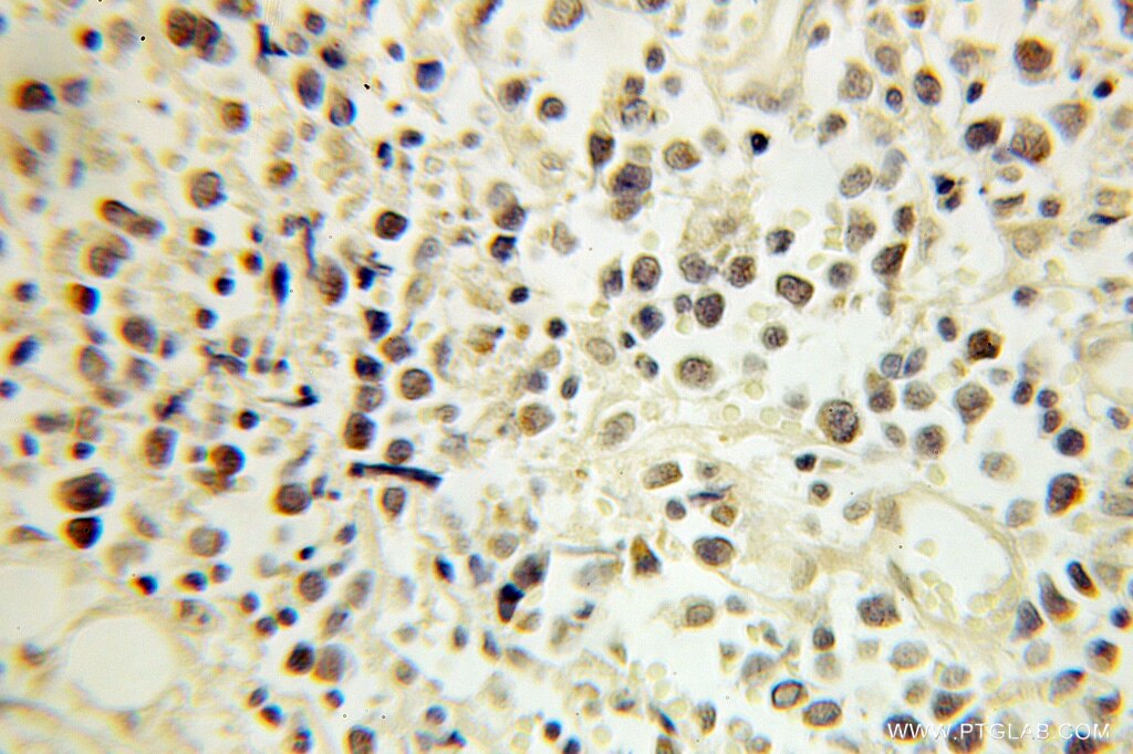
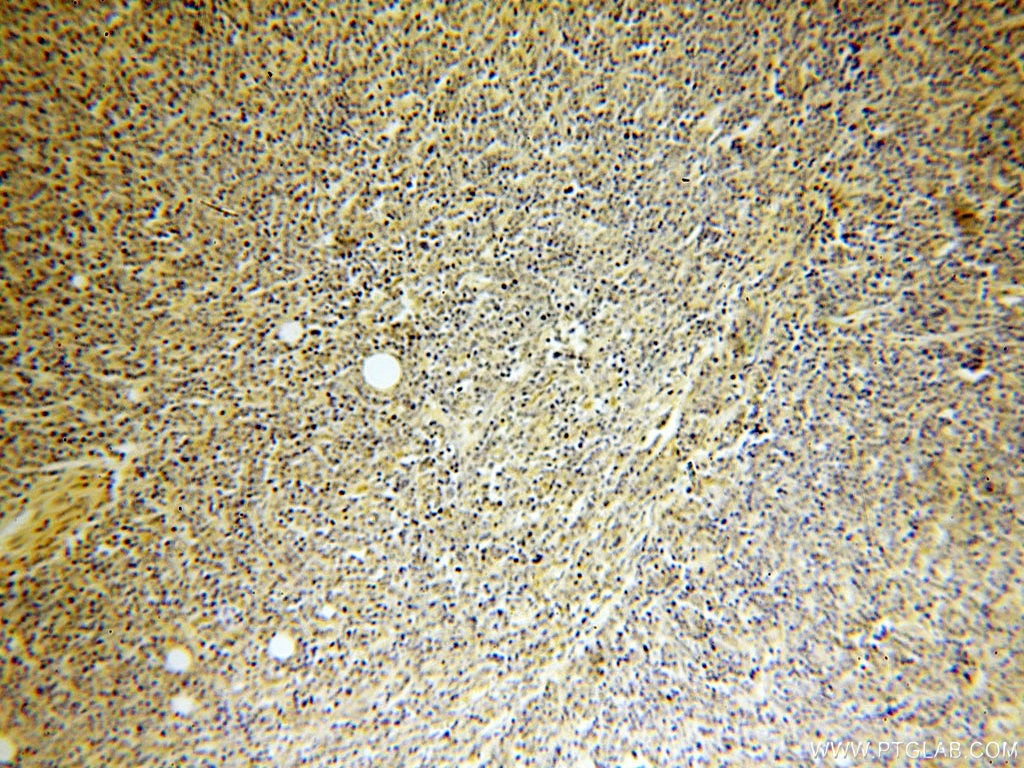
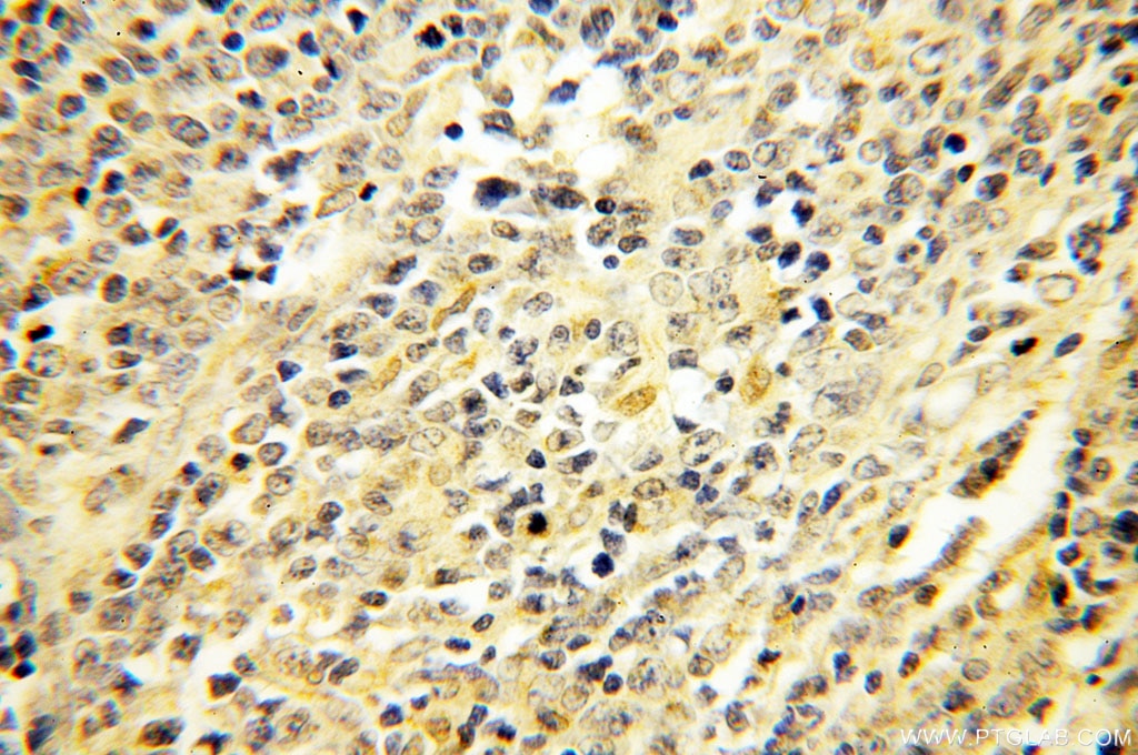
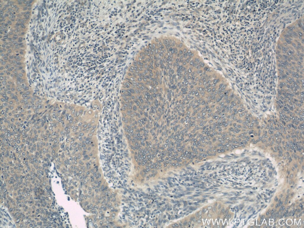
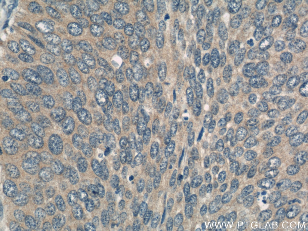
| Erfolgreiche Detektion in IHC | humanes Lymphomgewebe, humanes Zervixkarzinomgewebe Hinweis: Antigendemaskierung mit TE-Puffer pH 9,0 empfohlen. (*) Wahlweise kann die Antigendemaskierung auch mit Citratpuffer pH 6,0 erfolgen. |
Empfohlene Verdünnung
| Anwendung | Verdünnung |
|---|---|
| Western Blot (WB) | WB : 1:500-1:1000 |
| Immunpräzipitation (IP) | IP : 0.5-4.0 ug for 1.0-3.0 mg of total protein lysate |
| Immunhistochemie (IHC) | IHC : 1:20-1:200 |
| It is recommended that this reagent should be titrated in each testing system to obtain optimal results. | |
| Sample-dependent, check data in validation data gallery | |
Veröffentlichte Anwendungen
| WB | See 10 publications below |
| IHC | See 6 publications below |
| IF | See 1 publications below |
Produktinformation
10818-1-AP bindet in WB, IHC, IF, IP, ELISA ERCC2 und zeigt Reaktivität mit human
| Getestete Reaktivität | human |
| In Publikationen genannte Reaktivität | human, Maus, Ratte |
| Wirt / Isotyp | Kaninchen / IgG |
| Klonalität | Polyklonal |
| Typ | Antikörper |
| Immunogen | ERCC2 fusion protein Ag1038 |
| Vollständiger Name | excision repair cross-complementing rodent repair deficiency, complementation group 2 |
| Berechnetes Molekulargewicht | 87 kDa |
| Beobachtetes Molekulargewicht | 80 kDa |
| GenBank-Zugangsnummer | BC008346 |
| Gene symbol | ERCC2 |
| Gene ID (NCBI) | 2068 |
| Konjugation | Unkonjugiert |
| Form | Liquid |
| Reinigungsmethode | Antigen-Affinitätsreinigung |
| Lagerungspuffer | PBS with 0.02% sodium azide and 50% glycerol |
| Lagerungsbedingungen | Bei -20°C lagern. Nach dem Versand ein Jahr lang stabil Aliquotieren ist bei -20oC Lagerung nicht notwendig. 20ul Größen enthalten 0,1% BSA. |
Hintergrundinformationen
Excision-repair complementing defective in Chinese hamster 2(ERCC2), belongs to the nucleotide excision repair pathway, is a component of the TFIIH transcription complex and required for the association of the CDK-activating kinase(CAK) subcomplex with TFIIH. Since TFIIH is involved in both DNA repair and cell cycle progression, ERCC2 is implicated to play an integral role in the nucleotide excision repair pathway. Also it involves in the regulation of vitamin-D receptor activity. As a part of the mitotic spindle-associated MMXD complex, ERCC2 has a role in chromosome segregation.
Protokolle
| PRODUKTSPEZIFISCHE PROTOKOLLE | |
|---|---|
| WB protocol for ERCC2 antibody 10818-1-AP | Protokoll herunterladen |
| IHC protocol for ERCC2 antibody 10818-1-AP | Protokoll herunterladenl |
| IP protocol for ERCC2 antibody 10818-1-AP | Protokoll herunterladen |
| STANDARD-PROTOKOLLE | |
|---|---|
| Klicken Sie hier, um unsere Standardprotokolle anzuzeigen |
Publikationen
| Species | Application | Title |
|---|---|---|
Am J Cancer Res Predictive value of ERCC1, ERCC2, ERCC4, and glutathione S-Transferase Pi expression for the efficacy and safety of FOLFIRINOX in patients with unresectable pancreatic cancer. | ||
Methods Synchronous detection of miRNAs, their targets and downstream proteins in transferred FFPE sections: applications in clinical and basic research. | ||
Cancer Sci Very low prevalence of XPD K751Q polymorphism and its association with XPD expression and outcomes of FOLFOX-4 treatment in Asian patients with colorectal carcinoma. | ||
Biomarkers Aberrant methylation of nucleotide excision repair genes is associated with chronic arsenic poisoning | ||
J Transl Med Proteomic analysis predicts anti-angiogenic resistance in recurred glioblastoma |
Rezensionen
The reviews below have been submitted by verified Proteintech customers who received an incentive for providing their feedback.
FH Zizhao (Verified Customer) (08-15-2018) |
|
