- Featured Product
- KD/KO Validated
GPBP1 Polyklonaler Antikörper
GPBP1 Polyklonal Antikörper für WB, IHC, ELISA
Wirt / Isotyp
Kaninchen / IgG
Getestete Reaktivität
human, Maus, Ratte
Anwendung
WB, IP, IHC, ELISA
Konjugation
Unkonjugiert
Kat-Nr. : 21622-1-AP
Synonyme
 at dilution of 1:500 incubated at room temperature for 1.5 hours.")
 at dilution of 1:500 incubated at room temperature for 1.5 hours.")
 at dilution of 1:300 (under 10x lens).")
 at dilution of 1:300 (under 40x lens).")
 at dilution of 1:50.")
 at dilution of 1:50.")
Geprüfte Anwendungen
| Erfolgreiche Detektion in WB | HepG2-Zellen, L02-Zellen |
| Erfolgreiche Detektion in IHC | humanes Dünndarmgewebe, humanes Hodengewebe Hinweis: Antigendemaskierung mit TE-Puffer pH 9,0 empfohlen. (*) Wahlweise kann die Antigendemaskierung auch mit Citratpuffer pH 6,0 erfolgen. |
Empfohlene Verdünnung
| Anwendung | Verdünnung |
|---|---|
| Western Blot (WB) | WB : 1:500-1:2000 |
| Immunhistochemie (IHC) | IHC : 1:150-1:600 |
| It is recommended that this reagent should be titrated in each testing system to obtain optimal results. | |
| Sample-dependent, check data in validation data gallery | |
Veröffentlichte Anwendungen
| KD/KO | See 1 publications below |
| WB | See 1 publications below |
| IP | See 1 publications below |
Produktinformation
21622-1-AP bindet in WB, IP, IHC, ELISA GPBP1 und zeigt Reaktivität mit human, Maus, Ratten
| Getestete Reaktivität | human, Maus, Ratte |
| In Publikationen genannte Reaktivität | Maus |
| Wirt / Isotyp | Kaninchen / IgG |
| Klonalität | Polyklonal |
| Typ | Antikörper |
| Immunogen | GPBP1 fusion protein Ag14877 |
| Vollständiger Name | GC-rich promoter binding protein 1 |
| Berechnetes Molekulargewicht | 473 aa, 53 kDa |
| Beobachtetes Molekulargewicht | 45 kDa |
| GenBank-Zugangsnummer | BC000267 |
| Gene symbol | GPBP1 |
| Gene ID (NCBI) | 65056 |
| Konjugation | Unkonjugiert |
| Form | Liquid |
| Reinigungsmethode | Antigen-Affinitätsreinigung |
| Lagerungspuffer | PBS with 0.02% sodium azide and 50% glycerol |
| Lagerungsbedingungen | Bei -20°C lagern. Nach dem Versand ein Jahr lang stabil Aliquotieren ist bei -20oC Lagerung nicht notwendig. 20ul Größen enthalten 0,1% BSA. |
Protokolle
| PRODUKTSPEZIFISCHE PROTOKOLLE | |
|---|---|
| WB protocol for GPBP1 antibody 21622-1-AP | Protokoll herunterladen |
| IHC protocol for GPBP1 antibody 21622-1-AP | Protokoll herunterladenl |
| STANDARD-PROTOKOLLE | |
|---|---|
| Klicken Sie hier, um unsere Standardprotokolle anzuzeigen |
Rezensionen
The reviews below have been submitted by verified Proteintech customers who received an incentive for providing their feedback.
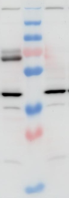
FH Joseph (Verified Customer) (04-30-2025) | Protein amount used for detection: 15–40 μg total protein per well Specificity: Validated with GPBP1 KO cells; some non-specific bands observed Molecular weight: Detects two bands around 70 kDa Blocking: 5% BSA in PBS + 0.1% Tween 20 (5% milk caused high background) Primary antibody: 1:1500, 1 h at RT Wash: 3–4 × 5 min with PBS + 0.1% Tween 20
 |

