- Featured Product
- KD/KO Validated
HDAC1 Polyklonaler Antikörper
HDAC1 Polyklonal Antikörper für WB, IHC, IF/ICC, IP, ELISA
Wirt / Isotyp
Kaninchen / IgG
Getestete Reaktivität
human, Maus, Ratte und mehr (1)
Anwendung
WB, IHC, IF/ICC, IP, CoIP, ChIP, ELISA
Konjugation
Unkonjugiert
Kat-Nr. : 10197-1-AP
Synonyme
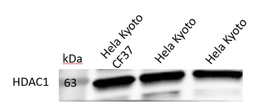
 at dilution of 1:4000 incubated at room temperature for 1.5 hours.")
 at dilution of 1:1000 incubated at room temperature for 1.5 hours.")
 at dilution of 1:15000 incubated at room temperature for 1.5 hours.")
 with HeLa cells lysate 1600 ug.")
 with HeLa cells lysate 1280 ug.")
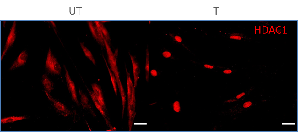
 at dilution of 1:50 (under 40x lens).")
 at dilution of 1:50 (under 10x lens).")
 at dilution of 1:50 (under 40x lens).")
 at dilution of 1:50 (under 10x lens).")
 fixed A431 cells using HDAC1 antibody (10197-1-AP) at dilution of 1:400 and Multi-rAb CoraLite ® Plus 488-Goat Anti-Rabbit Recombinant Secondary Antibody (H+L) (RGAR002).")
 fixed HeLa cells using HDAC1 antibody (10197-1-AP) at dilution of 1:200 and CoraLite®488-Conjugated AffiniPure Goat Anti-Rabbit IgG(H+L).")
Geprüfte Anwendungen
| Erfolgreiche Detektion in WB | C6-Zellen, HeLa-Zellen, Jurkat-Zellen, K-562-Zellen, Maushodengewebe, NIH/3T3-Zellen |
| Erfolgreiche IP | HeLa-Zellen |
| Erfolgreiche Detektion in IHC | humanes Pankreaskarzinomgewebe, humanes Hodengewebe Hinweis: Antigendemaskierung mit TE-Puffer pH 9,0 empfohlen. (*) Wahlweise kann die Antigendemaskierung auch mit Citratpuffer pH 6,0 erfolgen. |
| Erfolgreiche Detektion in IF/ICC | A431-Zellen, HeLa-Zellen |
Empfohlene Verdünnung
| Anwendung | Verdünnung |
|---|---|
| Western Blot (WB) | WB : 1:8000-1:20000 |
| Immunpräzipitation (IP) | IP : 0.5-4.0 ug for 1.0-3.0 mg of total protein lysate |
| Immunhistochemie (IHC) | IHC : 1:50-1:500 |
| Immunfluoreszenz (IF)/ICC | IF/ICC : 1:200-1:800 |
| It is recommended that this reagent should be titrated in each testing system to obtain optimal results. | |
| Sample-dependent, check data in validation data gallery | |
Veröffentlichte Anwendungen
| KD/KO | See 18 publications below |
| WB | See 113 publications below |
| IHC | See 9 publications below |
| IF | See 10 publications below |
| IP | See 10 publications below |
| CoIP | See 8 publications below |
| ChIP | See 4 publications below |
Produktinformation
10197-1-AP bindet in WB, IHC, IF/ICC, IP, CoIP, ChIP, ELISA HDAC1 und zeigt Reaktivität mit human, Maus, Ratten
| Getestete Reaktivität | human, Maus, Ratte |
| In Publikationen genannte Reaktivität | human, Hausschwein, Maus, Ratte |
| Wirt / Isotyp | Kaninchen / IgG |
| Klonalität | Polyklonal |
| Typ | Antikörper |
| Immunogen | HDAC1 fusion protein Ag0256 |
| Vollständiger Name | histone deacetylase 1 |
| Berechnetes Molekulargewicht | 55 kDa |
| Beobachtetes Molekulargewicht | 60 kDa |
| GenBank-Zugangsnummer | BC000301 |
| Gene symbol | HDAC1 |
| Gene ID (NCBI) | 3065 |
| Konjugation | Unkonjugiert |
| Form | Liquid |
| Reinigungsmethode | Antigen-Affinitätsreinigung |
| Lagerungspuffer | PBS with 0.02% sodium azide and 50% glycerol |
| Lagerungsbedingungen | Bei -20°C lagern. Nach dem Versand ein Jahr lang stabil Aliquotieren ist bei -20oC Lagerung nicht notwendig. 20ul Größen enthalten 0,1% BSA. |
Hintergrundinformationen
Background
Histone Deacetylase 1 (HDAC1) is a nuclear localized class I histone deacetylase that plays a role in the regulation of gene expression, mainly by repression of gene activity. Additionally, HDAC can mediate deacetylation of a subset of non-histone proteins, leading to their degradation. HDAC1 activity has an impact on cell growth, proliferation, and death.
1. What is the molecular weight of HDAC1?
The molecular size of HDAC1 is 60 kDa.
2. What is the subcellular localization of HDAC1?
Unlike some HDACs, HDAC1 is present entirely in the nucleus. Our HDAC1 antibody has been broadly tested for IF/ICC.
3. I cannot detect an HDAC1 specific signal in my sample during western blotting.
Make sure that you efficiently extract nuclear proteins during your sample preparation. Some lysis buffers (e.g., based on Triton X-100) may not extract nuclear proteins. We recommend using RIPA buffer (https://www.ptglab.com/support/protocols/). We highly recommend using nuclear loading control antibodies, such as lamin B1 or PCNA (https://www.ptglab.com/news/blog/loading-control-antibodies-for-western-blotting/). Alternatively, you may consider performing cell fractionation.
4. How do I perform chromatin immunoprecipitation (ChiP) with the HDAC1 antibody?
We recommend using our standard chromatin immunoprecipitation protocol (https://www.ptglab.com/media/2708/web_chip-protocol.pdf).
5. Is HDAC1 post-translationally modified?
HDAC1 is a protein deacetylase but itself can also be a subject of post-translational modifications, including phosphorylation, acetylation, ubiquitination, SUMOylation, nitrosylation, and carbonylation (PMID: 21197454).
6. What is the role of HDAC1 in cancer?
The acetylation and deacetylation of histones play an important part in the epigenetic regulation of gene expression. Alterations in the balance between these two opposing processes changes chromosome remodeling and increased deacetylation leads to gene silencing. HDAC1 levels are increased in many cancer cell types and the role of HDAC1 in cancer is connected not only to histone deacetylation but also to the deacetylation of other proteins, such as Rb family proteins, estrogen receptors, and p53 protein (PMID: 19383284).
Protokolle
| PRODUKTSPEZIFISCHE PROTOKOLLE | |
|---|---|
| WB protocol for HDAC1 antibody 10197-1-AP | Protokoll herunterladen |
| IHC protocol for HDAC1 antibody 10197-1-AP | Protokoll herunterladenl |
| IF protocol for HDAC1 antibody 10197-1-AP | Protokoll herunterladen |
| IP protocol for HDAC1 antibody 10197-1-AP | Protokoll herunterladen |
| STANDARD-PROTOKOLLE | |
|---|---|
| Klicken Sie hier, um unsere Standardprotokolle anzuzeigen |
Publikationen
| Species | Application | Title |
|---|---|---|
Acta Pharm Sin B Histone deacetylase inhibitors inhibit cervical cancer growth through Parkin acetylation-mediated mitophagy. | ||
J Clin Invest Acetaldehyde dehydrogenase 2 interactions with LDLR and AMPK regulate foam cell formation. | ||
Nat Commun PCGF6 controls neuroectoderm specification of human pluripotent stem cells by activating SOX2 expression. | ||
Nat Commun The methyltransferase METTL3 negatively regulates nonalcoholic steatohepatitis (NASH) progression. | ||
Hepatology PROX1 promotes hepatocellular carcinoma metastasis by way of up-regulating hypoxia-inducible factor 1α expression and protein stability. |
Rezensionen
The reviews below have been submitted by verified Proteintech customers who received an incentive for providing their feedback.
FH Sara (Verified Customer) (10-06-2024) | I tried WB and FC with mouse cells, works well. I still need to give it a try for immunofluorescence, but it seems to work as well based on other reviews.
|
FH Mi (Verified Customer) (06-25-2023) | Works pretty well in human adipocyte cells.
|
FH Christos (Verified Customer) (04-03-2023) | Separating Gel: 10% poly-acrylamide Transfer at 400mA for 2hr onto nitrocellulose membrane at 4oC Blocking and antibodies dilutions: 5% non-fat milk in PBS-Tween20 for 1hr Incubation with primary Abs: overnight at 4oC Incubation with secondary Abs: 1hr at 4oC
 |
FH Dipen (Verified Customer) (07-20-2020) | Great antibody for Western blotting.
 |
FH Uxoa (Verified Customer) (02-19-2020) | HDAC1 in human primary fibroblasts untreated vs treated. Cells fixed with 4% PFA 15 min RT. HDAC1 incubation 1:50 in PBST (0.1% triton) + 10% DS O/N 4ºC. Donkey anti-rabbit Alexa fluor 555 1:500 1h RT.
 |

