Tested Applications
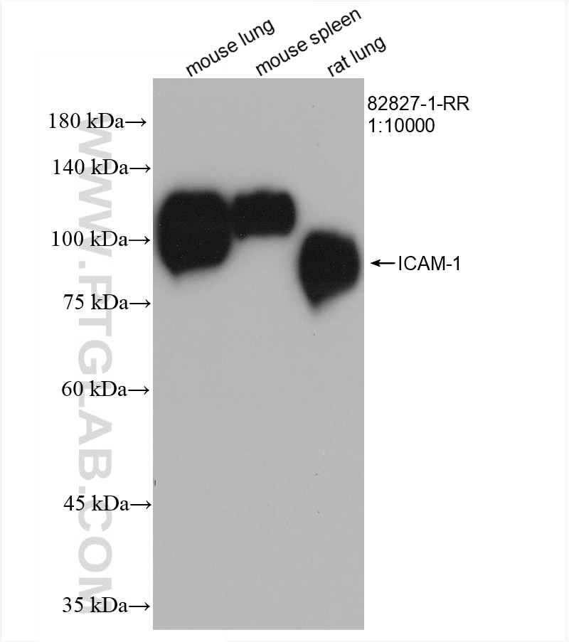
| Positive WB detected in | mouse lung tissue, mouse spleen tissue, rat lung tissue |
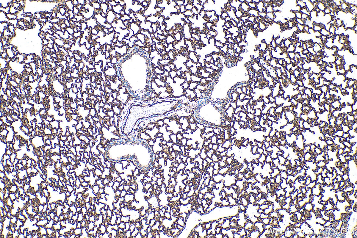
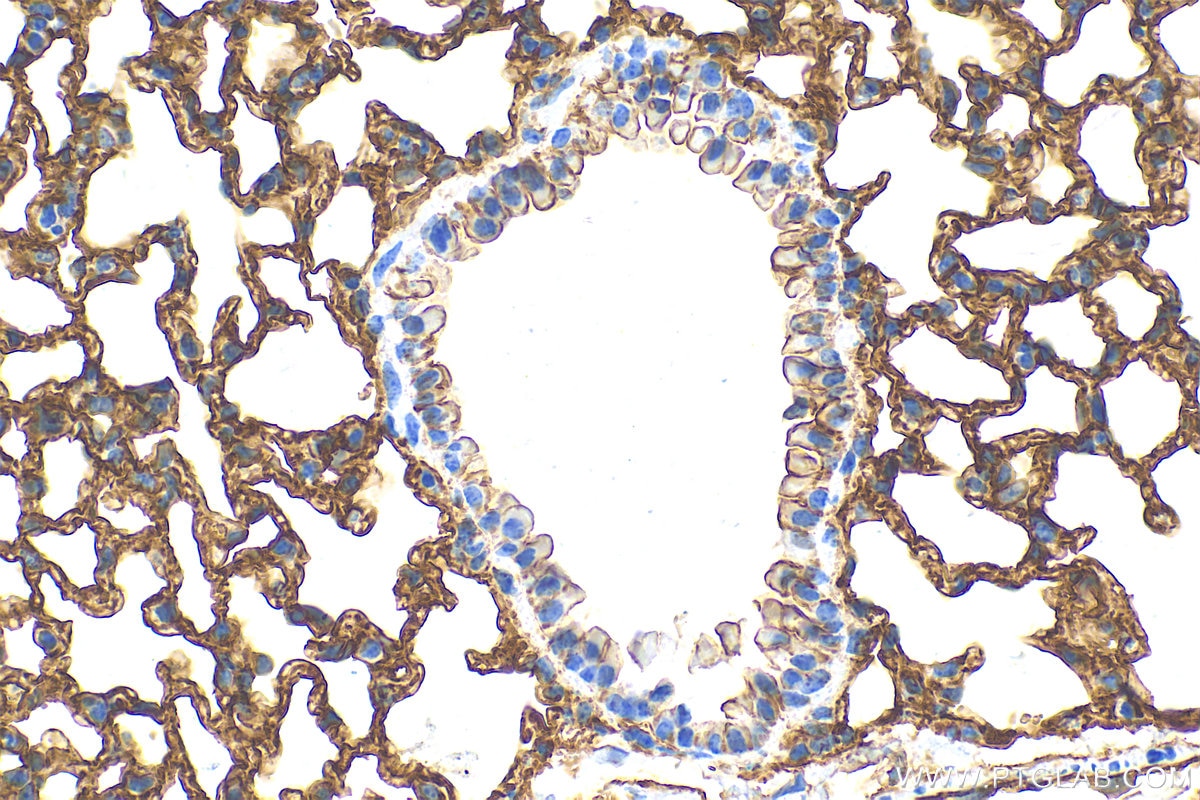
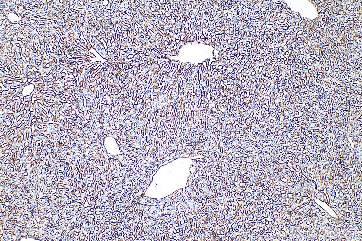
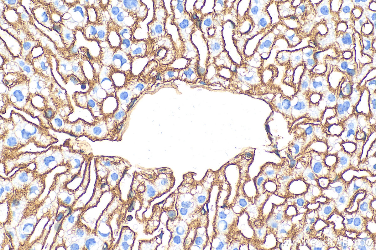
| Positive IHC detected in | mouse lung tissue, mouse liver tissue Note: suggested antigen retrieval with TE buffer pH 9.0; (*) Alternatively, antigen retrieval may be performed with citrate buffer pH 6.0 |
Recommended dilution
| Application | Dilution |
|---|---|
| Western Blot (WB) | WB : 1:5000-1:50000 |
| Immunohistochemistry (IHC) | IHC : 1:500-1:2000 |
| It is recommended that this reagent should be titrated in each testing system to obtain optimal results. | |
| Sample-dependent, Check data in validation data gallery. | |
Published Applications
| WB | See 2 publications below |
| IHC | See 1 publications below |
| IF | See 1 publications below |
Product Information
82827-1-RR targets ICAM-1/CD54 in WB, IHC, IF, ELISA applications and shows reactivity with mouse, rat samples.
| Tested Reactivity | mouse, rat |
| Cited Reactivity | mouse |
| Host / Isotype | Rabbit / IgG |
| Class | Recombinant |
| Type | Antibody |
| Immunogen |
CatNo: Eg0482 Product name: Recombinant mouse ICAM-1 protein Source: mammalian cells-derived, Pinfuse-IGSF8(A4) Tag: C-6*his Domain: 28-485 aa of NM_010493 Sequence: QVSIHPREAFLPQGGSVQVNCSSSCKEDLSLGLETQWLKDELESGPNWKLFELSEIGEDSSPLCFENCGTVQSSASATITVYSFPESVELRPLPAWQQVGKDLTLRCHVDGGAPRTQLSAVLLRGEEILSRQPVGGHPKDPKEITFTVLASRGDHGANFSCRTELDLRPQGLALFSNVSEARSLRTFDLPATIPKLDTPDLLEVGTQQKLFCSLEGLFPASEARIYLELGGQMPTQESTNSSDSVSATALVEVTEEFDRTLPLRCVLELADQILETQRTLTVYNFSAPVLTLSQLEVSEGSQVTVKCEAHSGSKVVLLSGVEPRPPTPQVQFTLNASSEDHKRSFFCSAALEVAGKFLFKNQTLELHVLYGPRLDETDCLGNWTWQEGSQQTLKCQAWGNPSPKMTCRRKADGALLPIGVVKSVKQEMNGTYVCHAFSSHGNVTRNVYLTVLYHSQNN Predict reactive species |
| Full Name | intercellular adhesion molecule 1 |
| Calculated Molecular Weight | 59 kDa |
| Observed Molecular Weight | 85-110 kDa |
| GenBank Accession Number | NM_010493 |
| Gene Symbol | ICAM-1 |
| Gene ID (NCBI) | 15894 |
| RRID | AB_3670566 |
| Conjugate | Unconjugated |
| Form | Liquid |
| Purification Method | Protein A purification |
| UNIPROT ID | P13597 |
| Storage Buffer | PBS with 0.02% sodium azide and 50% glycerol, pH 7.3. |
| Storage Conditions | Store at -20°C. Stable for one year after shipment. Aliquoting is unnecessary for -20oC storage. 20ul sizes contain 0.1% BSA. |
Background Information
ICAM-1 (CD54) is a transmembrane glycoprotein of the immunoglobulin superfamily and is critical for the firm attachment and transmigration of leukocytes out of blood vessels and into tissues (PMID: 19307690). ICAM-1 is expressed by several cell types, typically on endothelial cells and cells of the immune system, and its expression can be up-regulated by various stimuli, including TNF-α, INF-γ, IL-1 and thrombin (PMID: 3086451; 9694714; 15979056). It is a ligand for LFA-1 and Mac-1, serves as a receptor for rhinovirus, and is one of several receptors used by Plasmodium falciparum (PMID: 2566624; 2538244; 2475784).
Protocols
| Product Specific Protocols | |
|---|---|
| IHC protocol for ICAM-1/CD54 antibody 82827-1-RR | Download protocol |
| WB protocol for ICAM-1/CD54 antibody 82827-1-RR | Download protocol |
| Standard Protocols | |
|---|---|
| Click here to view our Standard Protocols |
Publications
| Species | Application | Title |
|---|---|---|
Microbiol Res Trojan horse strategy and TfR/ LDLR-Mediated transcytosis determine the dissemination of mycobacteria in tuberculous meningoencephalitis | ||
Lab Invest Aryl Hydrocarbon Receptor Deficiency Upregulates Intercellular Adhesion Molecule 1 in Retinal Pigment Epithelial Cells and Contributes to Retinal Inflammation | ||
Int J Biol Macromol Targeted gene delivery of shear-responsive forkhead box C1 using hyaluronic acid modified chitosan nanoparticles suppresses atherosclerosis through Hippo-YAP signaling pathway. |