Cytokeratin 10 Polyklonaler Antikörper
Cytokeratin 10 Polyklonal Antikörper für WB, IHC, IF/ICC, IF-P, FC (Intra), ELISA
Wirt / Isotyp
Kaninchen / IgG
Getestete Reaktivität
human, Maus, Ratte
Anwendung
WB, IHC, IF/ICC, IF-P, FC (Intra), ELISA
Konjugation
Unkonjugiert
Kat-Nr. : 18343-1-AP
Synonyme
 at dilution of 1:200000 incubated at room temperature for 1.5 hours.")
 at dilution of 1:20000 incubated at room temperature for 1.5 hours.")
 at dilution of 1:1000 (under 10x lens). Heat mediated antigen retrieval with Tris-EDTA buffer (pH 9.0).")
 at dilution of 1:200 (under 10x lens. Heat mediated antigen retrieval with Tris-EDTA buffer (pH 9.0).")
 at dilution of 1:200 (under 40x lens. Heat mediated antigen retrieval with Tris-EDTA buffer (pH 9.0).")
 at dilution of 1:1000 (under 40x lens). Heat mediated antigen retrieval with Tris-EDTA buffer (pH 9.0).")
 at dilution of 1:1000 (under 10x lens). Heat mediated antigen retrieval with Tris-EDTA buffer (pH 9.0).")
 at dilution of 1:1000 (under 40x lens). Heat mediated antigen retrieval with Tris-EDTA buffer (pH 9.0).")
 at dilution of 1:200 (under 10x lens).")
 at dilution of 1:200 (under 40x lens).")
 at dilution of 1:200 (under 40x lens).")
 at dilution of 1:200 (under 10x lens).")
 at dilution of 1:200 (under 10x lens).")
 at dilution of 1:200 (under 40x lens).")
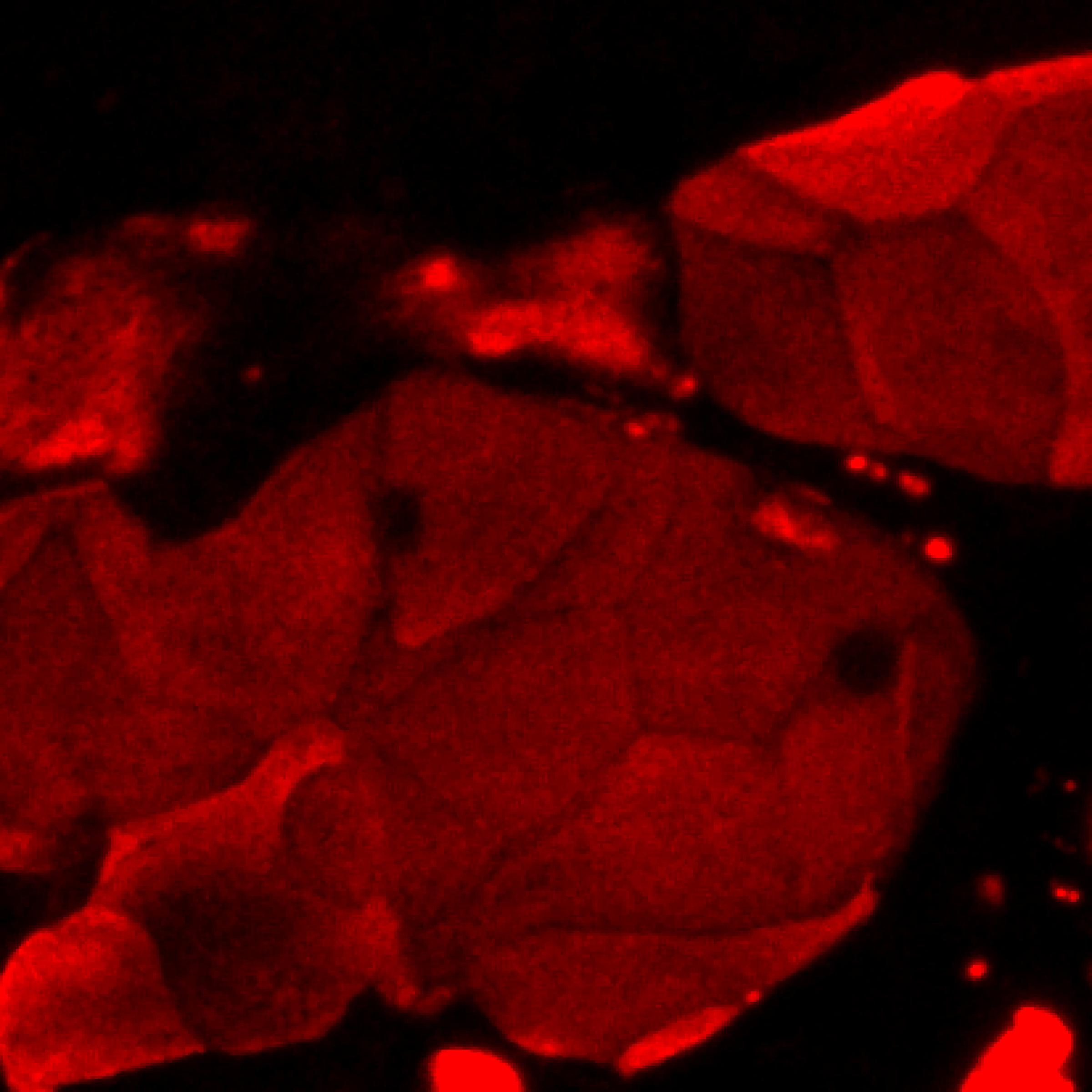
 fixed paraffin-embedded mouse skin tissue using Cytokeratin 10 antibody (18343-1-AP) at dilution of 1:200 and CoraLite®594-Conjugated Goat Anti-Rabbit IgG(H+L) (SA00013-4). Heat mediated antigen retrieval with Tris-EDTA buffer (pH 9.0).")
 fixed A431 cells using Cytokeratin 10 antibody (18343-1-AP) at dilution of 1:200 and CoraLite®488-Conjugated AffiniPure Goat Anti-Rabbit IgG(H+L) (SA00013-2).")
 and CoraLite®488-Conjugated AffiniPure Goat Anti-Rabbit IgG(H+L) at dilution 1:1000 (red), or 0.4 ug rabbit IgG isotype control (blue). Cells were fixed with 4% PFA and permeabilized with Flow Cytometry Perm Buffer (PF00011-C).")
Geprüfte Anwendungen
| Erfolgreiche Detektion in WB | Maushautgewebe, A431-Zellen, Rattenhautgewebe |
| Erfolgreiche Detektion in IHC | humanes Zervixkarzinomgewebe, humanes Mammakarzinomgewebe, humanes Lungenkarzinomgewebe, humanes Hautkrebsgewebe, Rattenhautgewebe Hinweis: Antigendemaskierung mit TE-Puffer pH 9,0 empfohlen. (*) Wahlweise kann die Antigendemaskierung auch mit Citratpuffer pH 6,0 erfolgen. |
| Erfolgreiche Detektion in IF-P | Maushautgewebe |
| Erfolgreiche Detektion in IF/ICC | A431-Zellen |
| Erfolgreiche Detektion in FC (Intra) | A431-Zellen |
Empfohlene Verdünnung
| Anwendung | Verdünnung |
|---|---|
| Western Blot (WB) | WB : 1:5000-1:20000 |
| Immunhistochemie (IHC) | IHC : 1:500-1:2000 |
| Immunfluoreszenz (IF)-P | IF-P : 1:50-1:500 |
| Immunfluoreszenz (IF)/ICC | IF/ICC : 1:50-1:500 |
| Durchflusszytometrie (FC) (INTRA) | FC (INTRA) : 0.40 ug per 10^6 cells in a 100 µl suspension |
| It is recommended that this reagent should be titrated in each testing system to obtain optimal results. | |
| Sample-dependent, check data in validation data gallery | |
Veröffentlichte Anwendungen
| WB | See 3 publications below |
| IHC | See 5 publications below |
| IF | See 11 publications below |
Produktinformation
18343-1-AP bindet in WB, IHC, IF/ICC, IF-P, FC (Intra), ELISA Cytokeratin 10 und zeigt Reaktivität mit human, Maus, Ratten
| Getestete Reaktivität | human, Maus, Ratte |
| In Publikationen genannte Reaktivität | human, Maus, Ratte |
| Wirt / Isotyp | Kaninchen / IgG |
| Klonalität | Polyklonal |
| Typ | Antikörper |
| Immunogen | Cytokeratin 10 fusion protein Ag13136 |
| Vollständiger Name | keratin 10 |
| Berechnetes Molekulargewicht | 584 aa, 59 kDa |
| Beobachtetes Molekulargewicht | 50-59 kDa |
| GenBank-Zugangsnummer | BC034697 |
| Gene symbol | Cytokeratin 10 |
| Gene ID (NCBI) | 3858 |
| Konjugation | Unkonjugiert |
| Form | Liquid |
| Reinigungsmethode | Antigen-Affinitätsreinigung |
| Lagerungspuffer | PBS with 0.02% sodium azide and 50% glycerol |
| Lagerungsbedingungen | Bei -20°C lagern. Nach dem Versand ein Jahr lang stabil Aliquotieren ist bei -20oC Lagerung nicht notwendig. 20ul Größen enthalten 0,1% BSA. |
Hintergrundinformationen
Keratins are a large family of proteins that form the intermediate filament cytoskeleton of epithelial cells, which are classified into two major sequence types. Type I keratins are a group of acidic intermediate filament proteins, including K9-K23, and the hair keratins Ha1-Ha8. Type II keratins are the basic or neutral courterparts to the acidic type I keratins, including K1-K8, and the hair keratins, Hb1-Hb6. As a type I keratin, keratin 10 is a suprabasal marker of differentiation in stratified squamous epithelia.
Protokolle
| PRODUKTSPEZIFISCHE PROTOKOLLE | |
|---|---|
| WB protocol for Cytokeratin 10 antibody 18343-1-AP | Protokoll herunterladen |
| IHC protocol for Cytokeratin 10 antibody 18343-1-AP | Protokoll herunterladenl |
| IF protocol for Cytokeratin 10 antibody 18343-1-AP | Protokoll herunterladen |
| FC protocol for Cytokeratin 10 antibody 18343-1-AP | Download protocol |
| STANDARD-PROTOKOLLE | |
|---|---|
| Klicken Sie hier, um unsere Standardprotokolle anzuzeigen |
Publikationen
| Species | Application | Title |
|---|---|---|
Cell Mol Biol Lett The role of NPY2R/NFATc1/DYRK1A regulatory axis in sebaceous glands for sebum synthesis | ||
Aging Cell Nociceptive transient receptor potential canonical 7 (TRPC7) mediates aging-associated tumorigenesis induced by ultraviolet B. | ||
Life Sci The potential of Diosgenin in treating psoriasis: Studies from HaCaT keratinocytes and imiquimod-induced murine model. | ||
mBio Commensal-Related Changes in the Epidermal Barrier Function Lead to Alterations in the Benzo[a]Pyrene Metabolite Profile and Its Distribution in 3D Skin. | ||
Eur Surg Res A novel combination of keratinocyte and autologous microskin grafting to repair full thickness skin loss | ||
Int J Biol Macromol Skin-adaptive film dressing with smart-release of growth factors accelerated diabetic wound healing |
Rezensionen
The reviews below have been submitted by verified Proteintech customers who received an incentive for providing their feedback.
FH Joshua (Verified Customer) (07-27-2019) | Cells differentiated at air-liquid interface. Cells fixed in 4% paraformaldehyde and stained at 4C overnight. Bright staining with minimal background.
 |

