Cytokeratin 6A Polyklonaler Antikörper
Cytokeratin 6A Polyklonal Antikörper für WB, IHC, IF/ICC, FC (Intra), ELISA
Wirt / Isotyp
Kaninchen / IgG
Getestete Reaktivität
human, Ratte und mehr (1)
Anwendung
WB, IHC, IF/ICC, FC (Intra), ELISA
Konjugation
Unkonjugiert
Kat-Nr. : 10590-1-AP
Synonyme
Geprüfte Anwendungen
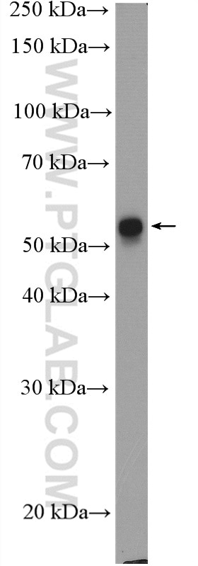
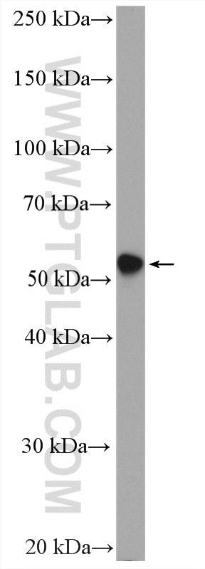
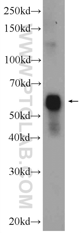
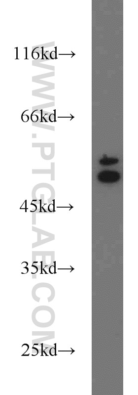
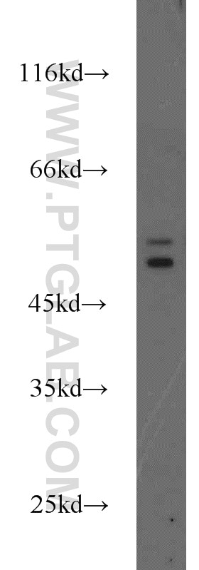
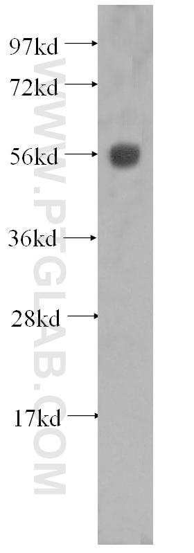
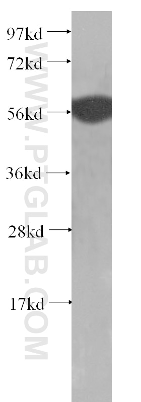
| Erfolgreiche Detektion in WB | A431-Zellen, A375-Zellen, HeLa-Zellen, MCF-7-Zellen, Rattenhautgewebe, U-251-Zellen |
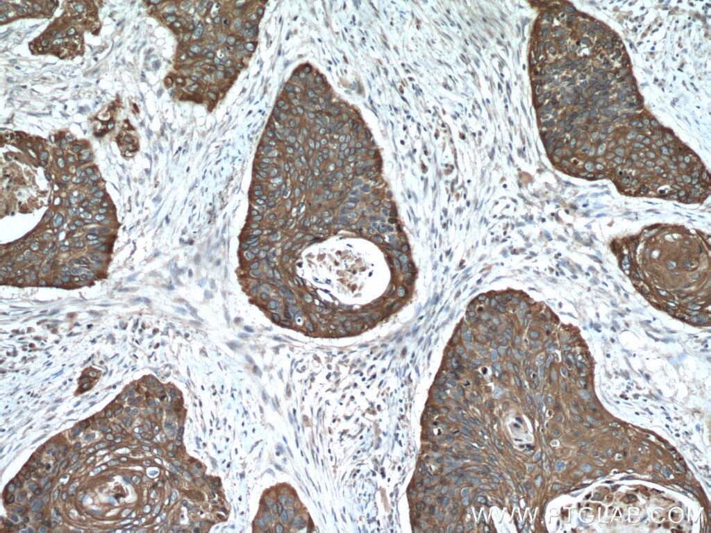
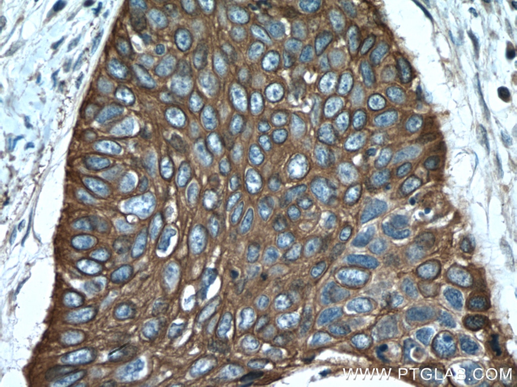
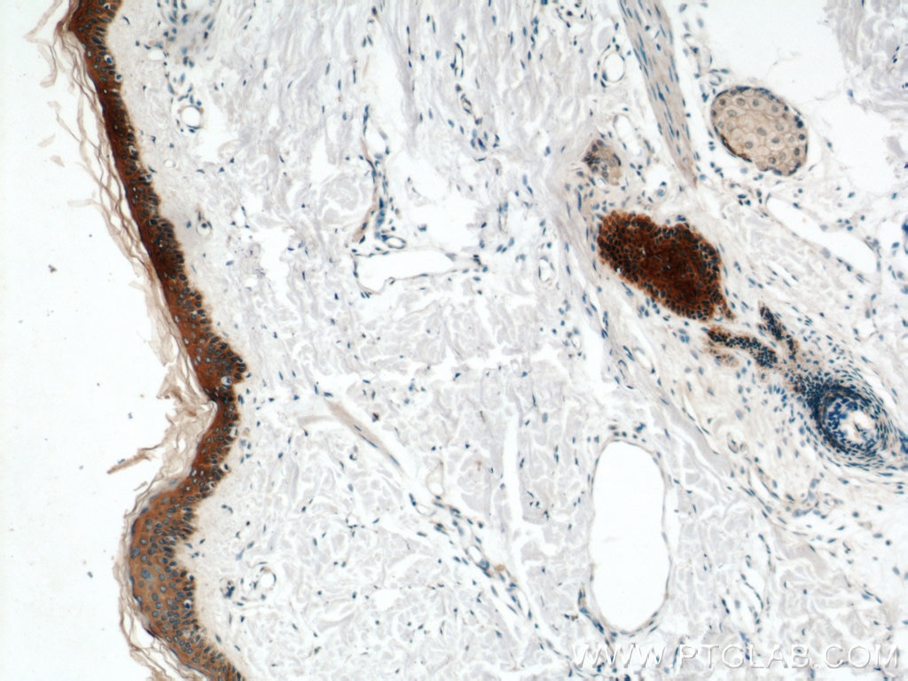
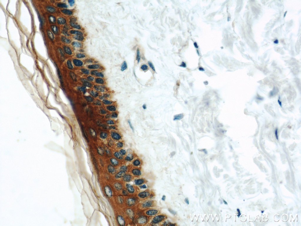
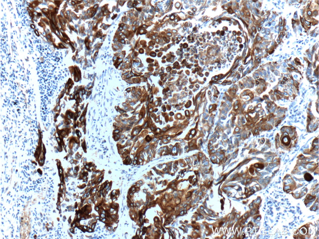
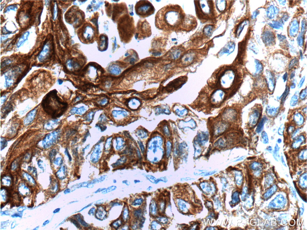
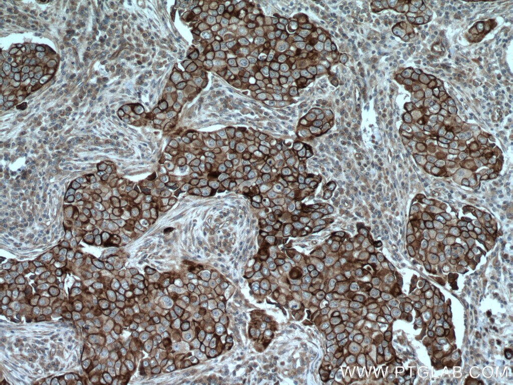
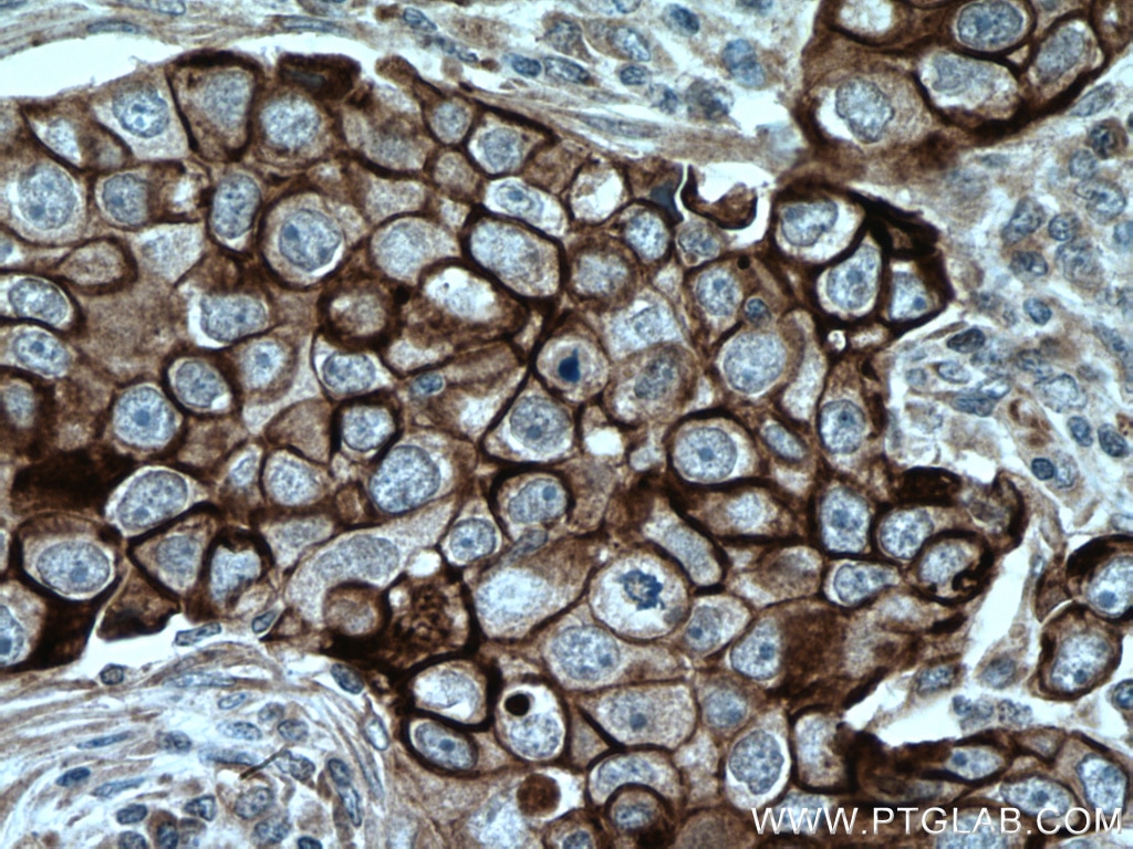
| Erfolgreiche Detektion in IHC | humanes Ösophaguskarzinomgewebe, humanes Mammakarzinomgewebe, humanes Lungenkarzinomgewebe, humanes Hautgewebe Hinweis: Antigendemaskierung mit TE-Puffer pH 9,0 empfohlen. (*) Wahlweise kann die Antigendemaskierung auch mit Citratpuffer pH 6,0 erfolgen. |
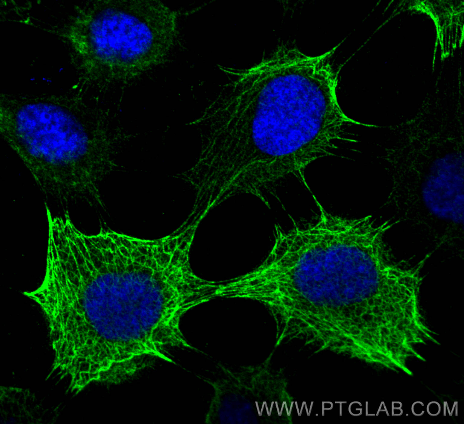
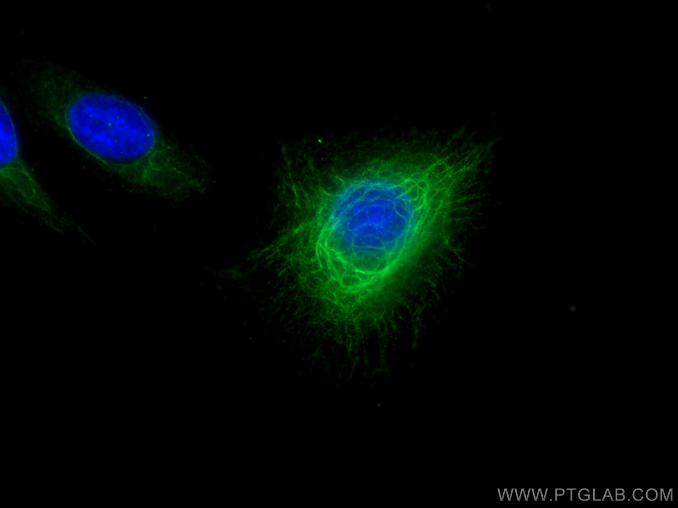
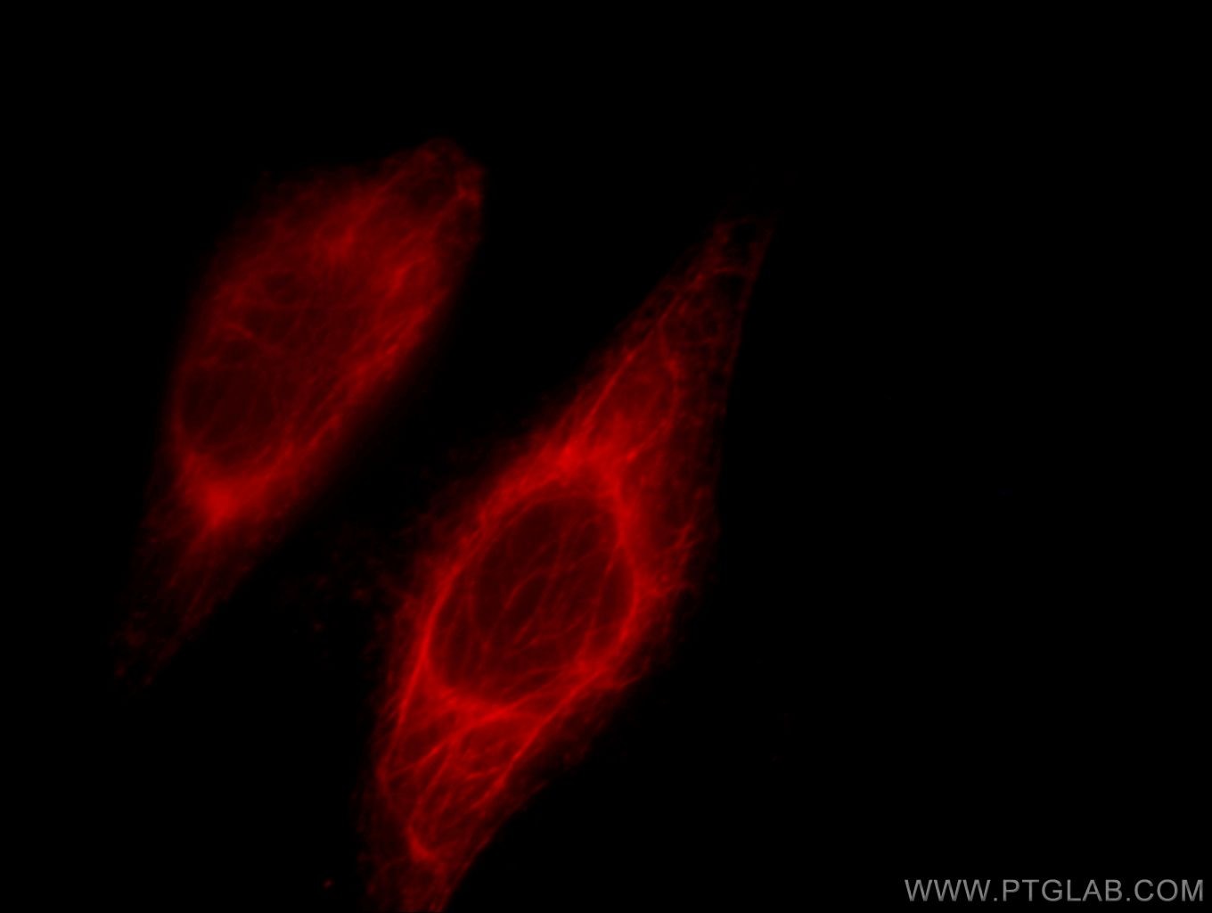
| Erfolgreiche Detektion in IF/ICC | A431-Zellen, HeLa-Zellen |
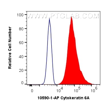
| Erfolgreiche Detektion in FC (Intra) | A431-Zellen |
Empfohlene Verdünnung
| Anwendung | Verdünnung |
|---|---|
| Western Blot (WB) | WB : 1:10000-1:50000 |
| Immunhistochemie (IHC) | IHC : 1:200-1:800 |
| Immunfluoreszenz (IF)/ICC | IF/ICC : 1:50-1:500 |
| Durchflusszytometrie (FC) (INTRA) | FC (INTRA) : 0.40 ug per 10^6 cells in a 100 µl suspension |
| It is recommended that this reagent should be titrated in each testing system to obtain optimal results. | |
| Sample-dependent, check data in validation data gallery | |
Veröffentlichte Anwendungen
| KD/KO | See 1 publications below |
| WB | See 13 publications below |
| IHC | See 11 publications below |
| IF | See 13 publications below |
Produktinformation
10590-1-AP bindet in WB, IHC, IF/ICC, FC (Intra), ELISA Cytokeratin 6A und zeigt Reaktivität mit human, Ratten
| Getestete Reaktivität | human, Ratte |
| In Publikationen genannte Reaktivität | human, Maus, Ratte |
| Wirt / Isotyp | Kaninchen / IgG |
| Klonalität | Polyklonal |
| Typ | Antikörper |
| Immunogen | Cytokeratin 6A fusion protein Ag0882 |
| Vollständiger Name | keratin 6A |
| Berechnetes Molekulargewicht | 60 kDa |
| Beobachtetes Molekulargewicht | 56 kDa |
| GenBank-Zugangsnummer | BC008807 |
| Gene symbol | Cytokeratin 6A |
| Gene ID (NCBI) | 3853 |
| Konjugation | Unkonjugiert |
| Form | Liquid |
| Reinigungsmethode | Antigen-Affinitätsreinigung |
| Lagerungspuffer | PBS with 0.02% sodium azide and 50% glycerol |
| Lagerungsbedingungen | Bei -20°C lagern. Nach dem Versand ein Jahr lang stabil Aliquotieren ist bei -20oC Lagerung nicht notwendig. 20ul Größen enthalten 0,1% BSA. |
Hintergrundinformationen
Keratins are a large family of proteins that form the intermediate filament cytoskeleton of epithelial cells, which are classified into two major sequence types. Type I keratins are a group of acidic intermediate filament proteins, including K9-K23, and the hair keratins Ha1-Ha8. Type II keratins are the basic or neutral courterparts to the acidic type I keratins, including K1-K8, and the hair keratins, Hb1-Hb6. Keratin 6 is a type II keratin. It is used as marker for epidermal hyperproliferation and differentiation. Three are three keratin 6 isoforms: ketatin 6A,6B and 6C. They share more than 99% identical DNA sequence.
Protokolle
| PRODUKTSPEZIFISCHE PROTOKOLLE | |
|---|---|
| WB protocol for Cytokeratin 6A antibody 10590-1-AP | Protokoll herunterladen |
| IHC protocol for Cytokeratin 6A antibody 10590-1-AP | Protokoll herunterladenl |
| IF protocol for Cytokeratin 6A antibody 10590-1-AP | Protokoll herunterladen |
| STANDARD-PROTOKOLLE | |
|---|---|
| Klicken Sie hier, um unsere Standardprotokolle anzuzeigen |
Publikationen
| Species | Application | Title |
|---|---|---|
Cancer Cell KMT2C deficiency drives transdifferentiation of double-negative prostate cancer and confer resistance to AR-targeted therapy | ||
Immunity Excessive Polyamine Generation in Keratinocytes Promotes Self-RNA Sensing by Dendritic Cells in Psoriasis. | ||
Cell Rep Collagen 1-mediated CXCL1 secretion in tumor cells activates fibroblasts to promote radioresistance of esophageal cancer | ||
Phytother Res Epigallocatechin-3-gallate promotes wound healing response in diabetic mice by activating keratinocytes and promoting re-epithelialization | ||
J Cell Mol Med Ozone therapy promotes the differentiation of basal keratinocytes via increasing Tp63-mediated transcription of KRT10 to improve psoriasis. |
