MYC tag Monoklonaler Antikörper
MYC tag Monoklonal Antikörper für WB, IF/ICC, FC (Intra), IP, ELISA
Wirt / Isotyp
Maus / IgG1
Getestete Reaktivität
rekombinanten Protein und mehr (2)
Anwendung
WB, IF/ICC, FC (Intra), IP, CoIP, ChIP, ELISA
Konjugation
Unkonjugiert
CloneNo.
1A5A2
Kat-Nr. : 60003-2-Ig
Synonyme
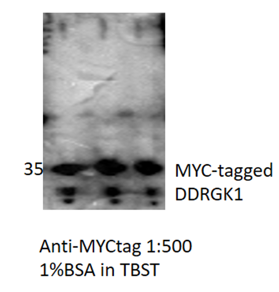
 at dilution of 1:10000 incubated at room temperature for 1.5 hours.")
 at various dilutions.")
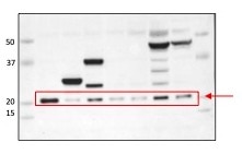
 with Transfected HEK-293 cells lysate 300ug.")
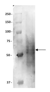
 fixed Transfected HEK-293 cells using MYC tag antibody (60003-2-Ig, Clone: 1A5A2 ) at dilution of 1:1000 and CoraLite®488-Conjugated AffiniPure Goat Anti-Mouse IgG(H+L).")
 and Multi-rAb CoraLite® Plus 647-Goat Anti-Mouse Recombinant Secondary Antibody (H+L)(RGAM005). Cells were fixed with 4% PFA and permeabilized with Flow Cytometry Perm Buffer (PF00011-C).")
"MYC tag Antibodies" Comparison
View side-by-side comparison of MYC tag antibodies from other vendors to find the one that best suits your research needs.
Geprüfte Anwendungen
| Erfolgreiche Detektion in WB | Transfected HEK-293T cells, recombinant proein |
| Erfolgreiche IP | Transfizierte HEK-293-Zellen |
| Erfolgreiche Detektion in IF/ICC | Transfizierte HEK-293-Zellen |
| Erfolgreiche Detektion in FC (Intra) | Transfizierte HEK-293-Zellen |
Empfohlene Verdünnung
| Anwendung | Verdünnung |
|---|---|
| Western Blot (WB) | WB : 1:5000-1:50000 |
| Immunpräzipitation (IP) | IP : 0.5-4.0 ug for 1.0-3.0 mg of total protein lysate |
| Immunfluoreszenz (IF)/ICC | IF/ICC : 1:500-1:2000 |
| Durchflusszytometrie (FC) (INTRA) | FC (INTRA) : 0.50 ug per 10^6 cells in a 100 µl suspension |
| It is recommended that this reagent should be titrated in each testing system to obtain optimal results. | |
| Sample-dependent, check data in validation data gallery | |
Veröffentlichte Anwendungen
| WB | See 226 publications below |
| IF | See 24 publications below |
| IP | See 76 publications below |
| CoIP | See 50 publications below |
| ChIP | See 1 publications below |
Produktinformation
60003-2-Ig bindet in WB, IF/ICC, FC (Intra), IP, CoIP, ChIP, ELISA MYC tag und zeigt Reaktivität mit rekombinanten Protein
| Getestete Reaktivität | rekombinanten Protein |
| In Publikationen genannte Reaktivität | human, Hausschwein |
| Wirt / Isotyp | Maus / IgG1 |
| Klonalität | Monoklonal |
| Typ | Antikörper |
| Immunogen | Peptid |
| Vollständiger Name | Myc tag |
| Gene symbol | Myc tag |
| Gene ID (NCBI) | 99 |
| Konjugation | Unkonjugiert |
| Form | Liquid |
| Reinigungsmethode | Protein-G-Reinigung |
| Lagerungspuffer | PBS with 0.02% sodium azide and 50% glycerol |
| Lagerungsbedingungen | Bei -20°C lagern. Nach dem Versand ein Jahr lang stabil Aliquotieren ist bei -20oC Lagerung nicht notwendig. 20ul Größen enthalten 0,1% BSA. |
Hintergrundinformationen
Protein tags are protein or peptide sequences located either on the C- or N- terminal of the target protein, which facilitates one or several of the following characteristics: solubility, detection, purification, localization and expression. The c-Myc tag corresponds to amino acid residues(EQKLISEEDL) of the human c-Myc protein. It can be used for affinity chromatography, then used to separate recombinant, overexpressed protein from wild type protein expressed by the host organism. It can also be used in the isolation of protein complexes with multiple subunits. Myc-Tag mouse mAb detects recombinant proteins containing the Myc tag. The antibody recognizes the Myc-tag EQKLISEEDL fused to either the amino- or carboxy- terminus of targeted proteins.
Protokolle
| PRODUKTSPEZIFISCHE PROTOKOLLE | |
|---|---|
| WB protocol for MYC tag antibody 60003-2-Ig | Protokoll herunterladen |
| IF protocol for MYC tag antibody 60003-2-Ig | Protokoll herunterladen |
| IP protocol for MYC tag antibody 60003-2-Ig | Protokoll herunterladen |
| FC protocol for MYC tag antibody 60003-2-Ig | Download protocol |
| STANDARD-PROTOKOLLE | |
|---|---|
| Klicken Sie hier, um unsere Standardprotokolle anzuzeigen |
Publikationen
| Species | Application | Title |
|---|---|---|
Cell Res Mannose antagonizes GSDME-mediated pyroptosis through AMPK activated by metabolite GlcNAc-6P | ||
Cell Discov Glc7/PP1 dephosphorylates histone H3T11 to regulate autophagy and telomere silencing in response to nutrient availability | ||
Nat Cancer Lymphatic endothelial-like cells promote glioblastoma stem cell growth through cytokine-driven cholesterol metabolism | ||
Protein Cell BiFC and FACS-based CRISPR screening revealed that QKI promotes PABPN1 LLPS in colorectal cancer cells | ||
Nat Commun Phosphoglycerate dehydrogenase activates PKM2 to phosphorylate histone H3T11 and attenuate cellular senescence |
Rezensionen
The reviews below have been submitted by verified Proteintech customers who received an incentive for providing their feedback.
FH VASUDEVARAO (Verified Customer) (07-23-2025) | The antibody works very well at the recommended dilutions and performs excellently in both immunoprecipitation and immunoblotting applications.
|
FH RASHMI (Verified Customer) (07-26-2024) | Used this product, highly recommended
|
FH Sarah (Verified Customer) (10-27-2023) | works well at low dilution for WB and worked for immunoprecipitation
|
FH manohar (Verified Customer) (07-10-2023) | Used Nitrocellulose membrane with 5% milk as blocking, antibody diluted in 1% milk.
|
FH Amy (Verified Customer) (03-05-2023) | Clean bands for detecting over expressed Myc-tagged proteins in HEK293T cell lysates.
 |
FH Isabelle Cristine (Verified Customer) (01-14-2022) | The antibody works generally well for the detection of MYC-tagged proteins.
 |
FH Tom (Verified Customer) (02-10-2021) | HEK293T cells were transfected with a Myc-tagged construct. The 60003-2-Ig antibody (1:2000) resulted in a lot of background. An alternative antibody was used alongside this one, with a stronger band and less background being achieved.
 |
FH Alinda (Verified Customer) (10-09-2019) | This antibody was used at 1:1000 in HEK cells transfected with a myc-tagged insert cloned into an overxpression. Cells were blocked for 1 hr at room temp in 5%goat serum PBST.This antibody has a lot of background; another antibody from cell signalling was tested in parallel and performed much better
|

