- Featured Product
- KD/KO Validated
ODF2 Polyklonaler Antikörper
ODF2 Polyklonal Antikörper für WB, IHC, IF/ICC, IP, ELISA
Wirt / Isotyp
Kaninchen / IgG
Getestete Reaktivität
human, Maus, Ratte
Anwendung
WB, IHC, IF/ICC, IP, ELISA
Konjugation
Unkonjugiert
Kat-Nr. : 12058-1-AP
Synonyme
 at dilution of 1:500 incubated at room temperature for 1.5 hours.")
 at dilution of 1:400 incubated at room temperature for 1.5 hours.")
 with mouse testis tissue lysate 8000ug.")
 at dilution of 1:100 (under 10x lens. Heat mediated antigen retrieval with Tris-EDTA buffer (pH 9.0).")
 at dilution of 1:100 (under 40x lens. Heat mediated antigen retrieval with Tris-EDTA buffer (pH 9.0).")
 at dilution of 1:25 and Alexa Fluor 488-conjugated Goat Anti-Rabbit IgG(H+L).")
Geprüfte Anwendungen
| Erfolgreiche Detektion in WB | Maushodengewebe, Mauslungengewebe |
| Erfolgreiche IP | Maushodengewebe |
| Erfolgreiche Detektion in IHC | humanes Mammakarzinomgewebe Hinweis: Antigendemaskierung mit TE-Puffer pH 9,0 empfohlen. (*) Wahlweise kann die Antigendemaskierung auch mit Citratpuffer pH 6,0 erfolgen. |
| Erfolgreiche Detektion in IF/ICC | HeLa-Zellen |
Empfohlene Verdünnung
| Anwendung | Verdünnung |
|---|---|
| Western Blot (WB) | WB : 1:500-1:1000 |
| Immunpräzipitation (IP) | IP : 0.5-4.0 ug for 1.0-3.0 mg of total protein lysate |
| Immunhistochemie (IHC) | IHC : 1:50-1:500 |
| Immunfluoreszenz (IF)/ICC | IF/ICC : 1:10-1:100 |
| It is recommended that this reagent should be titrated in each testing system to obtain optimal results. | |
| Sample-dependent, check data in validation data gallery | |
Veröffentlichte Anwendungen
| KD/KO | See 2 publications below |
| WB | See 21 publications below |
| IHC | See 1 publications below |
| IF | See 26 publications below |
Produktinformation
12058-1-AP bindet in WB, IHC, IF/ICC, IP, ELISA ODF2 und zeigt Reaktivität mit human, Maus, Ratten
| Getestete Reaktivität | human, Maus, Ratte |
| In Publikationen genannte Reaktivität | human, Maus, Ratte |
| Wirt / Isotyp | Kaninchen / IgG |
| Klonalität | Polyklonal |
| Typ | Antikörper |
| Immunogen | ODF2 fusion protein Ag2693 |
| Vollständiger Name | outer dense fiber of sperm tails 2 |
| Berechnetes Molekulargewicht | 763 aa, 89 kDa |
| Beobachtetes Molekulargewicht | 67-100 kDa |
| GenBank-Zugangsnummer | BC010629 |
| Gene symbol | ODF2 |
| Gene ID (NCBI) | 4957 |
| Konjugation | Unkonjugiert |
| Form | Liquid |
| Reinigungsmethode | Antigen-Affinitätsreinigung |
| Lagerungspuffer | PBS with 0.02% sodium azide and 50% glycerol |
| Lagerungsbedingungen | Bei -20°C lagern. Nach dem Versand ein Jahr lang stabil Aliquotieren ist bei -20oC Lagerung nicht notwendig. 20ul Größen enthalten 0,1% BSA. |
Hintergrundinformationen
ODF2 (outer dense fiber 2) is the major cenexin protein of the cytoskeleton of the sperm tail. In somatic cells, it is a component of the centrosome in which it is located in the appendages of the mother centriole. The gene for the cenexin protein, which was first identified as a protein marker of the mother centriole, was subsequently found to be identical to ODF2 and encodes transcripts cenexin 1 and cenexin 2. ODF2 is ubiquitously expressed in many cell types as various splicing variants. Additionally, ODF2 localizes to centrioles, basal bodies, and primary cilia, which are all structurally and functionally interconnected. ODF2 has 10 isoforms with MW 67-100 kDa.
Protokolle
| PRODUKTSPEZIFISCHE PROTOKOLLE | |
|---|---|
| WB protocol for ODF2 antibody 12058-1-AP | Protokoll herunterladen |
| IHC protocol for ODF2 antibody 12058-1-AP | Protokoll herunterladenl |
| IF protocol for ODF2 antibody 12058-1-AP | Protokoll herunterladen |
| IP protocol for ODF2 antibody 12058-1-AP | Protokoll herunterladen |
| STANDARD-PROTOKOLLE | |
|---|---|
| Klicken Sie hier, um unsere Standardprotokolle anzuzeigen |
Publikationen
| Species | Application | Title |
|---|---|---|
Science A liquid-like spindle domain promotes acentrosomal spindle assembly in mammalian oocytes. | ||
Cell Structurally distinct ca(2+) signaling domains of sperm flagella orchestrate tyrosine phosphorylation and motility. | ||
Nat Commun Microtubule asters anchored by FSD1 control axoneme assembly and ciliogenesis. | ||
J Exp Med A DNAH17 missense variant causes flagella destabilization and asthenozoospermia. |
Rezensionen
The reviews below have been submitted by verified Proteintech customers who received an incentive for providing their feedback.
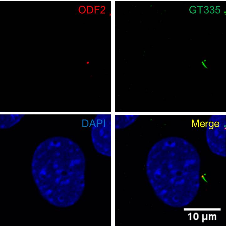
FH Jay (Verified Customer) (12-02-2019) | Methanol Fixation.Immunostaining result shows that Odf2 specifically marks the mother centriole in ciliated NIH3T3 cell (Odf2;red, GT335;green for primary cilia, DAPI;blue for nucleus).
 |

