- Featured Product
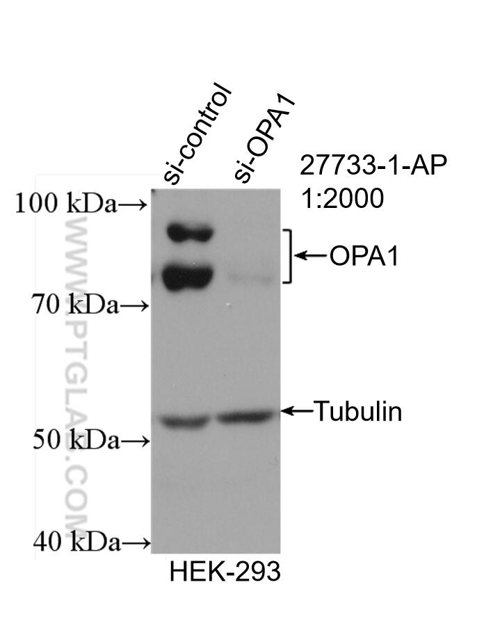
- KD/KO Validated
OPA1 Polyklonaler Antikörper
OPA1 Polyklonal Antikörper für WB, IHC, IP, ELISA
Wirt / Isotyp
Kaninchen / IgG
Getestete Reaktivität
human, Maus, Ratte und mehr (7)
Anwendung
WB, IHC, IF, IP, CoIP, ELISA
Konjugation
Unkonjugiert
Kat-Nr. : 27733-1-AP
Synonyme
Geprüfte Anwendungen
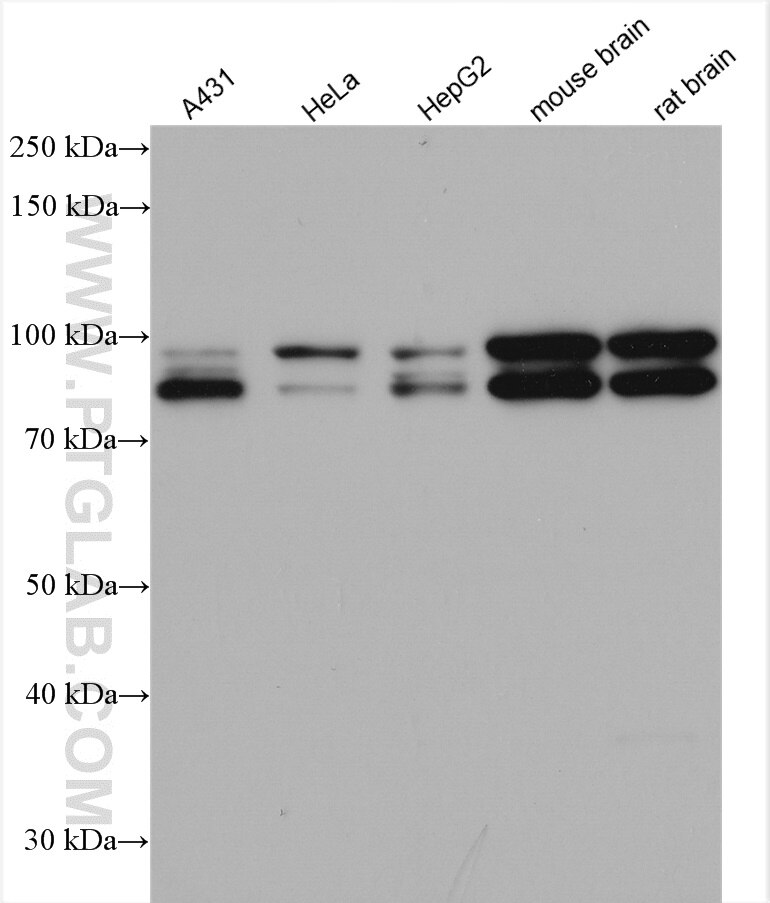
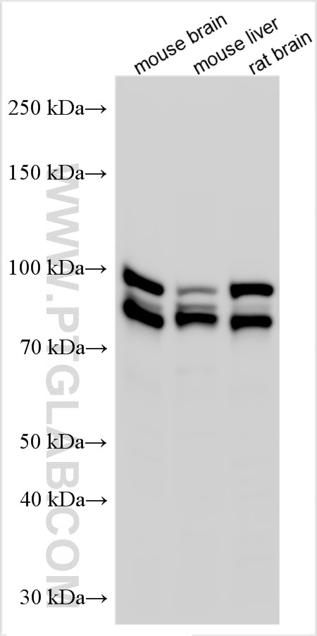
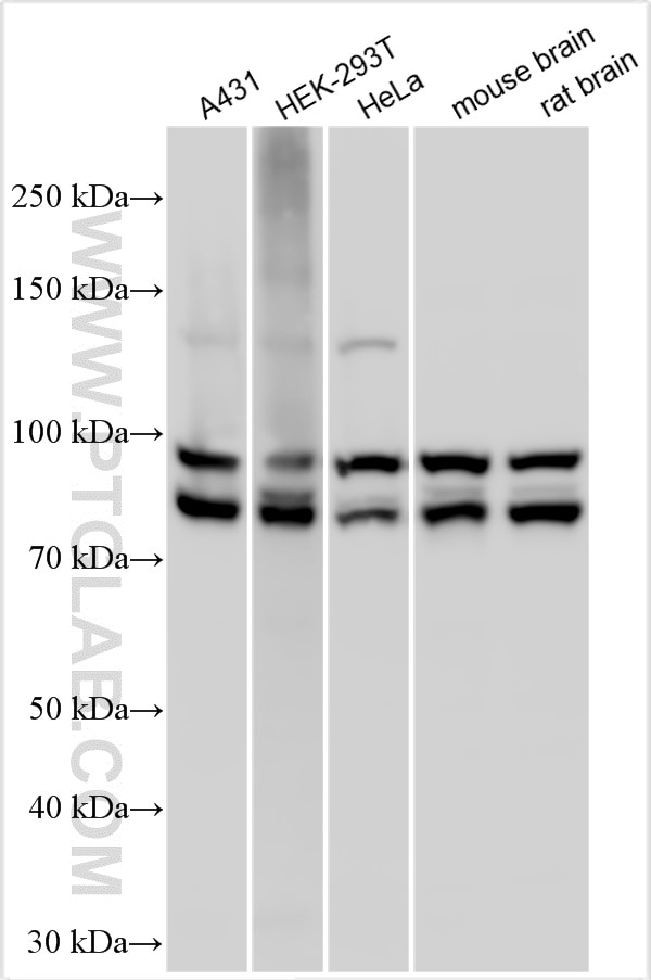
| Erfolgreiche Detektion in WB | A431-Zellen, HEK-293-Zellen, HEK-293T-Zellen, HeLa-Zellen, HepG2-Zellen, L02-Zellen, Maushirngewebe, Mauslebergewebe, Rattenhirngewebe |
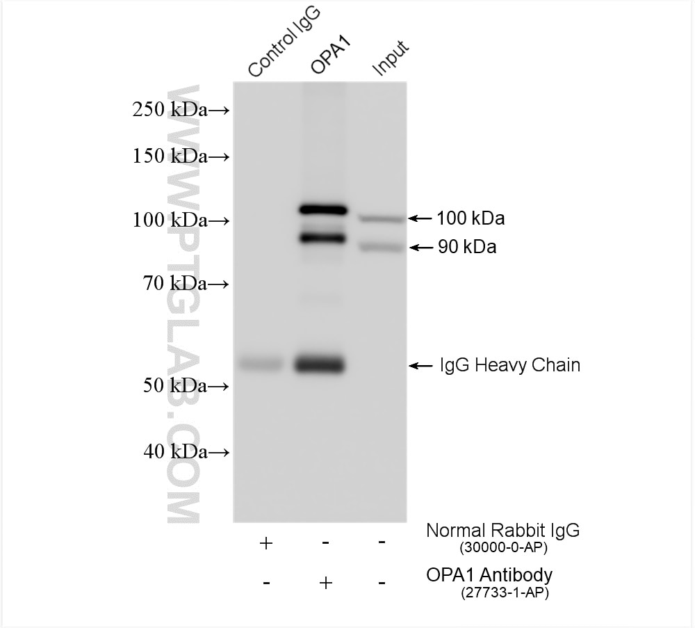
| Erfolgreiche IP | Maushirngewebe |
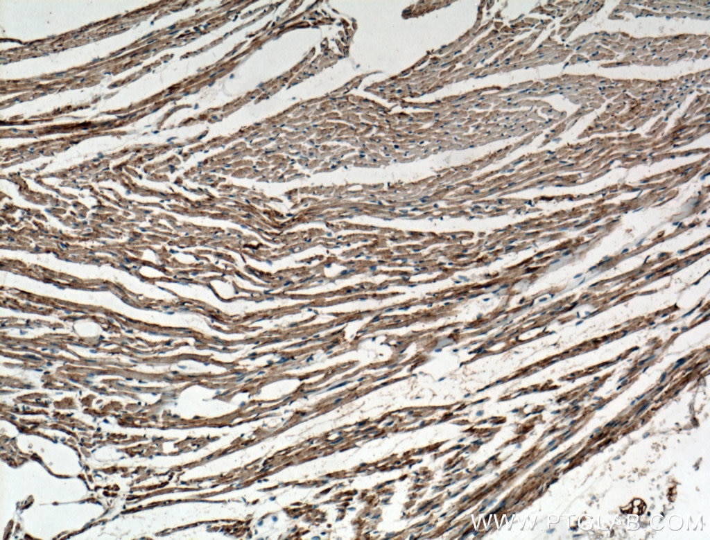
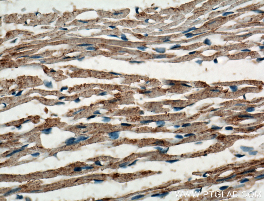
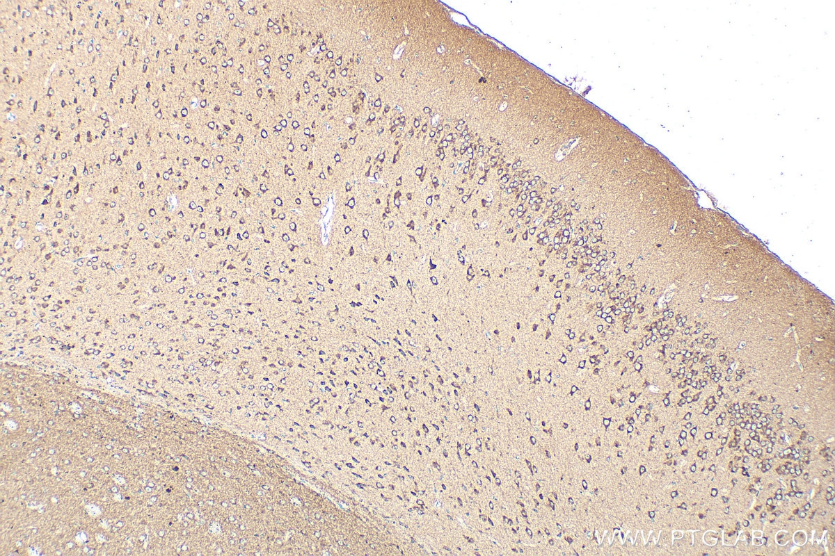
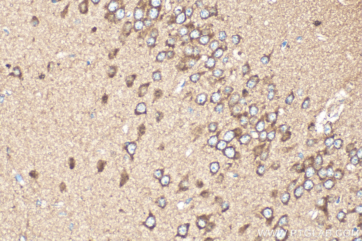
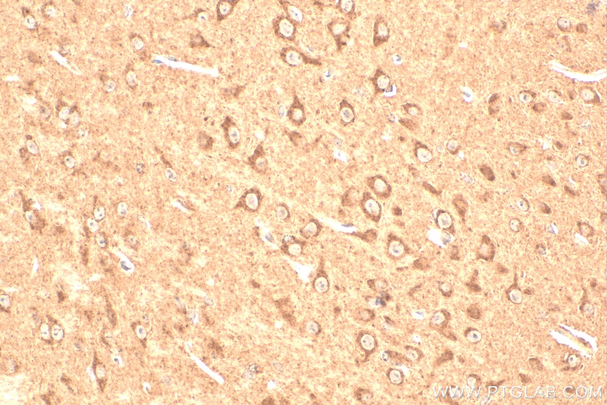
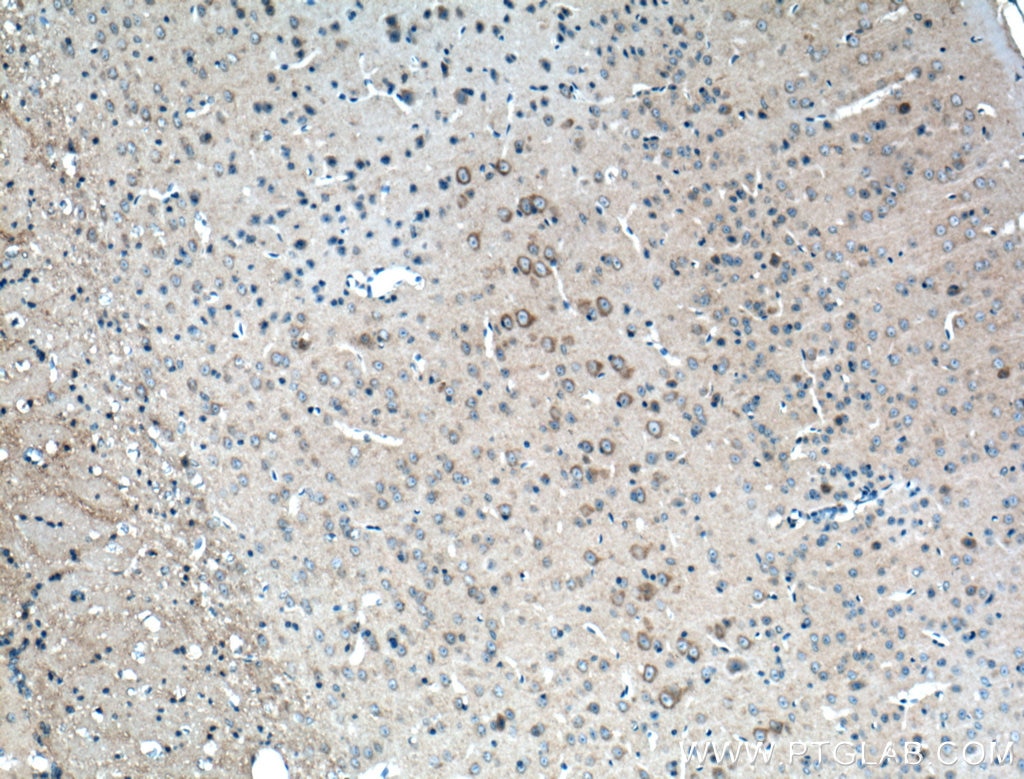
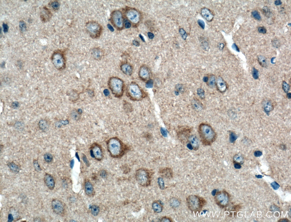
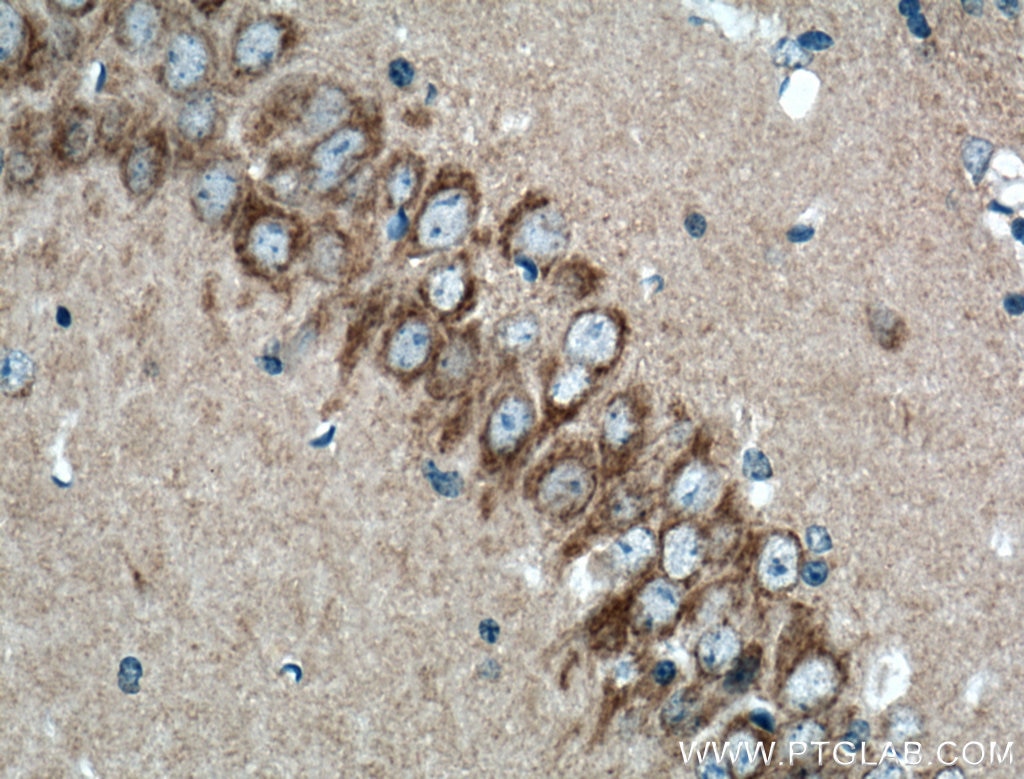
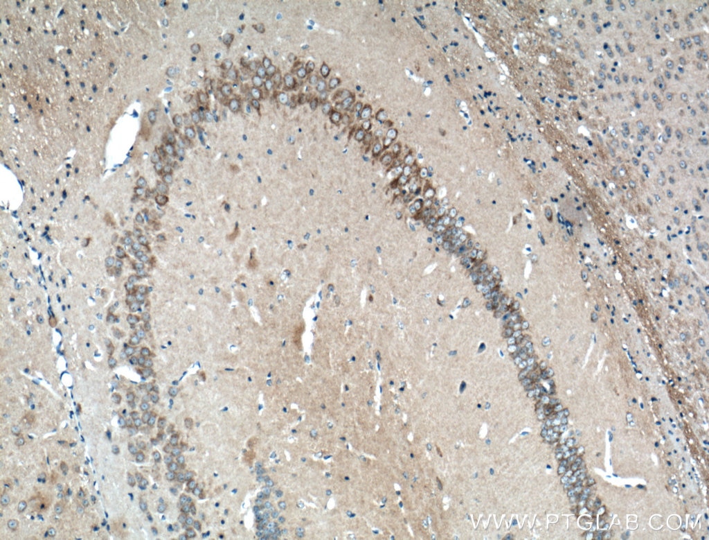
| Erfolgreiche Detektion in IHC | Mausherzgewebe, Maushirngewebe Hinweis: Antigendemaskierung mit TE-Puffer pH 9,0 empfohlen. (*) Wahlweise kann die Antigendemaskierung auch mit Citratpuffer pH 6,0 erfolgen. |
Empfohlene Verdünnung
| Anwendung | Verdünnung |
|---|---|
| Western Blot (WB) | WB : 1:5000-1:50000 |
| Immunpräzipitation (IP) | IP : 0.5-4.0 ug for 1.0-3.0 mg of total protein lysate |
| Immunhistochemie (IHC) | IHC : 1:50-1:500 |
| It is recommended that this reagent should be titrated in each testing system to obtain optimal results. | |
| Sample-dependent, check data in validation data gallery | |
Veröffentlichte Anwendungen
| KD/KO | See 4 publications below |
| WB | See 194 publications below |
| IHC | See 8 publications below |
| IF | See 12 publications below |
| IP | See 1 publications below |
| CoIP | See 1 publications below |
Produktinformation
27733-1-AP bindet in WB, IHC, IF, IP, CoIP, ELISA OPA1 und zeigt Reaktivität mit human, Maus, Ratten
| Getestete Reaktivität | human, Maus, Ratte |
| In Publikationen genannte Reaktivität | human, Ente, hamster, Hausschwein, Huhn, Maus, Ratte, Rind, Zebrafisch, Ziege |
| Wirt / Isotyp | Kaninchen / IgG |
| Klonalität | Polyklonal |
| Typ | Antikörper |
| Immunogen | OPA1 fusion protein Ag26887 |
| Vollständiger Name | optic atrophy 1 (autosomal dominant) |
| Berechnetes Molekulargewicht | 960 aa, 112 kDa |
| Beobachtetes Molekulargewicht | 80-100 kDa |
| GenBank-Zugangsnummer | BC075805 |
| Gene symbol | OPA1 |
| Gene ID (NCBI) | 4976 |
| Konjugation | Unkonjugiert |
| Form | Liquid |
| Reinigungsmethode | Antigen-Affinitätsreinigung |
| Lagerungspuffer | PBS with 0.02% sodium azide and 50% glycerol |
| Lagerungsbedingungen | Bei -20°C lagern. Nach dem Versand ein Jahr lang stabil Aliquotieren ist bei -20oC Lagerung nicht notwendig. 20ul Größen enthalten 0,1% BSA. |
Hintergrundinformationen
OPA1 is a nuclear-encoded mitochondrial protein with similarity to dynamin-related GTPases. OPA1 localizes to the inner mitochondrial membrane and helps regulate mitochondrial stability and energy output. This protein also sequesters cytochrome c. OPA1 is associated with the inner membrane and protects cells from apoptosis by regulating inner membrane dynamics. Mutation of OPA1 causes the disease dominant optic atrophy, a degeneration of the retinal ganglion cells. OPA1 undergoes complex posttranscriptional regulation and posttranslational proteolysis. OPA1 is regulated by proteolytic cleavage, which degrades long OPA1 isoforms into short isoforms. The gene OPA1 can be cleaved into some chains with MW 100 kDa and 80-90 kDa.
Protokolle
| PRODUKTSPEZIFISCHE PROTOKOLLE | |
|---|---|
| WB protocol for OPA1 antibody 27733-1-AP | Protokoll herunterladen |
| IHC protocol for OPA1 antibody 27733-1-AP | Protokoll herunterladenl |
| IP protocol for OPA1 antibody 27733-1-AP | Protokoll herunterladen |
| STANDARD-PROTOKOLLE | |
|---|---|
| Klicken Sie hier, um unsere Standardprotokolle anzuzeigen |
Publikationen
| Species | Application | Title |
|---|---|---|
Cell Res Mitochondria-localized cGAS suppresses ferroptosis to promote cancer progression | ||
ACS Cent Sci Macrophage Inactivation by Small Molecule Wedelolactone via Targeting sEH for the Treatment of LPS-Induced Acute Lung Injury | ||
Mol Cell β-hydroxybutyrate facilitates mitochondrial-derived vesicle biogenesis and improves mitochondrial functions | ||
Mol Cell Serine synthesis sustains macrophage IL-1β production via NAD+-dependent protein acetylation | ||
Mol Cell Filamentous GLS1 promotes ROS-induced apoptosis upon glutamine deprivation via insufficient asparagine synthesis.
| ||
Acta Pharm Sin B Honokiol alleviated neurodegeneration by reducing oxidative stress and improving mitochondrial function in mutant SOD1 cellular and mouse models of amyotrophic lateral sclerosis |
Rezensionen
The reviews below have been submitted by verified Proteintech customers who received an incentive for providing their feedback.
FH Kahimbi (Verified Customer) (01-31-2025) | It was used as a primary antibody for a western blot and the bands were clearly visible. The antibody worked well.
|
FH P (Verified Customer) (09-23-2024) | excellent!
 |
FH Mi (Verified Customer) (02-21-2023) | Works well in human adipocyte cells, we got clean bands at the expected size.
|

