- Featured Product
- KD/KO Validated
PDI Monoklonaler Antikörper
PDI Monoklonal Antikörper für WB, IHC, IF/ICC, ELISA
Wirt / Isotyp
Maus / IgG2b
Getestete Reaktivität
Hausschwein, human, Maus, Ratte und mehr (1)
Anwendung
WB, IHC, IF/ICC, ELISA
Konjugation
Unkonjugiert
CloneNo.
2E6A11
Kat-Nr. : 66422-1-Ig
Synonyme
 at dilution of 1:50000 incubated at room temperature for 1.5 hours.")
 with sh-Control and sh-PDI transfected HEK-293 cells.")
 at dilution of 1:50000 incubated at room temperature for 1.5 hours.")
 at dilution of 1:200 (under 10x lens).")
 at dilution of 1:200 (under 40x lens).")
 at dilution of 1:200 (under 10x lens).")
 at dilution of 1:200 (under 40x lens).")
 fixed HepG2 cells using PDI antibody (66422-1-Ig, Clone: 2E6A11 ) at dilution of 1:400 and CoraLite®488-Conjugated AffiniPure Goat Anti-Mouse IgG(H+L).")
Geprüfte Anwendungen
| Erfolgreiche Detektion in WB | HEK-293-Zellen, COLO 320-Zellen, HepG2-Zellen, L02-Zellen, Rattenhirngewebe |
| Erfolgreiche Detektion in IHC | humanes Lebergewebe, humanes Dünndarmgewebe Hinweis: Antigendemaskierung mit TE-Puffer pH 9,0 empfohlen. (*) Wahlweise kann die Antigendemaskierung auch mit Citratpuffer pH 6,0 erfolgen. |
| Erfolgreiche Detektion in IF/ICC | HepG2-Zellen |
Empfohlene Verdünnung
| Anwendung | Verdünnung |
|---|---|
| Western Blot (WB) | WB : 1:10000-1:100000 |
| Immunhistochemie (IHC) | IHC : 1:100-1:400 |
| Immunfluoreszenz (IF)/ICC | IF/ICC : 1:200-1:800 |
| It is recommended that this reagent should be titrated in each testing system to obtain optimal results. | |
| Sample-dependent, check data in validation data gallery | |
Veröffentlichte Anwendungen
| WB | See 5 publications below |
| IF | See 16 publications below |
Produktinformation
66422-1-Ig bindet in WB, IHC, IF/ICC, ELISA PDI und zeigt Reaktivität mit Hausschwein, human, Maus, Ratten
| Getestete Reaktivität | Hausschwein, human, Maus, Ratte |
| In Publikationen genannte Reaktivität | human, Maus, Rind |
| Wirt / Isotyp | Maus / IgG2b |
| Klonalität | Monoklonal |
| Typ | Antikörper |
| Immunogen | PDI fusion protein Ag1747 |
| Vollständiger Name | prolyl 4-hydroxylase, beta polypeptide |
| Berechnetes Molekulargewicht | 57 kDa |
| Beobachtetes Molekulargewicht | 57 kDa |
| GenBank-Zugangsnummer | BC014504 |
| Gene symbol | PDI |
| Gene ID (NCBI) | 5034 |
| Konjugation | Unkonjugiert |
| Form | Liquid |
| Reinigungsmethode | Protein-A-Reinigung |
| Lagerungspuffer | PBS with 0.02% sodium azide and 50% glycerol |
| Lagerungsbedingungen | Bei -20°C lagern. Nach dem Versand ein Jahr lang stabil Aliquotieren ist bei -20oC Lagerung nicht notwendig. 20ul Größen enthalten 0,1% BSA. |
Hintergrundinformationen
PDIA1(Protein disulfide-isomerase) is also named as ERBA2L, PDI, P4HB, PO4DB. It is a multifunctional protein that catalyzes the formation, breakage and rearrangement of disulfide bonds. In some cell types, it seems to be secreted or associated with the plasma membrane, where it undergoes constant shedding and replacement from intracellular sources.It can exsit as homodimer and monomers and homotetramers may also occur(PMID:12095988).
Protokolle
| PRODUKTSPEZIFISCHE PROTOKOLLE | |
|---|---|
| WB protocol for PDI antibody 66422-1-Ig | Protokoll herunterladen |
| IHC protocol for PDI antibody 66422-1-Ig | Protokoll herunterladenl |
| IF protocol for PDI antibody 66422-1-Ig | Protokoll herunterladen |
| STANDARD-PROTOKOLLE | |
|---|---|
| Klicken Sie hier, um unsere Standardprotokolle anzuzeigen |
Publikationen
| Species | Application | Title |
|---|---|---|
J Agric Food Chem Acylated Ghrelin Activates PI3K/mTOR Signaling Pathway by Promoting ThPOK Acetylation to Promote Milk Fat Synthesis in Bovine Mammary Epithelial Cells | ||
Biochim Biophys Acta Mol Cell Res SLC35A2 deficiency reduces protein levels of core 1 β-1,3-galactosyltransferase 1 (C1GalT1) and its chaperone Cosmc and affects their subcellular localization | ||
J Cell Sci A general role for TANGO1, encoded by MIA3, in secretory pathway organization and function | ||
Mol Pharm Highly Efficient Method for Intracellular Delivery of Proteins Mediated by Cholera Toxin-Induced Protein Internalization. | ||
Cell Signal HRD1-mediated PTEN degradation promotes cell proliferation and hepatocellular carcinoma progression. | ||
Biochim Biophys Acta Gen Subj Expression of GALNT8 and O-glycosylation of BMP receptor 1A suppress breast cancer cell proliferation by upregulating ERα levels |
Rezensionen
The reviews below have been submitted by verified Proteintech customers who received an incentive for providing their feedback.
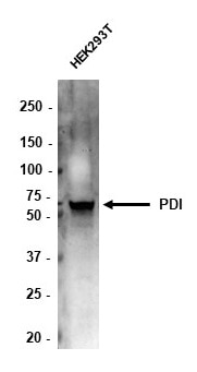
FH Tom (Verified Customer) (12-15-2020) | 10ug total protein of HEK293T lysate loaded. Membrane blocked in 5% BSA. Antibody (1:5,000) incubated overnight in block at 4 degrees. Anti-mouse HRP used at 1 in 10,000 to detect band.
 |

