- Featured Product
- KD/KO Validated
PHLDA1 Polyklonaler Antikörper
PHLDA1 Polyklonal Antikörper für WB, IHC, IF/ICC, ELISA
Wirt / Isotyp
Kaninchen / IgG
Getestete Reaktivität
human, Maus
Anwendung
WB, IHC, IF/ICC, ELISA
Konjugation
Unkonjugiert
Kat-Nr. : 18263-1-AP
Synonyme
 at dilution of 1:4000 incubated at room temperature for 1.5 hours.")
 at dilution of 1:600 incubated at room temperature for 1.5 hours.")
 at dilution of 1:800 (under 20x lens). Heat mediated antigen retrieval with Tris-EDTA buffer (pH 9.0).")
 fixed A375 cells using PHLDA1 antibody (18263-1-AP) at dilution of 1:400 and CoraLite®488-Conjugated AffiniPure Goat Anti-Rabbit IgG(H+L).")
Geprüfte Anwendungen
| Erfolgreiche Detektion in WB | A375-Zellen, BxPC-3-Zellen, HeLa-Zellen, MDA-MB-231-Zellen, Maushirngewebe |
| Erfolgreiche Detektion in IHC | humanes Hautkrebsgewebe Hinweis: Antigendemaskierung mit TE-Puffer pH 9,0 empfohlen. (*) Wahlweise kann die Antigendemaskierung auch mit Citratpuffer pH 6,0 erfolgen. |
| Erfolgreiche Detektion in IF/ICC | A375-Zellen |
Empfohlene Verdünnung
| Anwendung | Verdünnung |
|---|---|
| Western Blot (WB) | WB : 1:1000-1:6000 |
| Immunhistochemie (IHC) | IHC : 1:400-1:1600 |
| Immunfluoreszenz (IF)/ICC | IF/ICC : 1:200-1:800 |
| It is recommended that this reagent should be titrated in each testing system to obtain optimal results. | |
| Sample-dependent, check data in validation data gallery | |
Veröffentlichte Anwendungen
| KD/KO | See 6 publications below |
| WB | See 9 publications below |
| IHC | See 2 publications below |
| IF | See 2 publications below |
Produktinformation
18263-1-AP bindet in WB, IHC, IF/ICC, ELISA PHLDA1 und zeigt Reaktivität mit human, Maus
| Getestete Reaktivität | human, Maus |
| In Publikationen genannte Reaktivität | human, Maus |
| Wirt / Isotyp | Kaninchen / IgG |
| Klonalität | Polyklonal |
| Typ | Antikörper |
| Immunogen | PHLDA1 fusion protein Ag13125 |
| Vollständiger Name | pleckstrin homology-like domain, family A, member 1 |
| Berechnetes Molekulargewicht | 45 kDa |
| Beobachtetes Molekulargewicht | 40-45 kDa |
| GenBank-Zugangsnummer | BC018929 |
| Gene symbol | PHLDA1 |
| Gene ID (NCBI) | 22822 |
| Konjugation | Unkonjugiert |
| Form | Liquid |
| Reinigungsmethode | Antigen-Affinitätsreinigung |
| Lagerungspuffer | PBS with 0.02% sodium azide and 50% glycerol |
| Lagerungsbedingungen | Bei -20°C lagern. Nach dem Versand ein Jahr lang stabil Aliquotieren ist bei -20oC Lagerung nicht notwendig. 20ul Größen enthalten 0,1% BSA. |
Hintergrundinformationen
PHLDA1, also known as PHRIP and TDAG51, is a multifunctional protein involved in various biological processes. It can induce apoptosis in various cell types, including T cells, hippocampal cells, endothelial cells, melanoma cells, and mouse embryonic fibroblasts(PMID: 30207029). PHLDA1 plays a role in inhibiting growth factor signaling and has been implicated in tumor suppression through its ability to repress Akt activity by binding to phosphatidylinositol (PIP) lipids(PMID: 36142223).
Protokolle
| PRODUKTSPEZIFISCHE PROTOKOLLE | |
|---|---|
| WB protocol for PHLDA1 antibody 18263-1-AP | Protokoll herunterladen |
| IHC protocol for PHLDA1 antibody 18263-1-AP | Protokoll herunterladenl |
| IF protocol for PHLDA1 antibody 18263-1-AP | Protokoll herunterladen |
| STANDARD-PROTOKOLLE | |
|---|---|
| Klicken Sie hier, um unsere Standardprotokolle anzuzeigen |
Publikationen
| Species | Application | Title |
|---|---|---|
Brain Behav Immun PHLDA1 promotes microglia-mediated neuroinflammation via regulating K63-linked ubiquitination of TRAF6.
| ||
Inflammation Multiple Machine Learning Identifies Key Gene PHLDA1 Suppressing NAFLD Progression | ||
Life Sci PHLDA1 is a new therapeutic target of oxidative stress and ischemia reperfusion-induced myocardial injury.
| ||
Inflammation TDAG51-Deficiency Podocytes are Protected from High-Glucose-Induced Damage Through Nrf2 Activation via the AKT-GSK-3β Pathway.
| ||
Hum Exp Toxicol Loss of PHLDA1 has a protective role in OGD/R-injured neurons via regulation of the GSK-3β/Nrf2 pathway.
| ||
Neuroreport Egr1 promotes Nlrc4-dependent neuronal pyroptosis through phlda1 in an in-vitro model of intracerebral hemorrhage
|
Rezensionen
The reviews below have been submitted by verified Proteintech customers who received an incentive for providing their feedback.
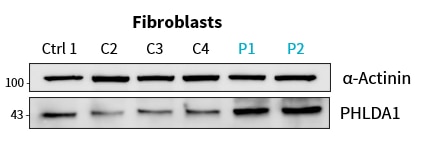
FH Lukas (Verified Customer) (01-31-2025) | worked very well for Western blot in patient primary fibroblasts and cell lines (in 1% milk at +4°C overnight)
 |

