eqRFP630/eqRFP611 Monoklonaler Antikörper
eqRFP630/eqRFP611 Monoklonal Antikörper für WB, ELISA
Wirt / Isotyp
Maus / IgG2b
Getestete Reaktivität
rekombinanten Protein und mehr (3)
Anwendung
WB, IF, ELISA
Konjugation
Unkonjugiert
CloneNo.
1E3C5
Kat-Nr. : 67378-1-Ig
Synonyme
Geprüfte Anwendungen
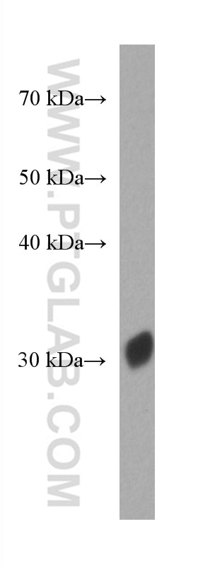
| Erfolgreiche Detektion in WB | ag27669 |
Empfohlene Verdünnung
| Anwendung | Verdünnung |
|---|---|
| Western Blot (WB) | WB : 1:5000-1:100000 |
| It is recommended that this reagent should be titrated in each testing system to obtain optimal results. | |
| Sample-dependent, check data in validation data gallery | |
Veröffentlichte Anwendungen
| WB | See 6 publications below |
| IF | See 1 publications below |
Produktinformation
67378-1-Ig bindet in WB, IF, ELISA eqRFP630/eqRFP611 und zeigt Reaktivität mit rekombinanten Protein
| Getestete Reaktivität | rekombinanten Protein |
| In Publikationen genannte Reaktivität | human, Hefe, Maus |
| Wirt / Isotyp | Maus / IgG2b |
| Klonalität | Monoklonal |
| Typ | Antikörper |
| Immunogen | eqRFP630/eqRFP611 fusion protein Ag27669 |
| Vollständiger Name | eqRFP630/eqRFP611 |
| Berechnetes Molekulargewicht | 26 kDa |
| Gene symbol | |
| Gene ID (NCBI) | |
| Konjugation | Unkonjugiert |
| Form | Liquid |
| Reinigungsmethode | Protein-A-Reinigung |
| Lagerungspuffer | PBS with 0.02% sodium azide and 50% glycerol |
| Lagerungsbedingungen | Bei -20°C lagern. Nach dem Versand ein Jahr lang stabil Aliquotieren ist bei -20oC Lagerung nicht notwendig. 20ul Größen enthalten 0,1% BSA. |
Hintergrundinformationen
Red fluorescent proteins (RFPs) is a collective term referring to a heterogenous group of red chromophore-carrying proteins, originating from various species and forming different protein lineages. The original RFP (dsRed) is a 225 amino acid fluorescent protein (25.9 kDa) derived from Discosoma sp.. It emits red light with a peak wavelength of 593 nm upon excitation by green light (excitation peak at 558 nm). When fused with other proteins, RFP serves as a versatile reporter protein e.g. for quantifying expression levels or facilitates visualization of subcellular localization through fluorescence microscopy. RFP611 and RFP 630 is a basic (constitutively fluorescent) red fluorescent protein derived from Entacmaea quadricolor. This antibody is a mouse (IgG2b) monoclonal antibody raised against RFP (eqFP611), it can also recognize eqFP630. This antibody does not recognize DsRed or mRFP.
Protokolle
| PRODUKTSPEZIFISCHE PROTOKOLLE | |
|---|---|
| WB protocol for eqRFP630/eqRFP611 antibody 67378-1-Ig | Protokoll herunterladen |
| STANDARD-PROTOKOLLE | |
|---|---|
| Klicken Sie hier, um unsere Standardprotokolle anzuzeigen |
Publikationen
| Species | Application | Title |
|---|---|---|
bioRxiv Membrane binding of endocytic myosin-1s is inhibited by a class of ankyrin repeat proteins | ||
PNAS Nexus Ribosome stalling during c-myc translation presents actionable cancer cell vulnerability | ||
Autophagy The Valsa mali effector Vm1G-1794 protects the aggregated MdEF-Tu from autophagic degradation to promote infection in apple | ||
EMBO Rep Myddosome clustering in IL-1 receptor signaling regulates the formation of an NF-kB activating signalosome | ||
Mol Biol Cell Membrane binding of endocytic myosin-1s is inhibited by a class of ankyrin repeat proteins |
Rezensionen
The reviews below have been submitted by verified Proteintech customers who received an incentive for providing their feedback.
FH April (Verified Customer) (08-28-2025) | Product worked well for detecting RFP in mouse tissues with cells containing RFP.
|
FH Christine (Verified Customer) (08-05-2025) | Tried on total cell lysates of HEK293T transfected with a mCHERRY tagged protein. This RFP antibody was not very good at detecting the mCHERRY tag (mCHERRY antibody from proteintech 68088-1-ig) was better.
|


