TBR1 Polyklonaler Antikörper
TBR1 Polyklonal Antikörper für WB, IHC, IF-P, IP, ELISA
Wirt / Isotyp
Kaninchen / IgG
Getestete Reaktivität
human, Maus, Ratte
Anwendung
WB, IHC, IF-P, IP, ELISA
Konjugation
Unkonjugiert
Kat-Nr. : 20932-1-AP
Synonyme
 at dilution of 1:500 incubated at room temperature for 1.5 hours.")
 with mouse brain tissue lysate 4000ug.")
 at dilution of 1:100 (under 10x lens).")
 at dilution of 1:100 (under 40x lens).")
 at dilution of 1:400 (under 10x lens). Heat mediated antigen retrieval with Tris-EDTA buffer (pH 9.0).")
 at dilution of 1:400 (under 40x lens). Heat mediated antigen retrieval with Tris-EDTA buffer (pH 9.0).")
 at dilution of 1:200 (under 10x lens. Heat mediated antigen retrieval with Tris-EDTA buffer (pH 9.0).")
 at dilution of 1:200 (under 40x lens. Heat mediated antigen retrieval with Tris-EDTA buffer (pH 9.0).")
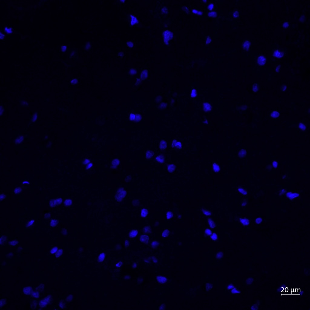
 fixed mouse brain tissue using TBR1 antibody (20932-1-AP) at dilution of 1:200 and CoraLite®488-Conjugated AffiniPure Goat Anti-Rabbit IgG(H+L).")
 fixed paraffin-embedded mouse brain tissue using TBR1 antibody (20932-1-AP) at dilution of 1:400 and Multi-rAb CoraLite ® Plus 488-Goat Anti-Rabbit Recombinant Secondary Antibody (H+L) (RGAR002). Heat mediated antigen retrieval with Tris-EDTA buffer (pH 9.0).")
Geprüfte Anwendungen
| Erfolgreiche Detektion in WB | Maushirngewebe |
| Erfolgreiche IP | Maushirngewebe |
| Erfolgreiche Detektion in IHC | humanes Hirngewebe, Maushirngewebe, Rattenhirngewebe Hinweis: Antigendemaskierung mit TE-Puffer pH 9,0 empfohlen. (*) Wahlweise kann die Antigendemaskierung auch mit Citratpuffer pH 6,0 erfolgen. |
| Erfolgreiche Detektion in IF-P | Maushirngewebe |
Empfohlene Verdünnung
| Anwendung | Verdünnung |
|---|---|
| Western Blot (WB) | WB : 1:500-1:1000 |
| Immunpräzipitation (IP) | IP : 0.5-4.0 ug for 1.0-3.0 mg of total protein lysate |
| Immunhistochemie (IHC) | IHC : 1:20-1:200 |
| Immunfluoreszenz (IF)-P | IF-P : 1:200-1:800 |
| It is recommended that this reagent should be titrated in each testing system to obtain optimal results. | |
| Sample-dependent, check data in validation data gallery | |
Veröffentlichte Anwendungen
| WB | See 9 publications below |
| IHC | See 5 publications below |
| IF | See 24 publications below |
Produktinformation
20932-1-AP bindet in WB, IHC, IF-P, IP, ELISA TBR1 und zeigt Reaktivität mit human, Maus, Ratten
| Getestete Reaktivität | human, Maus, Ratte |
| In Publikationen genannte Reaktivität | human, Maus, Ratte |
| Wirt / Isotyp | Kaninchen / IgG |
| Klonalität | Polyklonal |
| Typ | Antikörper |
| Immunogen | TBR1 fusion protein Ag14935 |
| Vollständiger Name | T-box, brain, 1 |
| Berechnetes Molekulargewicht | 682 aa, 74 kDa |
| Beobachtetes Molekulargewicht | 74 kDa |
| GenBank-Zugangsnummer | BC104844 |
| Gene symbol | TBR1 |
| Gene ID (NCBI) | 10716 |
| Konjugation | Unkonjugiert |
| Form | Liquid |
| Reinigungsmethode | Antigen-Affinitätsreinigung |
| Lagerungspuffer | PBS with 0.02% sodium azide and 50% glycerol |
| Lagerungsbedingungen | Bei -20°C lagern. Nach dem Versand ein Jahr lang stabil Aliquotieren ist bei -20oC Lagerung nicht notwendig. 20ul Größen enthalten 0,1% BSA. |
Hintergrundinformationen
TBR1, also named as T-box brain protein 1, is a 682 amino acid protein, which contains one T-box DNA-binding domain and localizes in the nucleus. TBR1 is expressed in the brain and as a transcriptional regulator is involved in developmental processes. TBR1 is required for normal brain development.
Protokolle
| PRODUKTSPEZIFISCHE PROTOKOLLE | |
|---|---|
| WB protocol for TBR1 antibody 20932-1-AP | Protokoll herunterladen |
| IHC protocol for TBR1 antibody 20932-1-AP | Protokoll herunterladenl |
| IF protocol for TBR1 antibody 20932-1-AP | Protokoll herunterladen |
| IP protocol for TBR1 antibody 20932-1-AP | Protokoll herunterladen |
| STANDARD-PROTOKOLLE | |
|---|---|
| Klicken Sie hier, um unsere Standardprotokolle anzuzeigen |
Publikationen
| Species | Application | Title |
|---|---|---|
Nat Neurosci A tau homeostasis signature is linked with the cellular and regional vulnerability of excitatory neurons to tau pathology. | ||
Nat Commun GRAMD1B is a regulator of lipid homeostasis, autophagic flux and phosphorylated tau | ||
Nat Commun Pathogenic POGZ mutation causes impaired cortical development and reversible autism-like phenotypes. | ||
Nat Commun Disrupted neuronal maturation in Angelman syndrome-derived induced pluripotent stem cells. | ||
Proc Natl Acad Sci U S A Human intermediate progenitor diversity during cortical development. |
Rezensionen
The reviews below have been submitted by verified Proteintech customers who received an incentive for providing their feedback.
FH Reyes (Verified Customer) (03-01-2024) | Used on human brain cortical sections, it marked nicely the nucleus of my neurons.
 |
FH Mandi (Verified Customer) (03-09-2020) | Good staining on iPSC-derived Cortical neurons. Needed a decent amount of optimization to prevent non-specific binding though. Make sure to run primary/secondary deletes.
|

