• Featured Product
  • KD/KO Validated

YY1 Monoklonaler Antikörper

YY1 Monoklonal Antikörper für WB, IHC, IF/ICC, IP, ELISA

Wirt / Isotyp

Maus / IgG2a

Getestete Reaktivität

Affe, human, Maus, Ratte und mehr (1)

Anwendung

WB, IHC, IF/ICC, IP, CoIP, ChIP, RIP, ELISA

Konjugation

Unkonjugiert

Publikationen(56)

CloneNo.

2E11C5

Kat-Nr. : 66281-1-Ig

Synonyme

DELTA, Delta transcription factor, INO80 complex subunit S, INO80S, NF E1, UCRBP, Yin and yang 1, YIN YANG 1, YY 1, YY1, YY1 transcription factor


Filter:
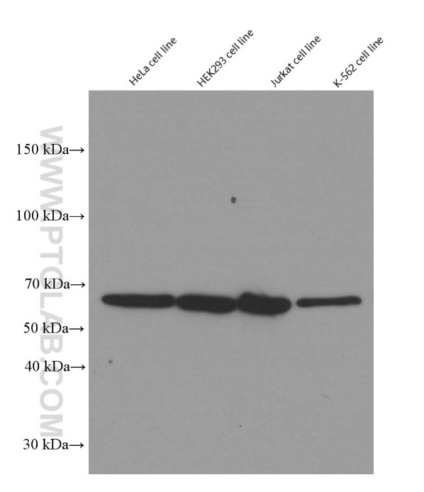
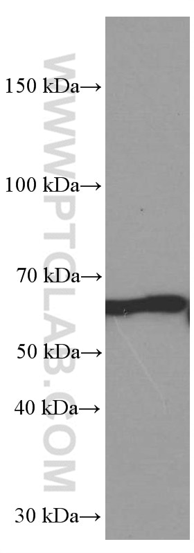
  • Western Blot (WB) analysis of HeLa cells using YY1 Monoclonal antibody (66281-1-Ig)

    WB analysis of HeLa using 66281-1-Ig

    Western blot of YY1 in various human cell lines with 66281-1-Ig at dilution of 1:50000 incubated at room temperature for 1.5 hours.

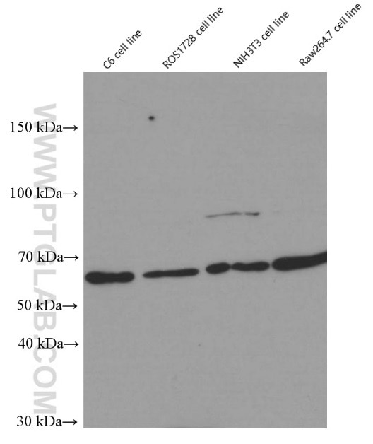
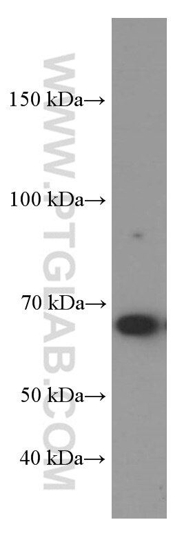
  • Western Blot (WB) analysis of C6 cells using YY1 Monoclonal antibody (66281-1-Ig)

    WB analysis of C6 using 66281-1-Ig

    Western blot of YY1 in rat (C6 and ROS1728) and mouse(NIH3T3 and Raw264.7) cell lines with 66281-1-Ig at dilution of 1:50000 incubated at room temperature for 1.5 hours.

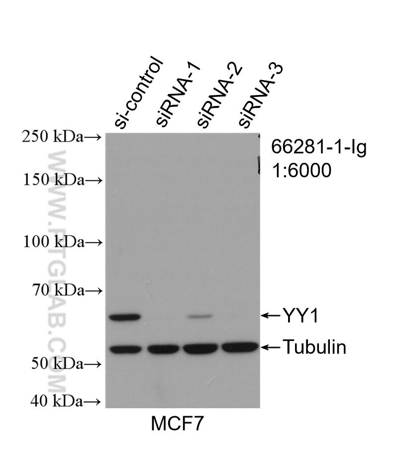
  • Western Blot (WB) analysis of MCF-7 cells using YY1 Monoclonal antibody (66281-1-Ig)

    WB analysis of MCF-7 using 66281-1-Ig

    WB result of YY1 antibody (66281-1-Ig; 1:6000; incubated at room temperature for 1.5 hours) with sh-Control and sh-YY1 transfected MCF-7 cells.

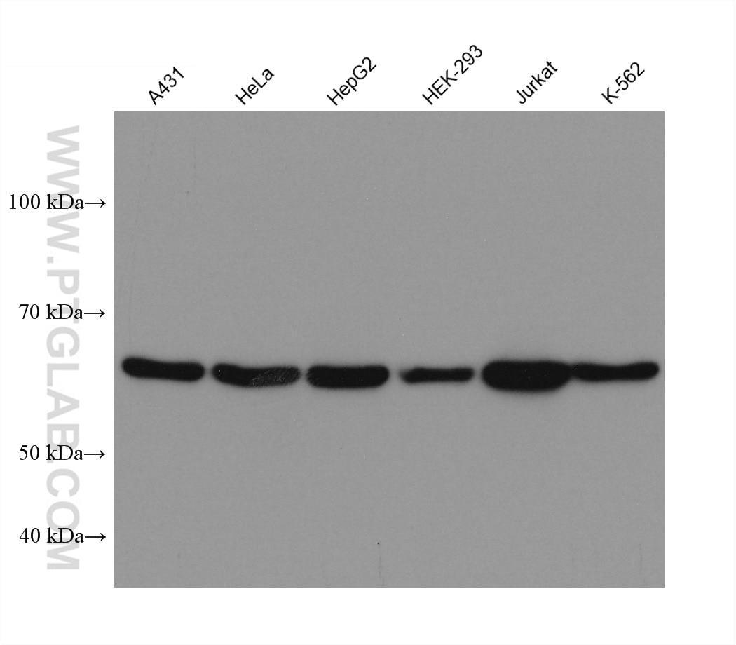
  • Western Blot (WB) analysis of various lysates using YY1 Monoclonal antibody (66281-1-Ig)

    WB analysis using 66281-1-Ig

    Various lysates were subjected to SDS PAGE followed by western blot with 66281-1-Ig (YY1 antibody) at dilution of 1:20000 incubated at room temperature for 1.5 hours.

  • Western Blot (WB) analysis of HeLa cells using YY1 Monoclonal antibody (66281-1-Ig)

    WB analysis of HeLa using 66281-1-Ig

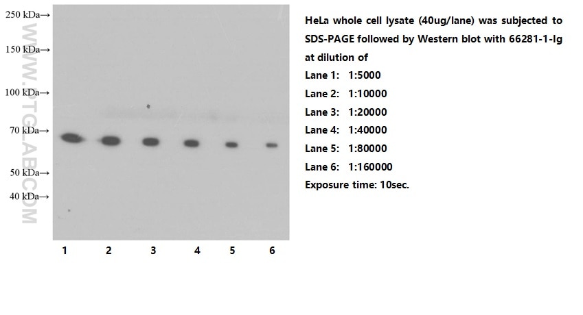
    Western blot of Hela cells with mouse anti-YY1 monoclonal antibody (66281-1-Ig) at various dilutions incubated at room temperature for 1.5 hours.

  • Western Blot (WB) analysis of COS-7 cells using YY1 Monoclonal antibody (66281-1-Ig)

    WB analysis of COS-7 using 66281-1-Ig

    COS-7 cells were subjected to SDS PAGE followed by western blot with 66281-1-Ig (YY1 Antibody) at dilution of 1:50000 incubated at room temperature for 1.5 hours.

  • Western Blot (WB) analysis of NIH/3T3 cells using YY1 Monoclonal antibody (66281-1-Ig)

    WB analysis of NIH/3T3 using 66281-1-Ig

    NIH/3T3 cells were subjected to SDS PAGE followed by western blot with 66281-1-Ig (YY1 Antibody) at dilution of 1:50000 incubated at room temperature for 1.5 hours.

  • Immunoprecipitation (IP) experiment of NIH/3T3 cells using YY1 Monoclonal antibody (66281-1-Ig)

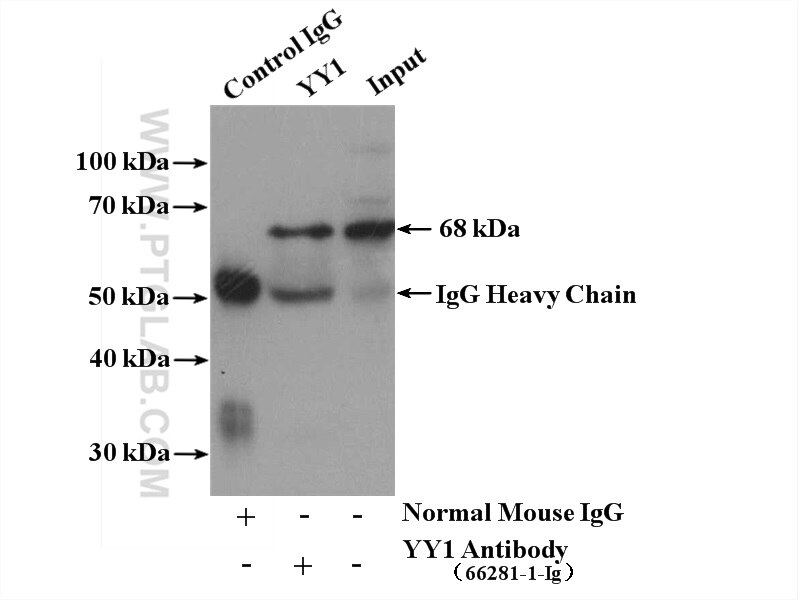
    IP experiment of NIH/3T3 using 66281-1-Ig

    IP result of anti-YY1 (IP:66281-1-Ig, 5ug; Detection:66281-1-Ig 1:1000) with NIH/3T3 cells lysate 4000ug.

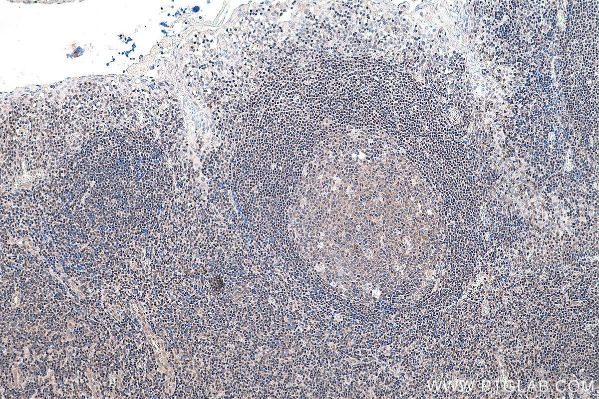
  • Immunohistochemistry (IHC) staining of human tonsillitis tissue using YY1 Monoclonal antibody (66281-1-Ig)

    IHC staining of human tonsillitis using 66281-1-Ig

    Immunohistochemical analysis of paraffin-embedded human tonsillitis tissue slide using 66281-1-Ig (YY1 antibody) at dilution of 1:5000 (under 10x lens). Heat mediated antigen retrieval with Tris-EDTA buffer (pH 9.0).

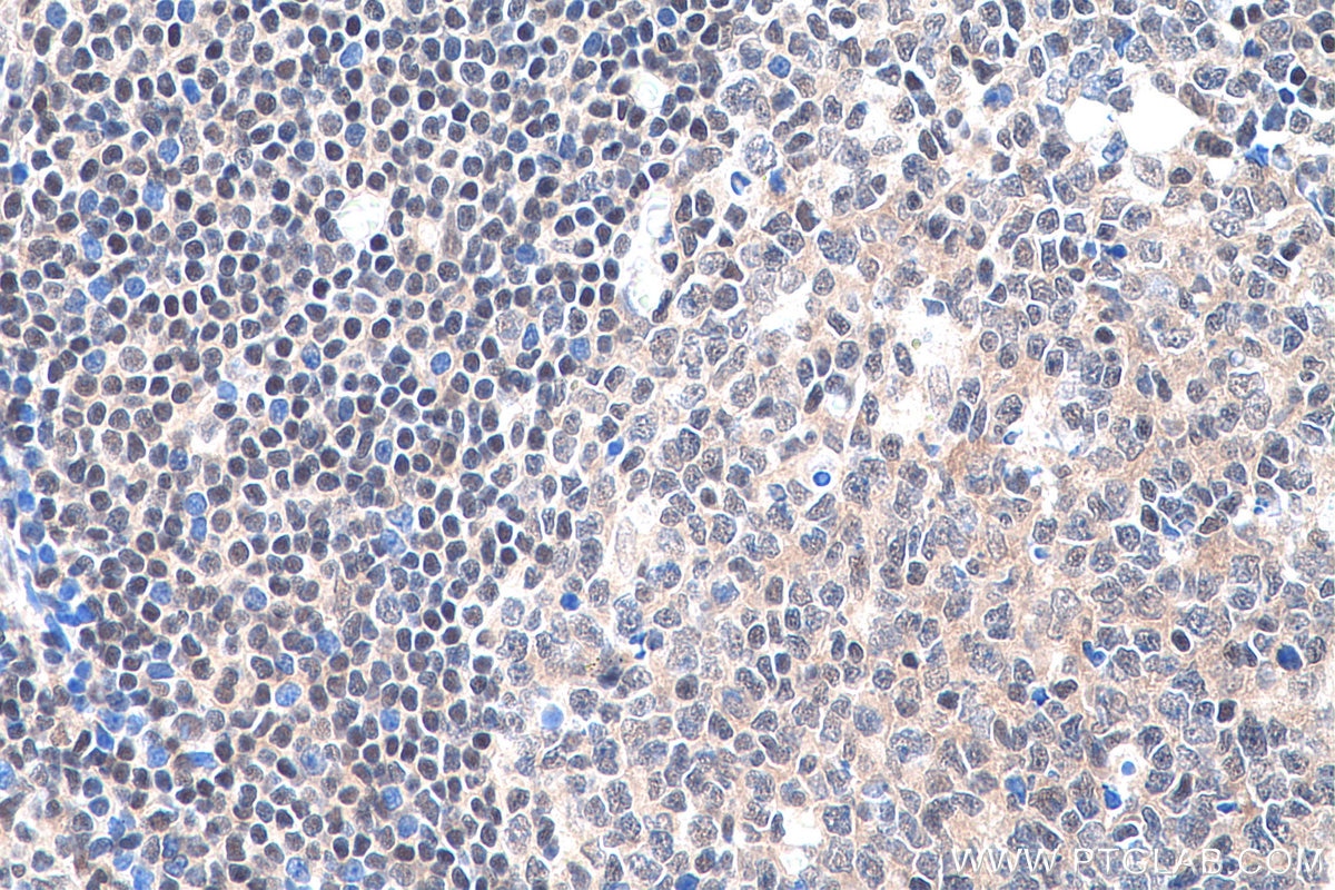
  • Immunohistochemistry (IHC) staining of human tonsillitis tissue using YY1 Monoclonal antibody (66281-1-Ig)

    IHC staining of human tonsillitis using 66281-1-Ig

    Immunohistochemical analysis of paraffin-embedded human tonsillitis tissue slide using 66281-1-Ig (YY1 antibody) at dilution of 1:5000 (under 40x lens). Heat mediated antigen retrieval with Tris-EDTA buffer (pH 9.0).

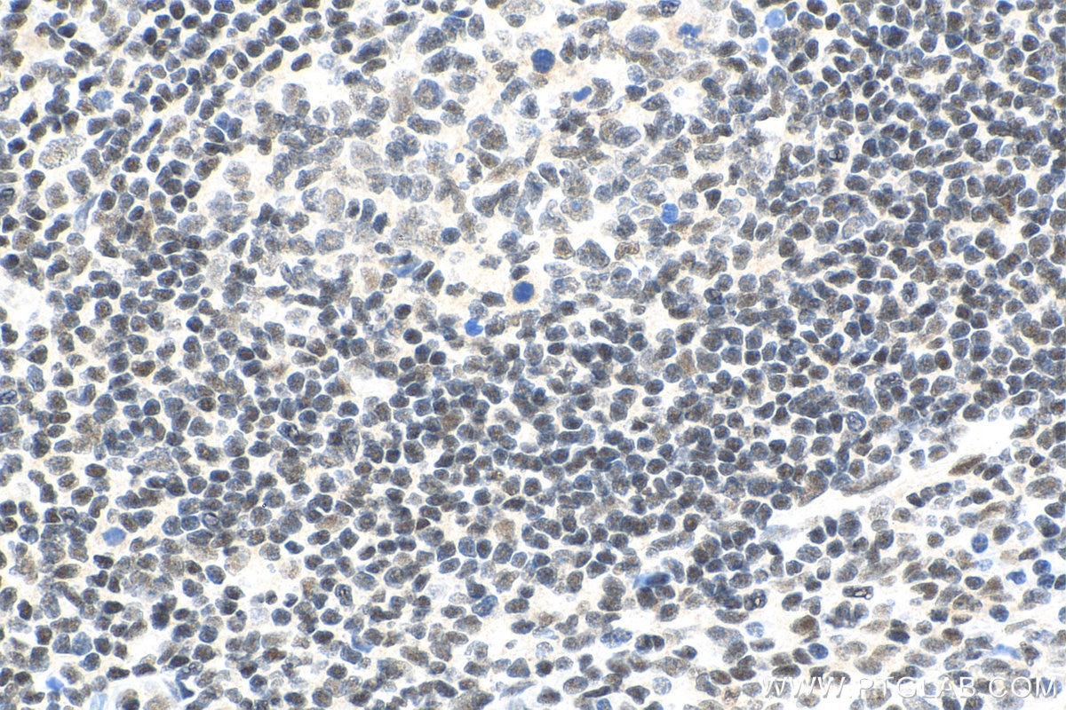
  • Immunohistochemistry (IHC) staining of human tonsillitis tissue using YY1 Monoclonal antibody (66281-1-Ig)

    IHC staining of human tonsillitis using 66281-1-Ig

    Immunohistochemical analysis of paraffin-embedded human tonsillitis tissue slide using 66281-1-Ig (YY1 antibody) at dilution of 1:10000 (under 40x lens). Heat mediated antigen retrieval with Tris-EDTA buffer (pH 9.0).

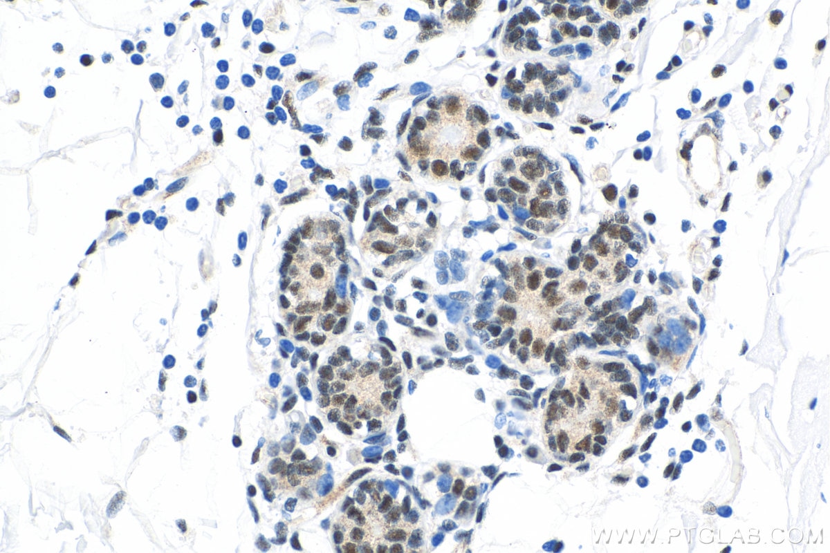
  • Immunohistochemistry (IHC) staining of human breast cancer tissue using YY1 Monoclonal antibody (66281-1-Ig)

    IHC staining of human breast cancer using 66281-1-Ig

    Immunohistochemical analysis of paraffin-embedded human breast cancer tissue slide using 66281-1-Ig (YY1 antibody) at dilution of 1:10000 (under 40x lens). Heat mediated antigen retrieval with Tris-EDTA buffer (pH 9.0).

  • Immunohistochemistry (IHC) staining of human colon cancer tissue using YY1 Monoclonal antibody (66281-1-Ig)

    IHC staining of human colon cancer using 66281-1-Ig

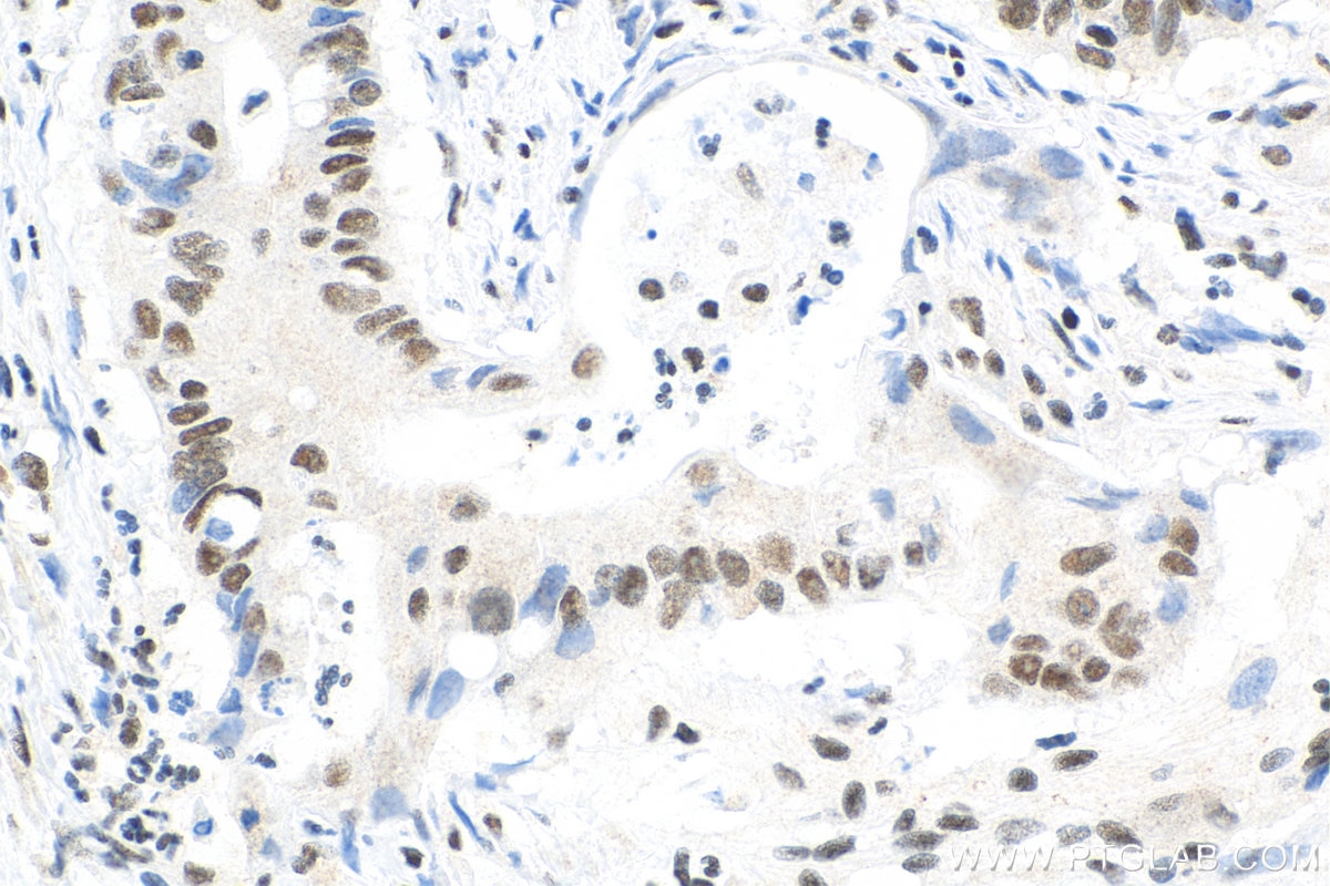
    Immunohistochemical analysis of paraffin-embedded human colon cancer tissue slide using 66281-1-Ig (YY1 antibody) at dilution of 1:10000 (under 40x lens). Heat mediated antigen retrieval with Tris-EDTA buffer (pH 9.0).

  • Immunohistochemistry (IHC) staining of human breast cancer tissue using YY1 Monoclonal antibody (66281-1-Ig)

    IHC staining of human breast cancer using 66281-1-Ig

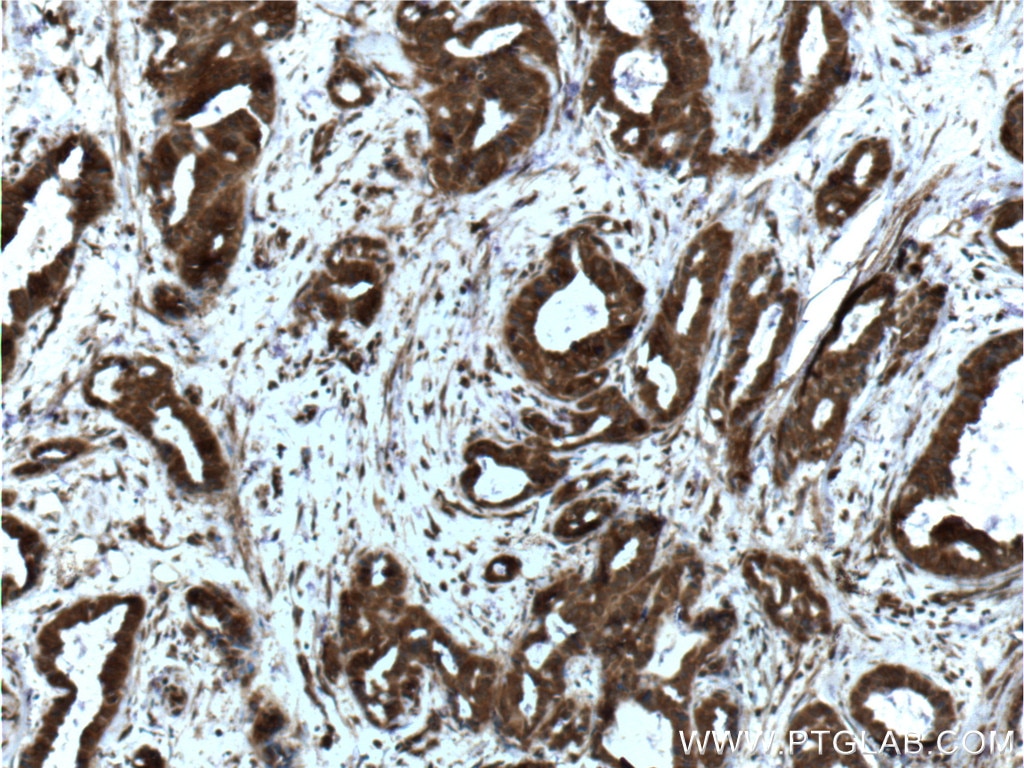
    Immunohistochemical analysis of paraffin-embedded human breast cancer tissue slide using 66281-1-Ig (YY1 Antibody) at dilution of 1:200 (under 10x lens). Heat mediated antigen retrieval with Tris-EDTA buffer (pH 9.0).

  • Immunohistochemistry (IHC) staining of human breast cancer tissue using YY1 Monoclonal antibody (66281-1-Ig)

    IHC staining of human breast cancer using 66281-1-Ig

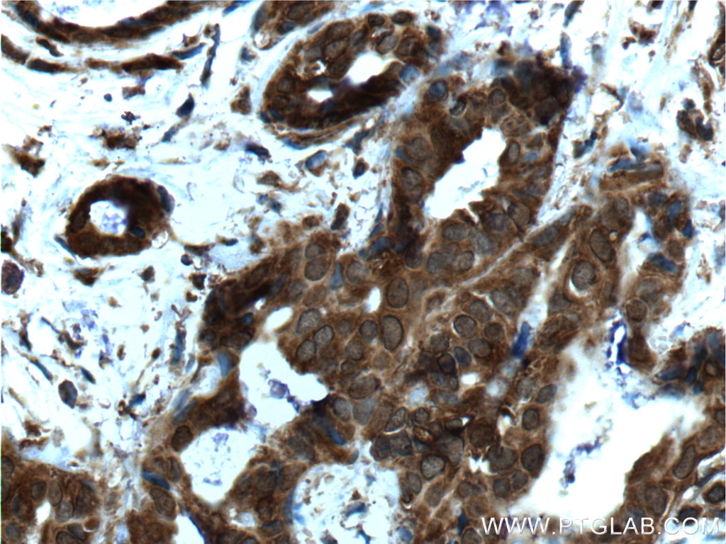
    Immunohistochemical analysis of paraffin-embedded human breast cancer tissue slide using 66281-1-Ig (YY1 Antibody) at dilution of 1:200 (under 40x lens). Heat mediated antigen retrieval with Tris-EDTA buffer (pH 9.0).

  • Immunofluorescence (IF) / fluorescent staining of HepG2 cells using YY1 Monoclonal antibody (66281-1-Ig)

    IF Staining of HepG2 using 66281-1-Ig

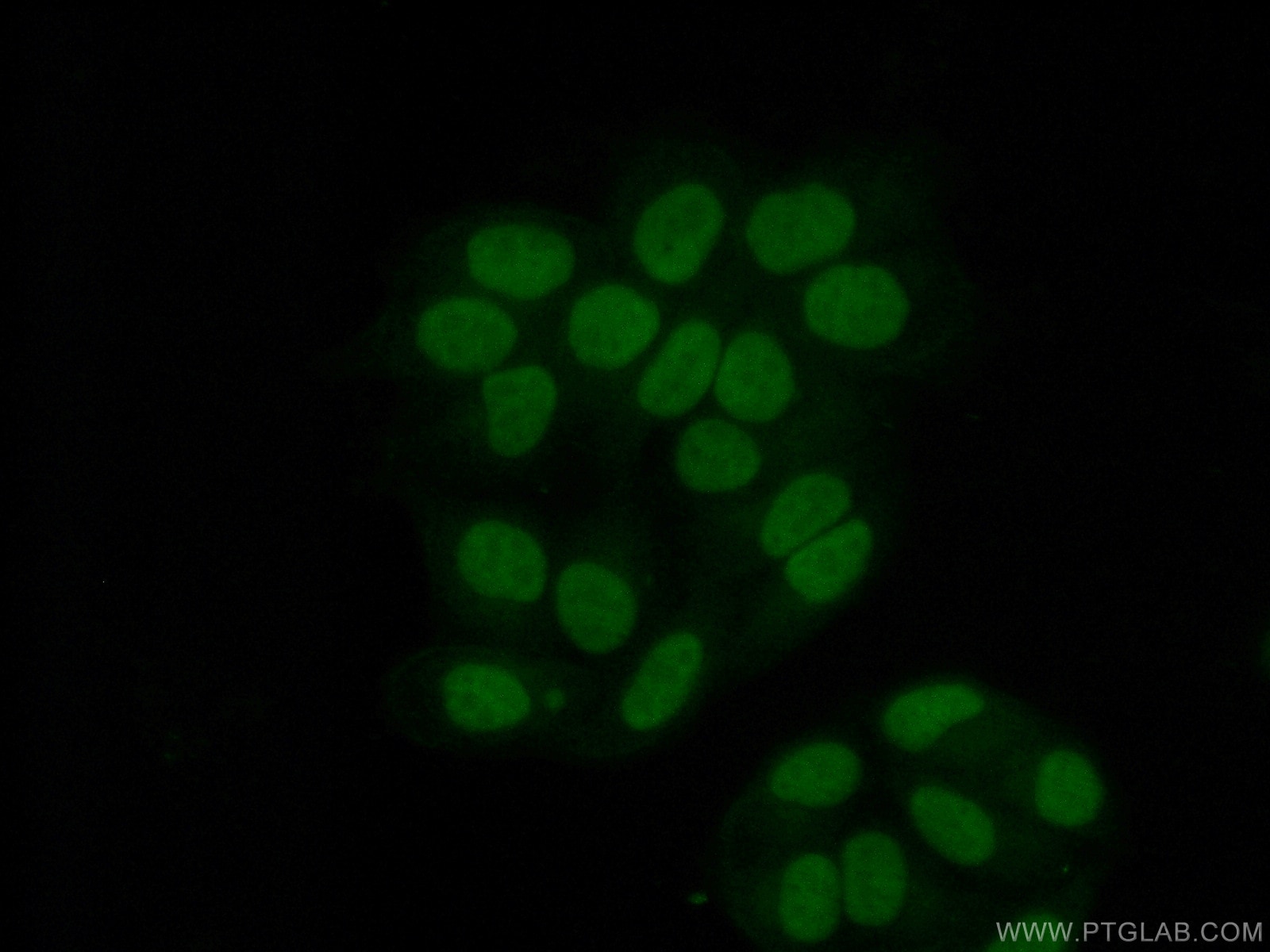
    Immunofluorescent analysis of (4% PFA) fixed HepG2 cells using 66281-1-Ig (YY1 antibody), at dilution of 1:400 and CoraLite®488-Conjugated Goat Anti-Mouse IgG(H+L).

  • Immunofluorescence (IF) / fluorescent staining of HepG2 cells using YY1 Monoclonal antibody (66281-1-Ig)

    IF Staining of HepG2 using 66281-1-Ig

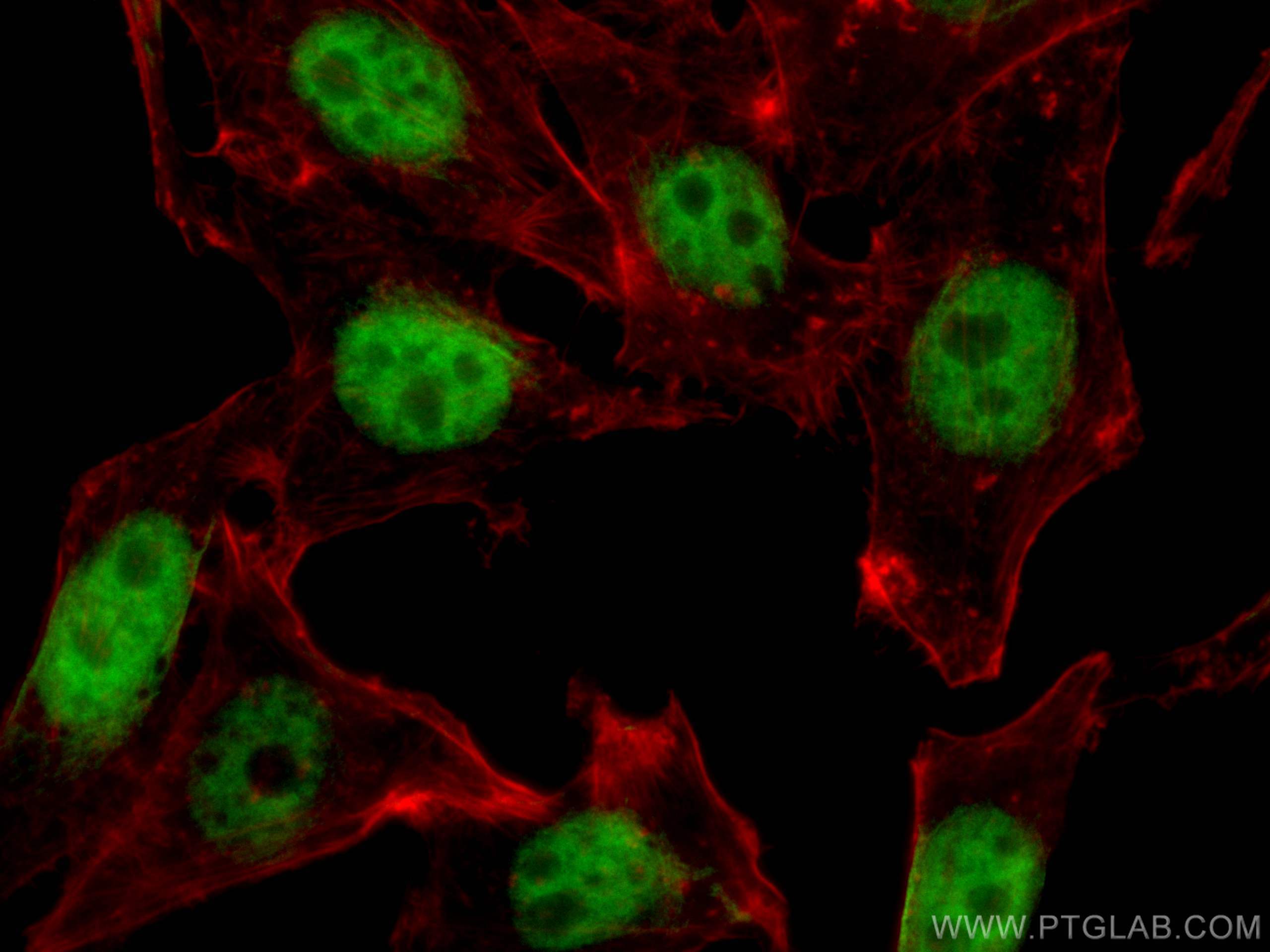
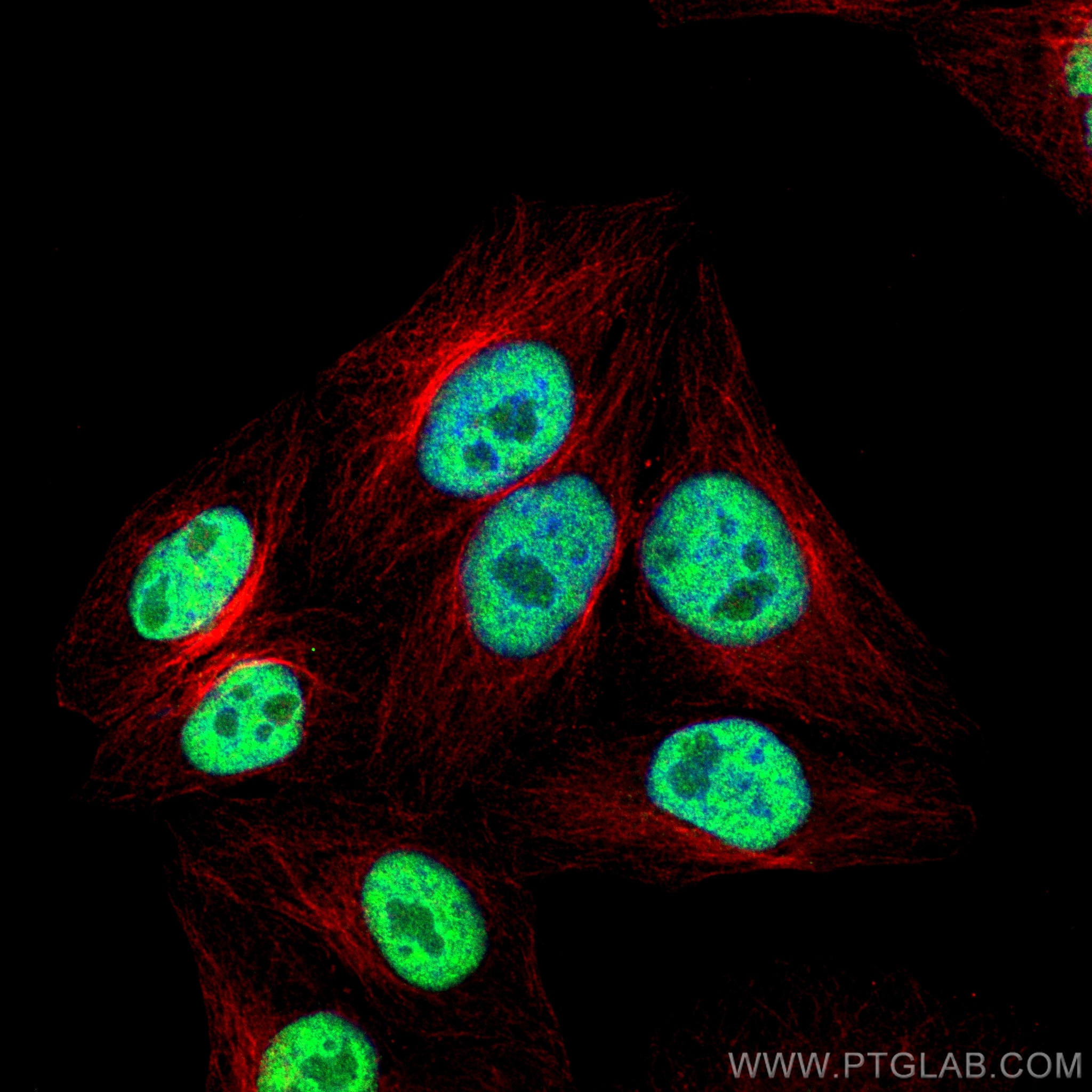
    Immunofluorescent analysis of (4% PFA) fixed HepG2 cells using YY1 antibody (66281-1-Ig, Clone: 2E11C5 ) at dilution of 1:1800 and Multi-rAb CoraLite ® Plus 488-Goat Anti-Mouse Recombinant Secondary Antibody (H+L) (RGAM002), Alpha Tubulin antibody (80762-1-RR, Clone: 1G7, red).

  • Immunofluorescence (IF) / fluorescent staining of HepG2 cells using YY1 Monoclonal antibody (66281-1-Ig)

    IF Staining of HepG2 using 66281-1-Ig

    Immunofluorescent analysis of (10% Formaldehyde ) fixed HepG2 cells using 66281-1-Ig(YY1 antibody) at dilution of 1:800 and Alexa Fluor 488-conjugated Goat Anti-Mouse IgG(H+L).

  • Immunofluorescence (IF) / fluorescent staining of HepG2 cells using YY1 Monoclonal antibody (66281-1-Ig)

    IF Staining of HepG2 using 66281-1-Ig

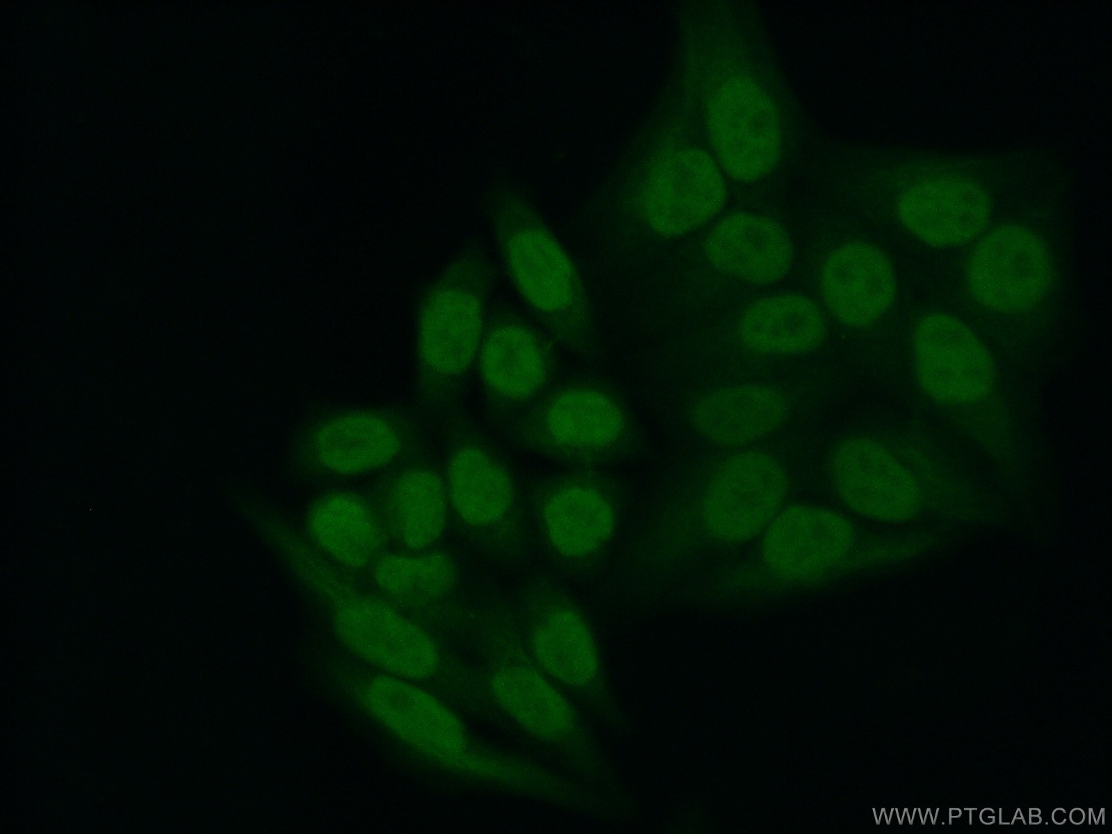
    Immunofluorescent analysis of (10% Formaldehyde ) fixed HepG2 cells using 66281-1-Ig(YY1 antibody) at dilution of 1:400 and Alexa Fluor 488-conjugated Goat Anti-Mouse IgG(H+L).

  • Immunofluorescence (IF) / fluorescent staining of HepG2 cells using YY1 Monoclonal antibody (66281-1-Ig)

    IF Staining of HepG2 using 66281-1-Ig

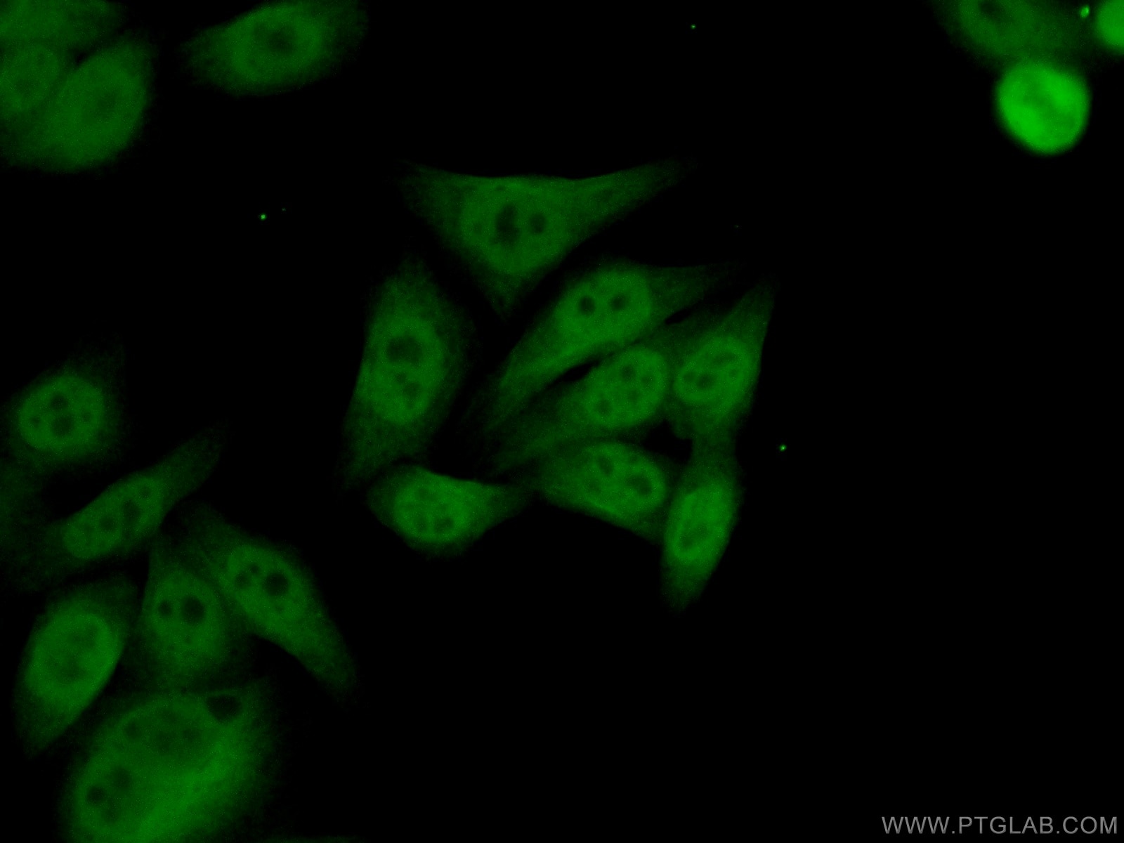
    Immunofluorescent analysis of (10% Formaldehyde ) fixed HepG2 cells using 66281-1-Ig(YY1 antibody) at dilution of 1:100 and Alexa Fluor 488-conjugated Goat Anti-Mouse IgG(H+L).

"YY1 Antibodies" Comparison

View side-by-side comparison of YY1 antibodies from other vendors to find the one that best suits your research needs.

Geprüfte Anwendungen

Erfolgreiche Detektion in WBC6-Zellen, A431-Zellen, COS-7-Zellen, HEK-293-Zellen, HeLa-Zellen, HepG2-Zellen, Jurkat-Zellen, K-562-Zellen, MCF-7-Zellen, NIH/3T3-Zellen
Erfolgreiche IPNIH/3T3-Zellen
Erfolgreiche Detektion in IHChumanes Mammakarzinomgewebe, humanes Kolonkarzinomgewebe, humanes Tonsillitisgewebe
Hinweis: Antigendemaskierung mit TE-Puffer pH 9,0 empfohlen. (*) Wahlweise kann die Antigendemaskierung auch mit Citratpuffer pH 6,0 erfolgen.
Erfolgreiche Detektion in IF/ICCHepG2-Zellen

Empfohlene Verdünnung

AnwendungVerdünnung
Western Blot (WB)WB : 1:5000-1:50000
Immunpräzipitation (IP)IP : 0.5-4.0 ug for 1.0-3.0 mg of total protein lysate
Immunhistochemie (IHC)IHC : 1:5000-1:20000
Immunfluoreszenz (IF)/ICCIF/ICC : 1:200-1:800
It is recommended that this reagent should be titrated in each testing system to obtain optimal results.
Sample-dependent, check data in validation data gallery

Produktinformation

66281-1-Ig bindet in WB, IHC, IF/ICC, IP, CoIP, ChIP, RIP, ELISA YY1 und zeigt Reaktivität mit Affe, human, Maus, Ratten

Getestete Reaktivität Affe, human, Maus, Ratte
In Publikationen genannte Reaktivitäthuman, Hausschwein, Maus, Ratte
Wirt / Isotyp Maus / IgG2a
Klonalität Monoklonal
Typ Antikörper
Immunogen YY1 fusion protein Ag17732
Vollständiger Name YY1 transcription factor
Berechnetes Molekulargewicht 414 aa, 45 kDa
Beobachtetes Molekulargewicht 65-70 kDa
GenBank-ZugangsnummerBC037308
Gene symbol YY1
Gene ID (NCBI) 7528
Konjugation Unkonjugiert
Form Liquid
Reinigungsmethode Protein-A-Reinigung
Lagerungspuffer PBS with 0.02% sodium azide and 50% glycerol
LagerungsbedingungenBei -20°C lagern. Nach dem Versand ein Jahr lang stabil Aliquotieren ist bei -20oC Lagerung nicht notwendig. 20ul Größen enthalten 0,1% BSA.

Hintergrundinformationen

YY1, also named as DELTA, INO80S and NF-E1, contains four C2H2-type zinc fingers and belongs to the YY transcription factor family. YY1 is a multifunctional transcription factor that exhibits positive and negative control on a large number of cellular and viral genes by binding to sites overlapping the transcription start site. YY1 may direct histone deacetylases and histone acetyltransferases to a promoter in order to activate or repress the promoter, thus implicating histone modification in the YY1. The open reading frame of the human YY1 cDNA encodes a protein of 414 amino acids with a predicted molecular weight of 44 kDa. However, YY1 migrates on SDS gels as a 65-68 kDa protein, probably due to the structure of the protein. It is a ubiquitously expressed transcription factor with fundamental roles in embryogenesis, differentiation, replication and proliferation.

Protokolle

PRODUKTSPEZIFISCHE PROTOKOLLE
WB protocol for YY1 antibody 66281-1-IgProtokoll herunterladen
IHC protocol for YY1 antibody 66281-1-IgProtokoll herunterladenl
IF protocol for YY1 antibody 66281-1-IgProtokoll herunterladen
IP protocol for YY1 antibody 66281-1-IgProtokoll herunterladen
STANDARD-PROTOKOLLE
Klicken Sie hier, um unsere Standardprotokolle anzuzeigen

Publikationen

SpeciesApplicationTitle
human

Cell

Pervasive Chromatin-RNA Binding Protein Interactions Enable RNA-Based Regulation of Transcription.

Authors - Rui Xiao
mouseWB

Nat Commun

miR-29b contributes to multiple types of muscle atrophy.

Authors - Jin Li
humanWB,IF,IP

Oncogene

AKIP1 accelerates glioblastoma progression through stabilizing EGFR expression

Authors - Sicheng Wan
mouseWB

Mol Ther Nucleic Acids

Angiotensin II-induced muscle atrophy via PPARγ suppression is mediated by miR-29b.

Authors - Jin Li
humanWB

Cell Death Dis

CircHIPK3 promotes colorectal cancer growth and metastasis by sponging miR-7.

Authors - Kaixuan Zeng
humanWB

Cancers (Basel)

MiR-302b as a Combinatorial Therapeutic Approach to Improve Cisplatin Chemotherapy Efficacy in Human Triple-Negative Breast Cancer.

Authors - Alessandra Cataldo