- Phare
- Validé par KD/KO
Anticorps Polyclonal de lapin anti-Beta Actin
Beta Actin Polyclonal Antibody for WB, IHC, IF/ICC, ELISA
Hôte / Isotype
Lapin / IgG
Réactivité testée
canin, Humain, rat, singe, souris et plus (3)
Applications
WB, IHC, IF/ICC, IP, CoIP, ELISA
Conjugaison
Non conjugué
N° de cat : 20536-1-AP
Synonymes
Galerie de données de validation
 at dilution of 1:5000 incubated at room temperature for 1.5 hours.")
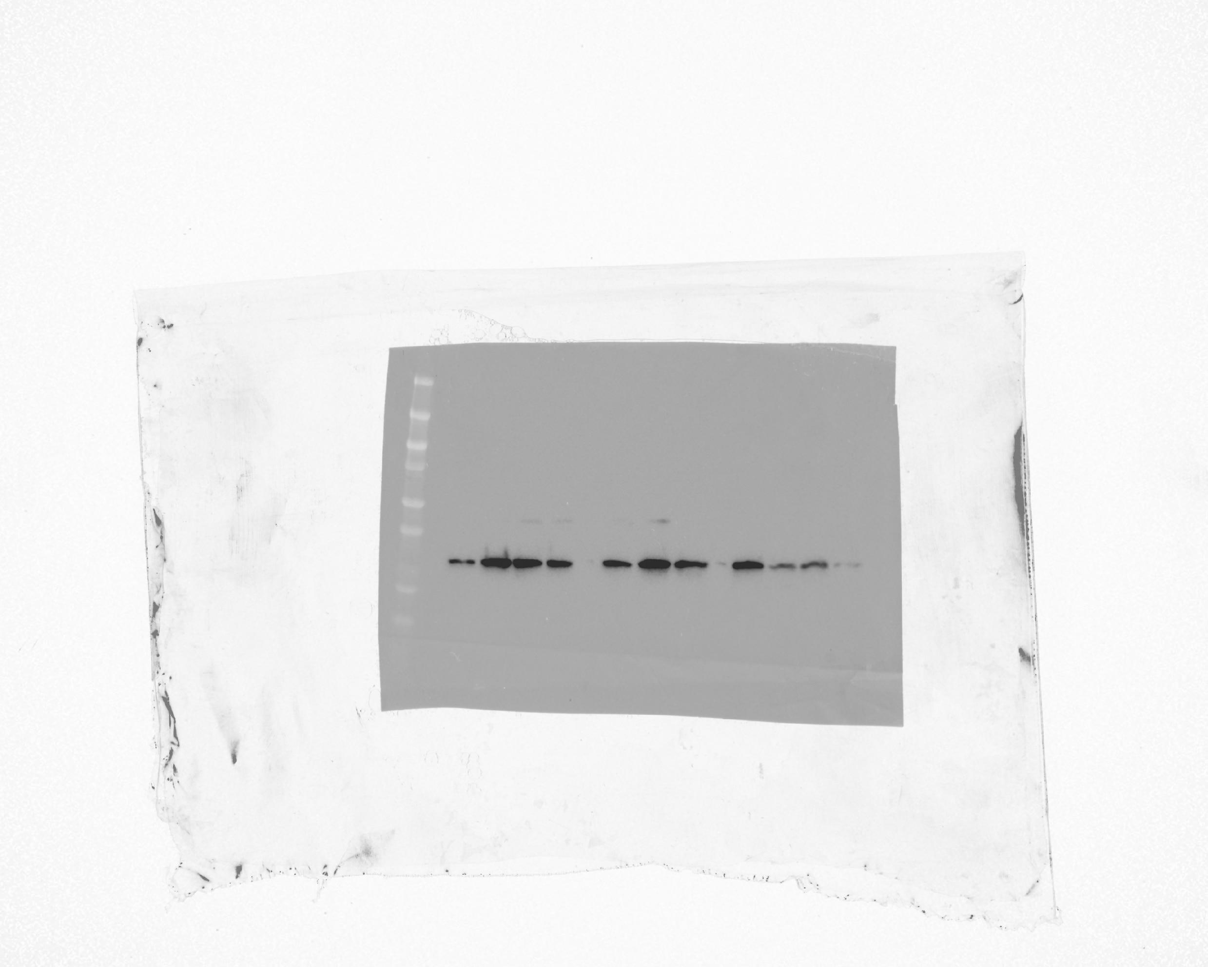
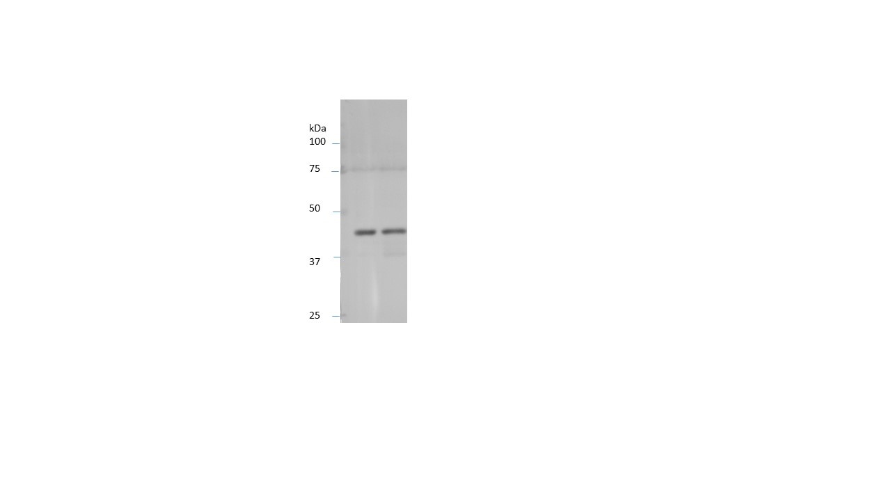
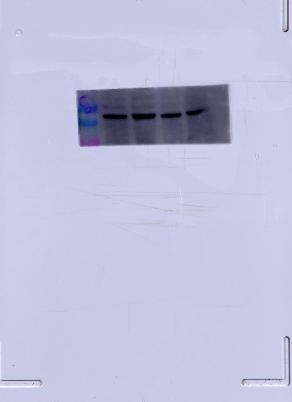
 were subjected to SDS PAGE followed by western blot with 20536-1-AP (ACTB antibody) at dilution of 1:500.")
 at dilution of 1:10000 incubated at room temperature for 1.5 hours.")
 at dilution of 1:8000 incubated at room temperature for 1.5 hours.")
 at dilution of 1:2000 incubated at room temperature for 1.5 hours.")
 at dilution of 1:2000 incubated at room temperature for 1.5 hours.")
 at dilution of 1:5000 incubated at room temperature for 1.5 hours.")
 at dilution of 1:200 (under 20x lens). Heat mediated antigen retrieval with Tris-EDTA buffer (pH 9.0).")
 at dilution of 1:200 (under 20x lens). Heat mediated antigen retrieval with Tris-EDTA buffer (pH 9.0).")
 at dilution of 1:200 (under 10x lens).")
 at dilution of 1:200 (under 40x lens).")
 fixed MDCK cells using Beta Actin antibody (20536-1-AP) at dilution of 1:400 and Multi-rAb CoraLite ® Plus 488-Goat Anti-Rabbit Recombinant Secondary Antibody (H+L) (RGAR002).")
Applications testées
| Résultats positifs en WB | cellules HEK-293, cellules A549, cellules C6, cellules Caco-2, cellules HeLa, cellules HepG2, cellules Jurkat, cellules NIH/3T3, cellules RAW 264.7, cellules SMMC-7721, tissu cérébral de rat, tissu cérébral de souris, tissu de côlon de souris, tissu hépatique de rat, tissu hépatique de souris, tissu rénal de rat, tissu splénique de rat |
| Résultats positifs en IHC | tissu de côlon humain, tissu rénal humain il est suggéré de démasquer l'antigène avec un tampon de TE buffer pH 9.0; (*) À défaut, 'le démasquage de l'antigène peut être 'effectué avec un tampon citrate pH 6,0. |
| Résultats positifs en IF/ICC | cellules MDCK, |
Dilution recommandée
| Application | Dilution |
|---|---|
| Western Blot (WB) | WB : 1:4000-1:10000 |
| Immunohistochimie (IHC) | IHC : 1:50-1:500 |
| Immunofluorescence (IF)/ICC | IF/ICC : 1:200-1:800 |
| It is recommended that this reagent should be titrated in each testing system to obtain optimal results. | |
| Sample-dependent, check data in validation data gallery | |
Applications publiées
| KD/KO | See 1 publications below |
| WB | See 4545 publications below |
| IHC | See 4 publications below |
| IF | See 15 publications below |
| IP | See 8 publications below |
| ELISA | See 1 publications below |
| CoIP | See 2 publications below |
Informations sur le produit
20536-1-AP cible Beta Actin dans les applications de WB, IHC, IF/ICC, IP, CoIP, ELISA et montre une réactivité avec des échantillons canin, Humain, rat, singe, souris
| Réactivité | canin, Humain, rat, singe, souris |
| Réactivité citée | rat, Chèvre, Humain, levure, poulet, souris |
| Hôte / Isotype | Lapin / IgG |
| Clonalité | Polyclonal |
| Type | Anticorps |
| Immunogène | Beta Actin Protéine recombinante Ag14521 |
| Nom complet | actin, beta |
| Masse moléculaire calculée | 375 aa, 42 kDa |
| Poids moléculaire observé | 42 kDa |
| Numéro d’acquisition GenBank | BC002409 |
| Symbole du gène | Beta Actin |
| Identification du gène (NCBI) | 60 |
| Conjugaison | Non conjugué |
| Forme | Liquide |
| Méthode de purification | Purification par affinité contre l'antigène |
| Tampon de stockage | PBS with 0.02% sodium azide and 50% glycerol |
| Conditions de stockage | Stocker à -20°C. Stable pendant un an après l'expédition. L'aliquotage n'est pas nécessaire pour le stockage à -20oC Les 20ul contiennent 0,1% de BSA. |
Informations générales
Beta Actin, also named as ACTB and F-Actin, belongs to the actin family. Actins are highly conserved globular proteins that are involved in various types of cell motility and are ubiquitously expressed in all eukaryotic cells. At least six isoforms of actins are known in mammals and other vertebrates: alpha (ACTC1, cardiac muscle 1), alpha 1 (ACTA1, skeletal muscle) and 2 (ACTA2, aortic smooth muscle), beta (ACTB), gamma 1 (ACTG1) and 2 (ACTG2, enteric smooth muscle). Beta and gamma 1 are two non-muscle actin proteins. Most actins consist of 376aa, while ACTG2 (rich in muscles) has 375aa and ACTG1(found in non-muscle cells) has only 374aa. Beta actin has been widely used as the internal control in RT-PCR and Western Blotting as a 42-kDa protein. However, the 37-40, 31, 15 kDa cleaved fragment of beta actin can be generated during apoptosis process. This antibody was generated against N-terminal region of human beta actin protein and can cross-react with other actins. (9173887, 11217076, 10229193 )
Protocole
| Product Specific Protocols | |
|---|---|
| WB protocol for Beta Actin antibody 20536-1-AP | Download protocol |
| IHC protocol for Beta Actin antibody 20536-1-AP | Download protocol |
| IF protocol for Beta Actin antibody 20536-1-AP | Download protocol |
| Standard Protocols | |
|---|---|
| Click here to view our Standard Protocols |
Publications
| Species | Application | Title |
|---|---|---|
Cancer Cell Targeting the immune privilege of tumor-initiating cells to enhance cancer immunotherapy | ||
Nature Aspm knockout ferret reveals an evolutionary mechanism governing cerebral cortical size. | ||
Signal Transduct Target Ther Identifying genetic targets in clinical subtypes of Parkinson's disease for optimizing pharmacological treatment strategies | ||
Cell Microglia jointly degrade fibrillar alpha-synuclein cargo by distribution through tunneling nanotubes. |
Avis
The reviews below have been submitted by verified Proteintech customers who received an incentive for providing their feedback.
FH Matthieu (Verified Customer) (09-24-2025) | Produces a clear band in WB at the expected molecular weight
|
FH Manon (Verified Customer) (09-17-2025) | Very good housekeeping antibody
|
FH YINGJIAN (Verified Customer) (05-27-2025) | This primary antibody is effective, even at a low concentration (1:10000).
 |
FH Julia (Verified Customer) (06-20-2023) | Our lab has been using Proteintech's beta actin polyclonal antibody for years. We use this antibody in all our western blot experiments for normalization. This antibody works consistently at a 1:1000 dilution for human and mouse retina and for HRECs. We have always gotten a beautiful result, with nice thick and specific bands. The only downside is that this antibody is hard to strip. Even after stripping, the membrane tends to have residual beta actin bands. Even so, we would highly recommend this product for routine western blots.
 |
FH Chun (Verified Customer) (06-19-2022) | This antibody worked very well.
|
FH Aaron (Verified Customer) (12-08-2020) | nice bands achieved
|
FH Wei (Verified Customer) (01-30-2020) | Strong, clear band for basal heart tissues.Good for internal control for heart diseases.
|
FH SCOTT (Verified Customer) (10-11-2019) | Used 1/1000 overnight 4 C. overnightHuman Grey matter cortex post mortem material
 |
FH Dan (Verified Customer) (08-19-2019) | The antibody works
|
FH Kishor (Verified Customer) (01-30-2019) | Working well for Western Blotting (1:5000)
 |

