Anticorps Polyclonal de lapin anti-AKT
AKT Polyclonal Antibody for WB, IHC, ELISA
Hôte / Isotype
Lapin / IgG
Réactivité testée
Humain, souris et plus (1)
Applications
WB, IHC, ELISA
Conjugaison
Non conjugué
N° de cat : 55230-1-AP
Synonymes
Galerie de données de validation
Applications testées
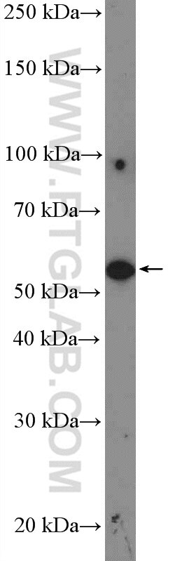
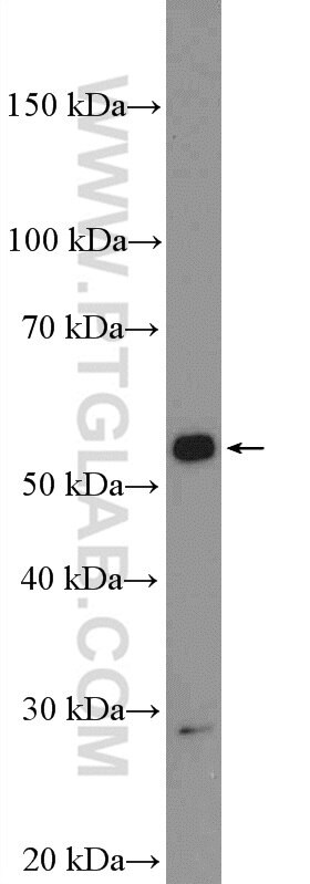
| Résultats positifs en WB | cellules HeLa, cellules A549 |
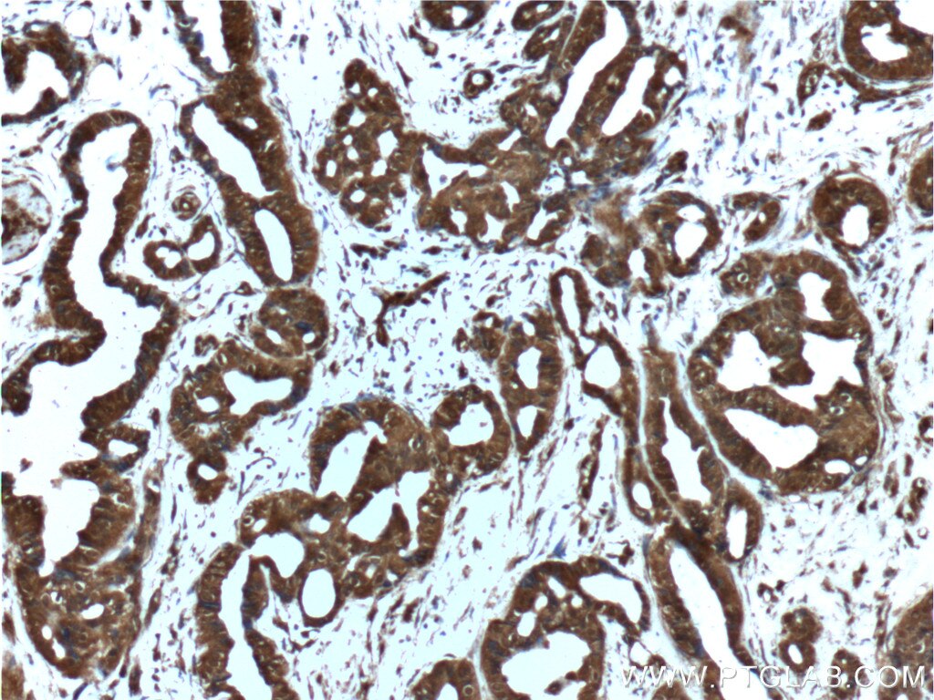
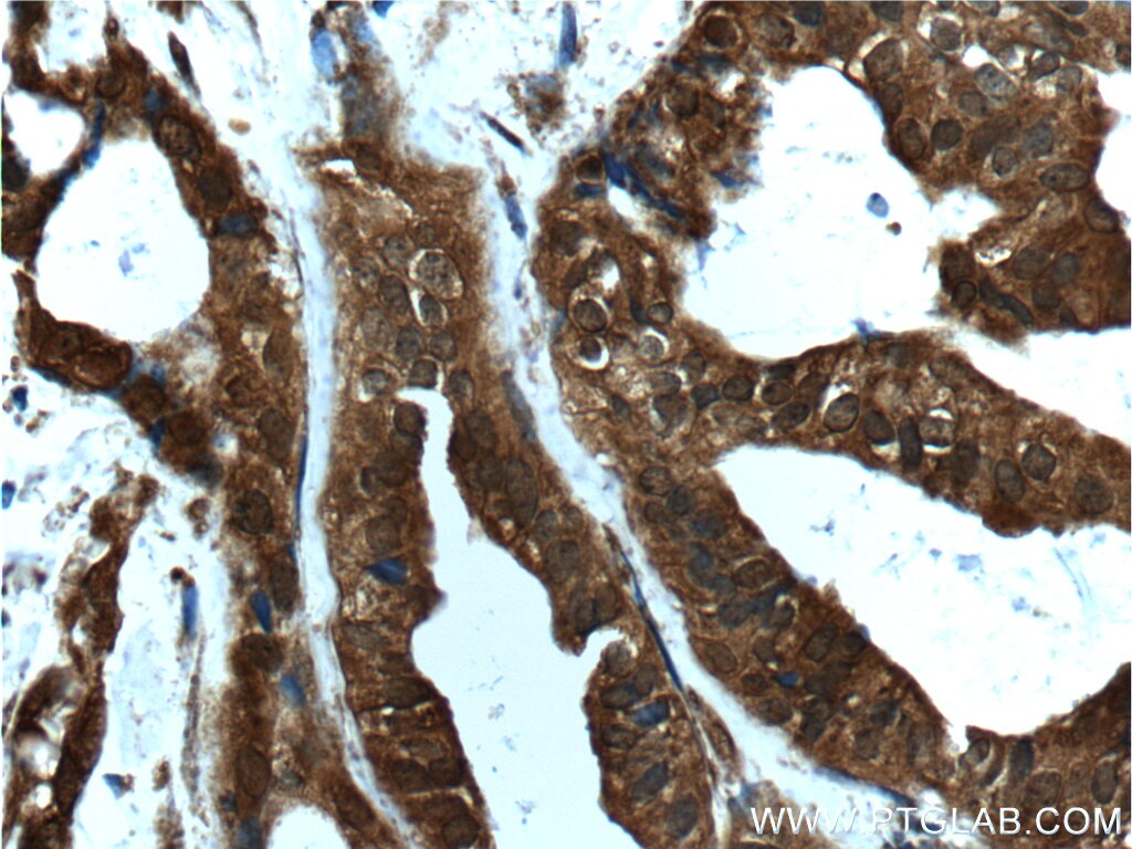
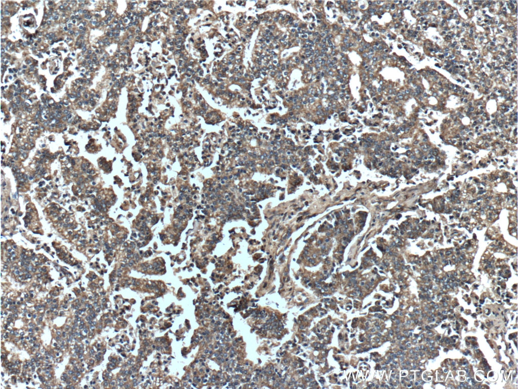
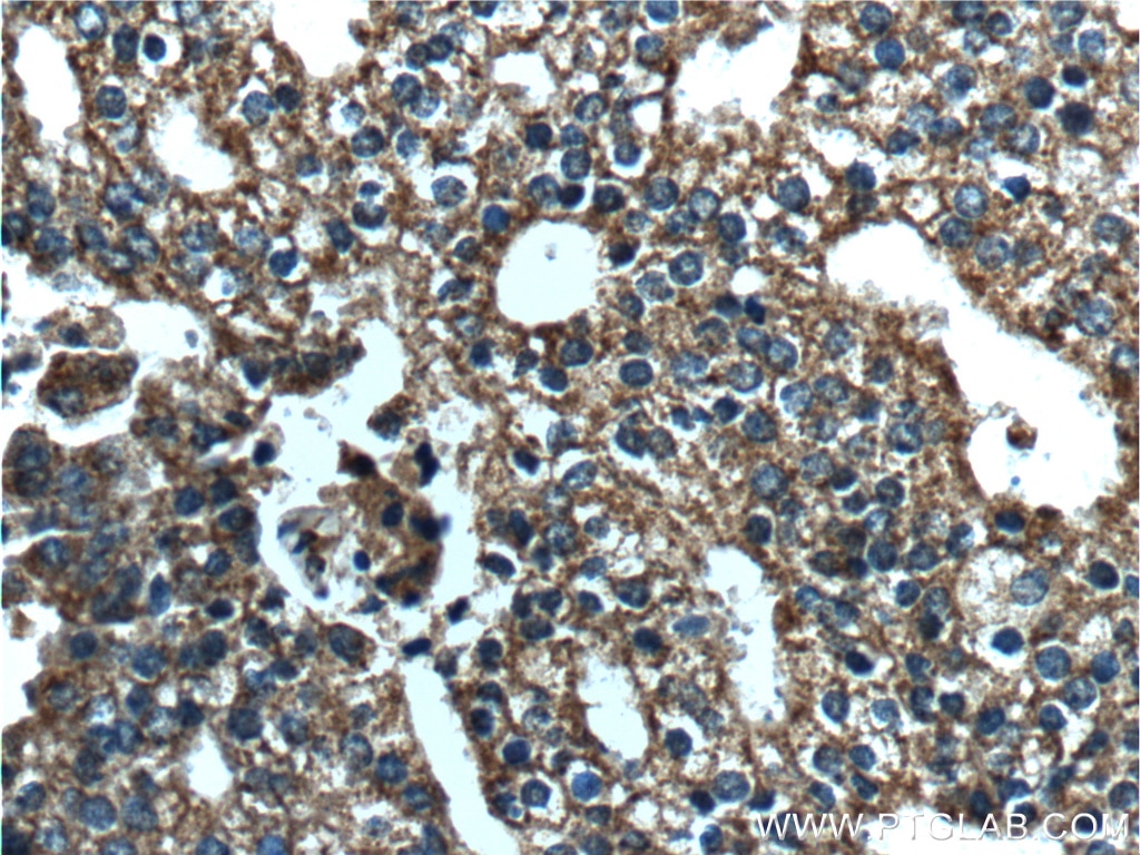
| Résultats positifs en IHC | tissu de cancer du sein humain, tissu de cancer de la prostate humain il est suggéré de démasquer l'antigène avec un tampon de TE buffer pH 9.0; (*) À défaut, 'le démasquage de l'antigène peut être 'effectué avec un tampon citrate pH 6,0. |
Dilution recommandée
| Application | Dilution |
|---|---|
| Western Blot (WB) | WB : 1:500-1:1000 |
| Immunohistochimie (IHC) | IHC : 1:50-1:500 |
| It is recommended that this reagent should be titrated in each testing system to obtain optimal results. | |
| Sample-dependent, check data in validation data gallery | |
Applications publiées
| WB | See 6 publications below |
| IHC | See 1 publications below |
Informations sur le produit
55230-1-AP cible AKT dans les applications de WB, IHC, ELISA et montre une réactivité avec des échantillons Humain, souris
| Réactivité | Humain, souris |
| Réactivité citée | rat, Humain, souris |
| Hôte / Isotype | Lapin / IgG |
| Clonalité | Polyclonal |
| Type | Anticorps |
| Immunogène | Peptide |
| Nom complet | v-akt murine thymoma viral oncogene homolog 1 |
| Masse moléculaire calculée | 56 kDa |
| Poids moléculaire observé | 56-62 kDa |
| Numéro d’acquisition GenBank | NM_001014431 |
| Symbole du gène | AKT1 |
| Identification du gène (NCBI) | 207 |
| Conjugaison | Non conjugué |
| Forme | Liquide |
| Méthode de purification | Purification par affinité contre l'antigène |
| Tampon de stockage | PBS with 0.02% sodium azide and 50% glycerol |
| Conditions de stockage | Stocker à -20 ℃. L'aliquotage n'est pas nécessaire pour le stockage à -20oC Les 20ul contiennent 0,1% de BSA. |
Informations générales
AKT1, also named as PKB and RAC, belongs to the protein kinase superfamily, AGC Ser/Thr protein kinase family and RAC subfamily. It plays a role as a key modulator of the AKT-mTOR signaling pathway controlling the tempo of the process of newborn neurons integration during adult neurogenesis, including correct neuron positioning, dendritic development and synapse formation. AKT1 promotes glycogen synthesis by mediating the insulin-induced activation of glycogen synthase. It plays a role in glucose transport by mediating insulin-induced translocation of the GLUT4 glucose transporter to the cell surface. AKT1 mediates the antiapoptotic effects of IGF-I. (PMID:16139227)This antibody is specific to AKT1. It has no cross reaction to AKT2 and AKT3.
Protocole
| Product Specific Protocols | |
|---|---|
| WB protocol for AKT antibody 55230-1-AP | Download protocol |
| IHC protocol for AKT antibody 55230-1-AP | Download protocol |
| Standard Protocols | |
|---|---|
| Click here to view our Standard Protocols |
Publications
| Species | Application | Title |
|---|---|---|
Mol Med Rep Matrine inhibits the invasive and migratory properties of human hepatocellular carcinoma by regulating epithelial‑mesenchymal transition. | ||
J Immunol The Intracellular Interaction of Porcine β-Defensin 2 with VASH1 Alleviates Inflammation via Akt Signaling Pathway. | ||
Int J Mol Sci Extracellular Calcium-Induced Calcium Transient Regulating the Proliferation of Osteoblasts through Glycolysis Metabolism Pathways | ||
J Ethnopharmacol Shenqu xiaoshi oral solution enhances digestive function and stabilizes the gastrointestinal microbiota of juvenile rats with infantile anorexia | ||
Acta Biochim Biophys Sin (Shanghai) Multiple circRNAs regulated by QKI5 conjointly spongemiR-214-3p to antagonize bisphenol A-inducedspermatocyte toxicity |
Avis
The reviews below have been submitted by verified Proteintech customers who received an incentive for providing their feedback.
FH Tanusree (Verified Customer) (12-18-2019) | Product worked well in WB at 1:500 dilution and 1:100 in IF
|
FH Tanusree (Verified Customer) (08-14-2019) | The antibody works good in mouse brain tissue in western blotting.
|
