- Phare
- Validé par KD/KO
Anticorps Monoclonal anti-Phospho-AKT (Ser473)
Phospho-AKT (Ser473) Monoclonal Antibody for WB, IHC, IF/ICC, FC (Intra), ELISA
Hôte / Isotype
Mouse / IgG1
Réactivité testée
Humain, rat, souris et plus (6)
Applications
WB, IHC, IF/ICC, FC (Intra), ELISA
Conjugaison
Non conjugué
CloneNo.
1C10B8
N° de cat : 66444-1-Ig
Synonymes
Galerie de données de validation
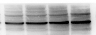
 antibody) at dilution of 1:5000 incubated at room temperature for 1.5 hours. The membrane was stripped and re-blotted with GAPDH antibody as loading control.")
 antibody) at dilution of 1:5000 incubated at room temperature for 1.5 hours. The membrane was stripped and re-blotted with GAPDH antibody as loading control..")
 antibody) at dilution of 1:10000 incubated at room temperature for 1.5 hours.")
 antibody) at dilution of 1:5000 incubated at room temperature for 1.5 hours.")
 antibody) at dilution of 1:5000 incubated at room temperature for 1.5 hours.")
 antibody) at dilution of 1:5000 incubated at room temperature for 1.5 hours. The membrane was stripped and re-blotted with GAPDH antibody as loading control.")
. Heat mediated antigen retrieval with Tris-EDTA buffer (pH 9.0).")
. Heat mediated antigen retrieval with Tris-EDTA buffer (pH 9.0).")
 or Calyculin A treated (right) Jurkat cells slide using 66444-1-Ig (Phospho-AKT (Ser473) antibody) at dilution of 1:8000 (under 10x lens). Heat mediated antigen retrieval with Tris-EDTA buffer (pH 9.0).")
 or Calyculin A treated (right) Jurkat cells slide using 66444-1-Ig (Phospho-AKT (Ser473) antibody) at dilution of 1:8000 (under 40x lens). Heat mediated antigen retrieval with Tris-EDTA buffer (pH 9.0).")
 antibody) at dilution of 1:2000 (under 10x lens). Heat mediated antigen retrieval with Tris-EDTA buffer (pH 9.0).")
 antibody) at dilution of 1:2000 (under 40x lens). Heat mediated antigen retrieval with Tris-EDTA buffer (pH 9.0).")
 fixed Calyculin A treated HeLa cells using Phospho-AKT (Ser473) antibody (66444-1-Ig, Clone: 1C10B8 ) at dilution of 1:400 and CoraLite®488-Conjugated Goat Anti-Mouse IgG(H+L) (SA00013-1).")
 or treated with Calyculin A (red) were intracellularly stained with 0.5 ug Anti-Human Phospho-AKT (Ser473) (66444-1-Ig, Clone:1C10B8) and CoraLite®488-Conjugated AffiniPure Goat Anti-Mouse IgG(H+L) at dilution 1:1000, or 0.5 ug Control Antibody (blue). Cells were fixed with 4% PFA and permeabilized with 90% MeOH.")
Applications testées
| Résultats positifs en WB | cellules PC-3 traitées à la calyculine A, cellules HEK-293T traitées à la calyculine A, cellules HSC-T6, cellules HSC-T6 traitées à la calyculine A, cellules Jurkat trai au TPA |
| Résultats positifs en IHC | tissu de cancer du sein humain, cellules Jurkat traitées à la calyculine A, tissu de cancer du côlon humain il est suggéré de démasquer l'antigène avec un tampon de TE buffer pH 9.0; (*) À défaut, 'le démasquage de l'antigène peut être 'effectué avec un tampon citrate pH 6,0. |
| Résultats positifs en IF/ICC | cellules HeLa traitées à la calyculine A, |
| Résultats positifs en FC (Intra) | cellules PC-3 traitées à la calyculine A, |
Dilution recommandée
| Application | Dilution |
|---|---|
| Western Blot (WB) | WB : 1:2000-1:10000 |
| Immunohistochimie (IHC) | IHC : 1:100-1:400 |
| Immunofluorescence (IF)/ICC | IF/ICC : 1:200-1:800 |
| Flow Cytometry (FC) (INTRA) | FC (INTRA) : 0.50 ug per 10^6 cells in a 100 µl suspension |
| It is recommended that this reagent should be titrated in each testing system to obtain optimal results. | |
| Sample-dependent, check data in validation data gallery | |
Applications publiées
| KD/KO | See 1 publications below |
| WB | See 1276 publications below |
| IHC | See 98 publications below |
| IF | See 23 publications below |
Informations sur le produit
66444-1-Ig cible Phospho-AKT (Ser473) dans les applications de WB, IHC, IF/ICC, FC (Intra), ELISA et montre une réactivité avec des échantillons Humain, rat, souris
| Réactivité | Humain, rat, souris |
| Réactivité citée | rat, Humain, Lapin, poisson-zèbre, porc, poulet, singe, souris, duck |
| Hôte / Isotype | Mouse / IgG1 |
| Clonalité | Monoclonal |
| Type | Anticorps |
| Immunogène | Peptide |
| Nom complet | v-akt murine thymoma viral oncogene homolog 1 |
| Poids moléculaire observé | 60-62 kDa |
| Numéro d’acquisition GenBank | NM_005163 |
| Symbole du gène | AKT1 |
| Identification du gène (NCBI) | 207 |
| Conjugaison | Non conjugué |
| Forme | Liquide |
| Méthode de purification | Purification par protéine G |
| Tampon de stockage | PBS with 0.02% sodium azide and 50% glycerol |
| Conditions de stockage | Stocker à -20°C. Stable pendant un an après l'expédition. L'aliquotage n'est pas nécessaire pour le stockage à -20oC Les 20ul contiennent 0,1% de BSA. |
Informations générales
1) What is AKT?
The serine/threonine kinase B AKT pathway (also known as the PI3K-Akt pathway) plays a vital role in the regulation of cellular processes, including cell proliferation, survival, and growth - processes that are essential for oncogenesis. Mutation of the regulator proteins PI3K and PTEN causes uncontrolled disruption within the PI3-kinase pathway, leading to the development of human cancers (1,2; see also AKT pathway poster for more details).
2) phospho-AKT and FAQs
A) What is the best way to normalize phosphorylated proteins analyzed by western blot?
Normalize phospho-AKT and total AKT with your loading control (e.g. Actin, tubulin), then calculate the phospho/total ratio using these normalized values.
Put more simply:
1. Calculate the ratio of band intensities of a phospho-AKT band: the loading control.
2. Calculate the ratio of band intensities of total AKT: loading control.
3. Divide ratio obtained #1 by #2 to obtain a normalized value for comparison among different conditions. This procedure allows one to distinguish between a change in AKT expression and a change in the ratio of phospho-AKT.
* If you are looking at the differences in a phospho-AKT expression resulting from an experimental condition (e.g., knockdown), you should also show the expression of total AKT to distinguish between a change in AKT expression (transcription/translation level) and a change in the AKT phosphorylation status.
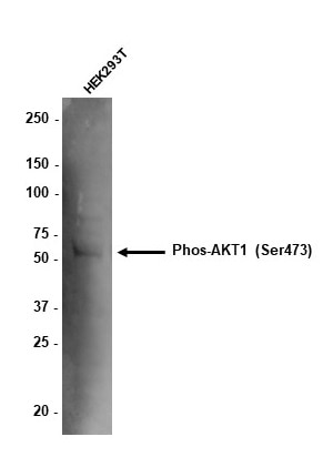
B) What is the observed molecular weight for AKT and phospho-AKT?
Molecular Weight AKT - 56 kDa
Molecular Weight phospho-AKT - 60 kDa (Figure 1)

Figure 1. WB: HEK-293 cell lysate was subjected to SDS PAGE followed by western blot with 60203-2-Ig (AKT antibody) and 66444-1-Ig (AKT-phospho-S473 antibody) at a dilution of 1:4000 incubated at room temperature for 1.5 hours.
C) Are there any special WB conditions to optimize staining of a phospho-AKT?
Since this is a phosphorylated protein, 5% BSA is recommended over non-fat milk as a blocking agent.
D) What are good positive and negative controls for a phospho-AKT?
- Positive Control: HEK293 cells
- Negative Control: Treatment with PI3K inhibitors (e.g. wortmannin)
E) What species does this antibody react with?
Our internal testing has confirmed that it reacts with the human and mouse forms of phospho-AKT.Reactivity with the human form is also supported by the literature's citations of this antibody.
References:
1. Perturbations of the AKT signaling pathway in human cancer.
2. Targeting the PI3K-Akt pathway in human cancer: rationale and promise.
Protocole
| Product Specific Protocols | |
|---|---|
| WB protocol for Phospho-AKT (Ser473) antibody 66444-1-Ig | Download protocol |
| IHC protocol for Phospho-AKT (Ser473) antibody 66444-1-Ig | Download protocol |
| IF protocol for Phospho-AKT (Ser473) antibody 66444-1-Ig | Download protocol |
| Standard Protocols | |
|---|---|
| Click here to view our Standard Protocols |
Publications
| Species | Application | Title |
|---|---|---|
Signal Transduct Target Ther Circulating tumor cells shielded with extracellular vesicle-derived CD45 evade T cell attack to enable metastasis | ||
Adv Mater Targeted Macrophage CRISPR-Cas13 Mrna Editing in Immunotherapy for Tendon Injury | ||
Cell Metab Disrupted methionine cycle triggers muscle atrophy in cancer cachexia through epigenetic regulation of REDD1 | ||
Cell Res Inhibiting Hv1 channel in peripheral sensory neurons attenuates chronic inflammatory pain and opioid side effects. | ||
Nat Commun Genome-wide enhancer-gene regulatory maps link causal variants to target genes underlying human cancer risk |
Avis
The reviews below have been submitted by verified Proteintech customers who received an incentive for providing their feedback.
FH Ana (Verified Customer) (06-17-2025) | The staining looks very good. It might be better to dilute a bit more than 1:2000 to reduce background noise.
 |
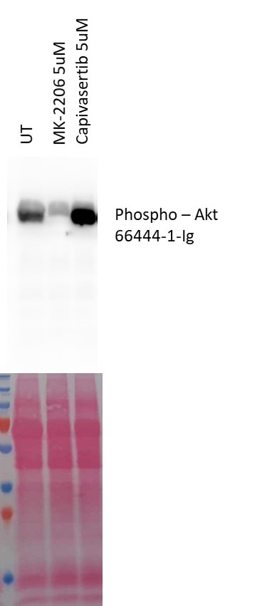
FH András (Verified Customer) (07-31-2023) | The Mk-2206 is an allosteric Akt inhibitor that prevents its recruitment to the membrane and consequently its phosphorylation. The Capivasertib is an Akt competitive inhibitor which induces its over phosphorylation.
 |
FH Jorge (Verified Customer) (07-26-2022) | Good signal. Unspecific band below 100 kDa. Used PageRuler Plus Prestained Protein Ladder and chemiluminescence was detected in the 70 kDa marker.
|
FH Tom (Verified Customer) (12-15-2020) | 10ug total protein of HEK293T lysate loaded. Membrane blocked in 5% BSA. Antibody (1:1,000) incubated overnight in block at 4 degrees. Anti-mouse HRP used at 1 in 10,000 to detect band.
 |

