


Tested Applications
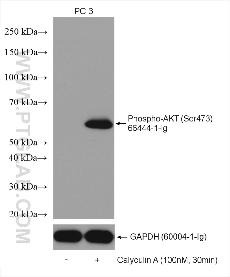
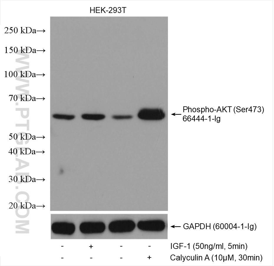
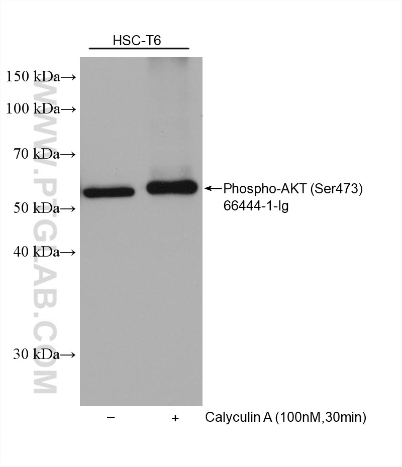
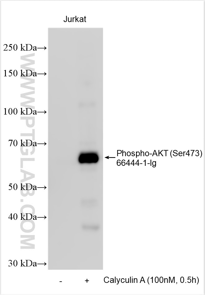
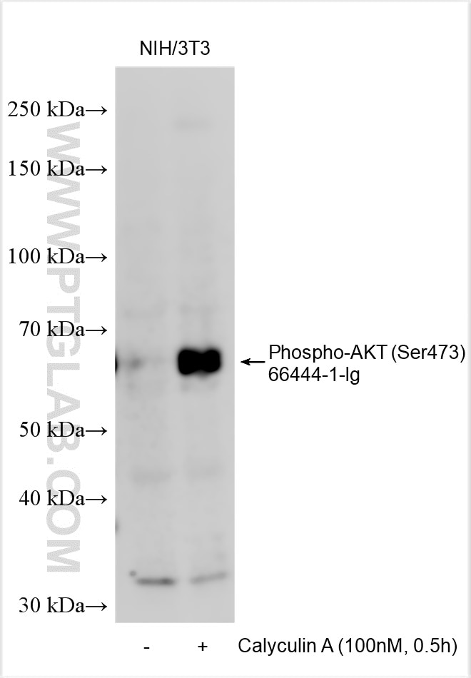
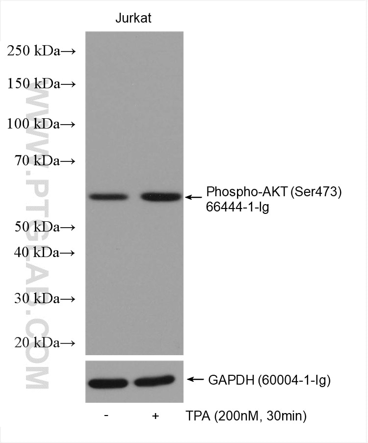
| Positive WB detected in | Calyculin A treated PC-3 cells, Calyculin A treated HEK-293T cells, HSC-T6 cells, TPA treated Jurkat cells, Calyculin A treated HSC-T6 cells |
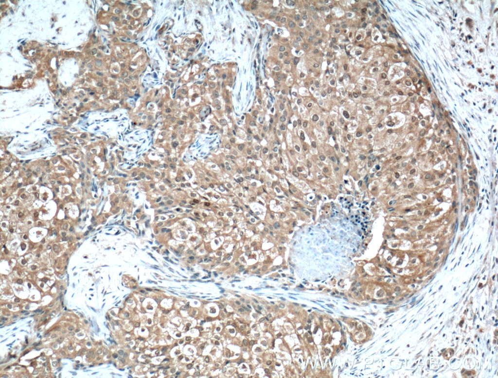
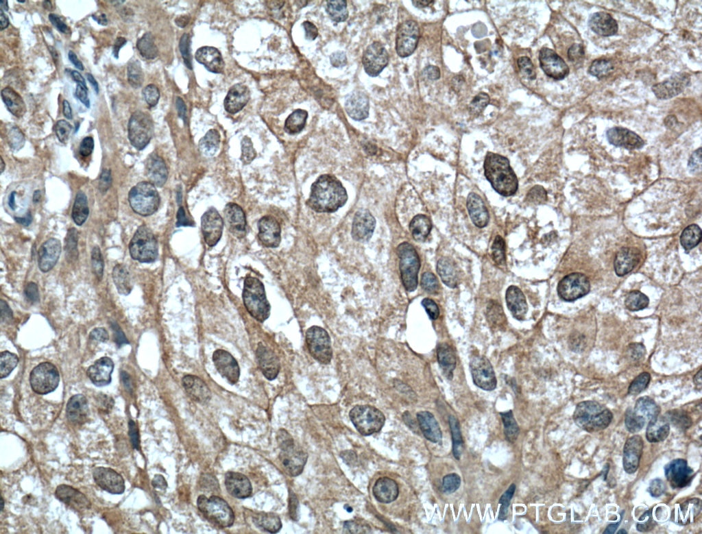
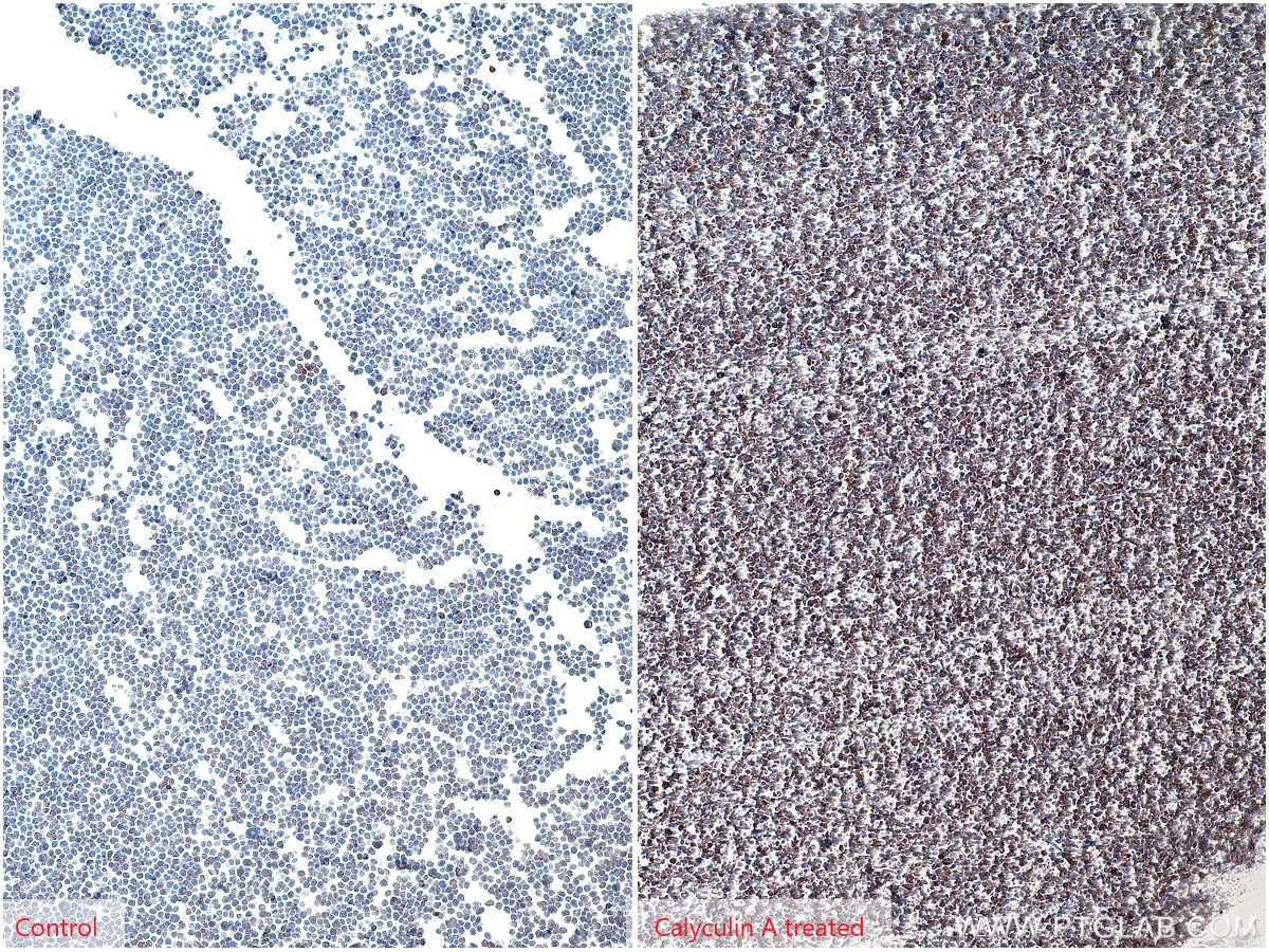
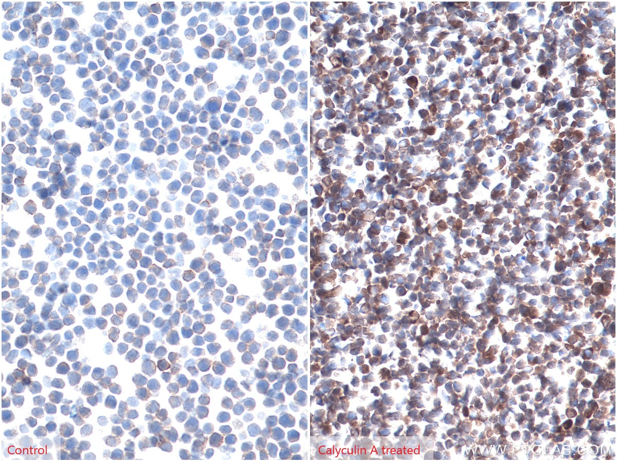
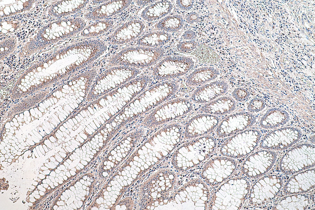
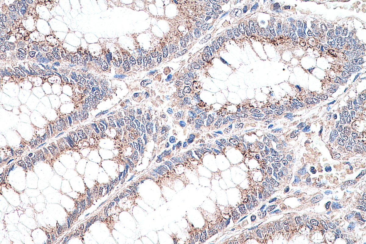
| Positive IHC detected in | human breast cancer tissue, Calyculin A treated Jurkat cells, human colon cancer tissue Note: suggested antigen retrieval with TE buffer pH 9.0; (*) Alternatively, antigen retrieval may be performed with citrate buffer pH 6.0 |
| Positive IF/ICC detected in | Calyculin A treated HeLa cells |
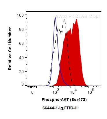
| Positive FC (Intra) detected in | Calyculin A treated PC-3 cells |
Recommended dilution
| Application | Dilution |
|---|---|
| Western Blot (WB) | WB : 1:2000-1:10000 |
| Immunohistochemistry (IHC) | IHC : 1:100-1:400 |
| Immunofluorescence (IF)/ICC | IF/ICC : 1:200-1:800 |
| Flow Cytometry (FC) (INTRA) | FC (INTRA) : 0.50 ug per 10^6 cells in a 100 µl suspension |
| It is recommended that this reagent should be titrated in each testing system to obtain optimal results. | |
| Sample-dependent, Check data in validation data gallery. | |
Product Information
66444-1-Ig targets Phospho-AKT (Ser473) in WB, IHC, IF/ICC, FC (Intra), IP, ELISA applications and shows reactivity with human, mouse, rat samples.
| Tested Reactivity | human, mouse, rat |
| Cited Reactivity | human, mouse, rat, pig, rabbit, monkey, chicken, zebrafish, sheep |
| Host / Isotype | Mouse / IgG1 |
| Class | Monoclonal |
| Type | Antibody |
| Immunogen |
Peptide Predict reactive species |
| Full Name | v-akt murine thymoma viral oncogene homolog 1 |
| Observed Molecular Weight | 60-62 kDa |
| GenBank Accession Number | NM_005163 |
| Gene Symbol | AKT1 |
| Gene ID (NCBI) | 207 |
| RRID | AB_2782958 |
| Conjugate | Unconjugated |
| Form | Liquid |
| Purification Method | Protein A purification |
| UNIPROT ID | P31749 |
| Storage Buffer | PBS with 0.02% sodium azide and 50% glycerol, pH 7.3. |
| Storage Conditions | Store at -20°C. Stable for one year after shipment. Aliquoting is unnecessary for -20oC storage. 20ul sizes contain 0.1% BSA. |
Background Information
1) What is AKT?
The serine/threonine kinase B AKT pathway (also known as the PI3K-Akt pathway) plays a vital role in the regulation of cellular processes, including cell proliferation, survival, and growth - processes that are essential for oncogenesis. Mutation of the regulator proteins PI3K and PTEN causes uncontrolled disruption within the PI3-kinase pathway, leading to the development of human cancers (1,2; see also AKT pathway poster for more details).
2) phospho-AKT and FAQs
A) What is the best way to normalize phosphorylated proteins analyzed by western blot?
Normalize phospho-AKT and total AKT with your loading control (e.g. Actin, tubulin), then calculate the phospho/total ratio using these normalized values.
Put more simply:
1. Calculate the ratio of band intensities of a phospho-AKT band: the loading control.
2. Calculate the ratio of band intensities of total AKT: loading control.
3. Divide ratio obtained #1 by #2 to obtain a normalized value for comparison among different conditions. This procedure allows one to distinguish between a change in AKT expression and a change in the ratio of phospho-AKT.
* If you are looking at the differences in a phospho-AKT expression resulting from an experimental condition (e.g., knockdown), you should also show the expression of total AKT to distinguish between a change in AKT expression (transcription/translation level) and a change in the AKT phosphorylation status.
B) What is the observed molecular weight for AKT and phospho-AKT?
Molecular Weight AKT - 56 kDa
Molecular Weight phospho-AKT - 60 kDa (Figure 1)

Figure 1. WB: HEK-293 cell lysate was subjected to SDS PAGE followed by western blot with 60203-2-Ig (AKT antibody) and 66444-1-Ig (AKT-phospho-S473 antibody) at a dilution of 1:4000 incubated at room temperature for 1.5 hours.
C) Are there any special WB conditions to optimize staining of a phospho-AKT?
Since this is a phosphorylated protein, 5% BSA is recommended over non-fat milk as a blocking agent.
D) What are good positive and negative controls for a phospho-AKT?
- Positive Control: HEK293 cells
- Negative Control: Treatment with PI3K inhibitors (e.g. wortmannin)
E) What species does this antibody react with?
Our internal testing has confirmed that it reacts with the human and mouse forms of phospho-AKT.Reactivity with the human form is also supported by the literature's citations of this antibody.
References:
1. Perturbations of the AKT signaling pathway in human cancer.
2. Targeting the PI3K-Akt pathway in human cancer: rationale and promise.
Protocols
| Product Specific Protocols | |
|---|---|
| IF protocol for Phospho-AKT (Ser473) antibody 66444-1-Ig | Download protocol |
| IHC protocol for Phospho-AKT (Ser473) antibody 66444-1-Ig | Download protocol |
| WB protocol for Phospho-AKT (Ser473) antibody 66444-1-Ig | Download protocol |
| Standard Protocols | |
|---|---|
| Click here to view our Standard Protocols |
Publications
What published studies show
The AKT antibody (Cat# 66444-1-Ig) has accumulated 1,972 citations. Publications appear in high-impact journals including Signal Transduction and Targeted Therapy, Nature Communications, Cell Metabolism, Cell Research, Advanced Science, Cancer Letters, Cell Death & Disease, Theranostics, and Phytomedicine. Research fields span oncology, immunology, neuroscience, metabolism, inflammation, autophagy, redox biology, tissue engineering, and pharmacology, with predominant applications in Western blot and immunohistochemistry across human, mouse, and rat models.
| Species | Application | Title |
|---|---|---|
Signal Transduct Target Ther Circulating tumor cells shielded with extracellular vesicle-derived CD45 evade T cell attack to enable metastasis | ||
Signal Transduct Target Ther Family with sequence similarity 114 member A1 orchestrates immune evasion in triple-negative breast cancer | ||
Signal Transduct Target Ther Overexpression of STX11 alleviates pulmonary fibrosis by inhibiting fibroblast activation via the PI3K/AKT/mTOR pathway | ||
Signal Transduct Target Ther Chimeric antigen receptor with novel intracellular modules improves antitumor performance of T cells | ||
J Hematol Oncol A novel lncRNA ROPM-mediated lipid metabolism governs breast cancer stem cell properties. | ||
J Hematol Oncol Long non-coding RNA-SNHG7 acts as a target of miR-34a to increase GALNT7 level and regulate PI3K/Akt/mTOR pathway in colorectal cancer progression. |
Reviews
What customers say
Users generally report strong performance for Western Blot, with clear detection even at low protein amounts. A common dilution is 1:2000, though one reviewer suggested increasing dilution to reduce background. Most applications used cell lines (HEK293T, pancreatic cancer, HCMECs) or primary cells (P19 neurons, white adipose tissue). One user noted a non-specific band below 100 kDa. Overall, the antibody is well-received for its sensitivity and signal quality in WB experiments.
The reviews below have been submitted by verified Proteintech customers who received an incentive for providing their feedback.
Areen (Verified Customer) (12-15-2025) | a good antibody, it detects a very low amount of protein/ lane in western blot
|
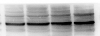
Ana (Verified Customer) (06-17-2025) | The staining looks very good. It might be better to dilute a bit more than 1:2000 to reduce background noise.
 |
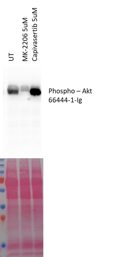
András (Verified Customer) (07-31-2023) | The Mk-2206 is an allosteric Akt inhibitor that prevents its recruitment to the membrane and consequently its phosphorylation. The Capivasertib is an Akt competitive inhibitor which induces its over phosphorylation.
 |
Jorge (Verified Customer) (07-26-2022) | Good signal. Unspecific band below 100 kDa. Used PageRuler Plus Prestained Protein Ladder and chemiluminescence was detected in the 70 kDa marker.
|
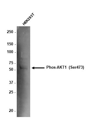
Tom (Verified Customer) (12-15-2020) | 10ug total protein of HEK293T lysate loaded. Membrane blocked in 5% BSA. Antibody (1:1,000) incubated overnight in block at 4 degrees. Anti-mouse HRP used at 1 in 10,000 to detect band.
 |















