Anticorps Polyclonal de lapin anti-AP1B1
AP1B1 Polyclonal Antibody for WB, IF/ICC, IP, ELISA
Hôte / Isotype
Lapin / IgG
Réactivité testée
Humain et plus (1)
Applications
WB, IF/ICC, IP, ELISA
Conjugaison
Non conjugué
N° de cat : 16932-1-AP
Synonymes
Galerie de données de validation
Applications testées
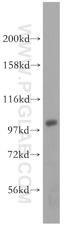
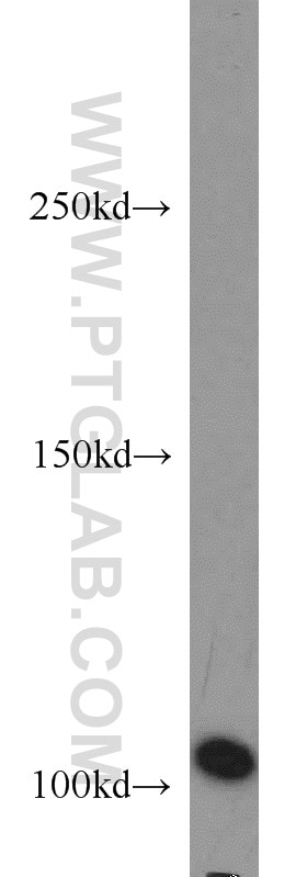
| Résultats positifs en WB | cellules HEK-293 |
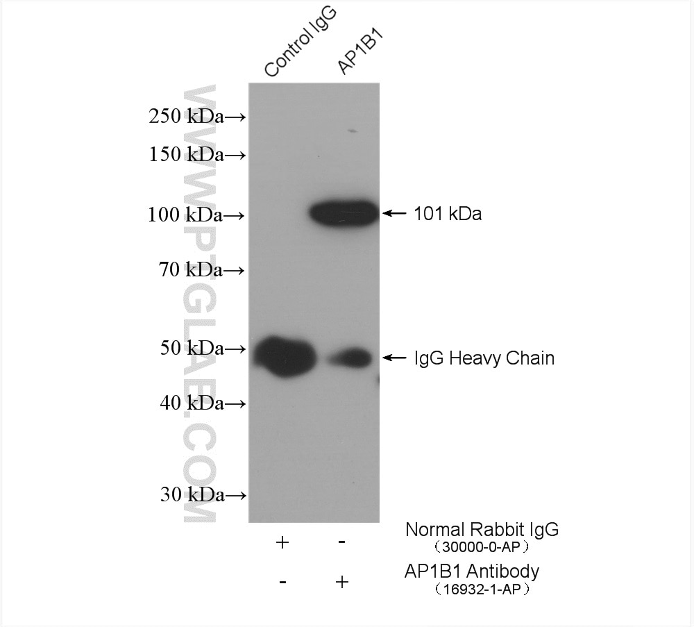
| Résultats positifs en IP | cellules HEK-293, |
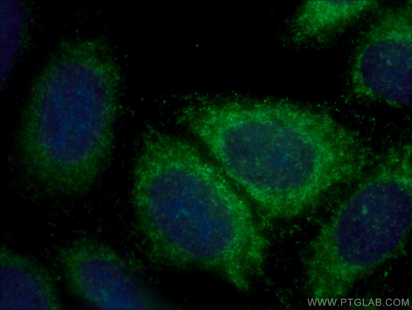
| Résultats positifs en IF/ICC | cellules HepG2, |
Dilution recommandée
| Application | Dilution |
|---|---|
| Western Blot (WB) | WB : 1:500-1:2400 |
| Immunoprécipitation (IP) | IP : 0.5-4.0 ug for 1.0-3.0 mg of total protein lysate |
| Immunofluorescence (IF)/ICC | IF/ICC : 1:50-1:500 |
| It is recommended that this reagent should be titrated in each testing system to obtain optimal results. | |
| Sample-dependent, check data in validation data gallery | |
Applications publiées
| WB | See 7 publications below |
Informations sur le produit
16932-1-AP cible AP1B1 dans les applications de WB, IF/ICC, IP, ELISA et montre une réactivité avec des échantillons Humain
| Réactivité | Humain |
| Réactivité citée | Humain, souris |
| Hôte / Isotype | Lapin / IgG |
| Clonalité | Polyclonal |
| Type | Anticorps |
| Immunogène | AP1B1 Protéine recombinante Ag10417 |
| Nom complet | adaptor-related protein complex 1, beta 1 subunit |
| Masse moléculaire calculée | 919 aa, 101 kDa |
| Poids moléculaire observé | 101 kDa |
| Numéro d’acquisition GenBank | BC046242 |
| Symbole du gène | AP1B1 |
| Identification du gène (NCBI) | 162 |
| Conjugaison | Non conjugué |
| Forme | Liquide |
| Méthode de purification | Purification par affinité contre l'antigène |
| Tampon de stockage | PBS with 0.02% sodium azide and 50% glycerol |
| Conditions de stockage | Stocker à -20°C. Stable pendant un an après l'expédition. L'aliquotage n'est pas nécessaire pour le stockage à -20oC Les 20ul contiennent 0,1% de BSA. |
Informations générales
AP1B1 is a subunit of the adaptor complex AP-1 and is a member of the adaptin protein family. Adaptor protein (AP) complexes are cytosolic heterotetramers that mediate the sorting of membrane proteins in the secretory and endocytic pathways. AP-1 is found at the cytoplasmic face of coated vesicles located at the Golgi complex, where it mediates both the recruitment of clathrin to the membrane and the recognition of sorting signals within the cytosolic tails of transmembrane receptors.
Protocole
| Product Specific Protocols | |
|---|---|
| WB protocol for AP1B1 antibody 16932-1-AP | Download protocol |
| IF protocol for AP1B1 antibody 16932-1-AP | Download protocol |
| IP protocol for AP1B1 antibody 16932-1-AP | Download protocol |
| Standard Protocols | |
|---|---|
| Click here to view our Standard Protocols |
Publications
| Species | Application | Title |
|---|---|---|
Nat Genet Bidirectional genome-wide CRISPR screens reveal host factors regulating SARS-CoV-2, MERS-CoV and seasonal HCoVs | ||
J Cell Biol Cargo-selective SNX-BAR proteins mediate retromer trimer independent retrograde transport. | ||
Mol Genet Metab Clinical, biochemical and cell biological characterization of KIDAR syndrome associated with a novel AP1B1 variant | ||
Nat Commun Synaptic vesicle-omics in mice captures signatures of aging and synucleinopathy |


