Tested Applications
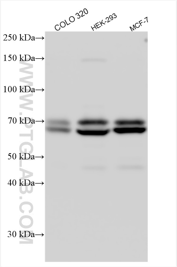
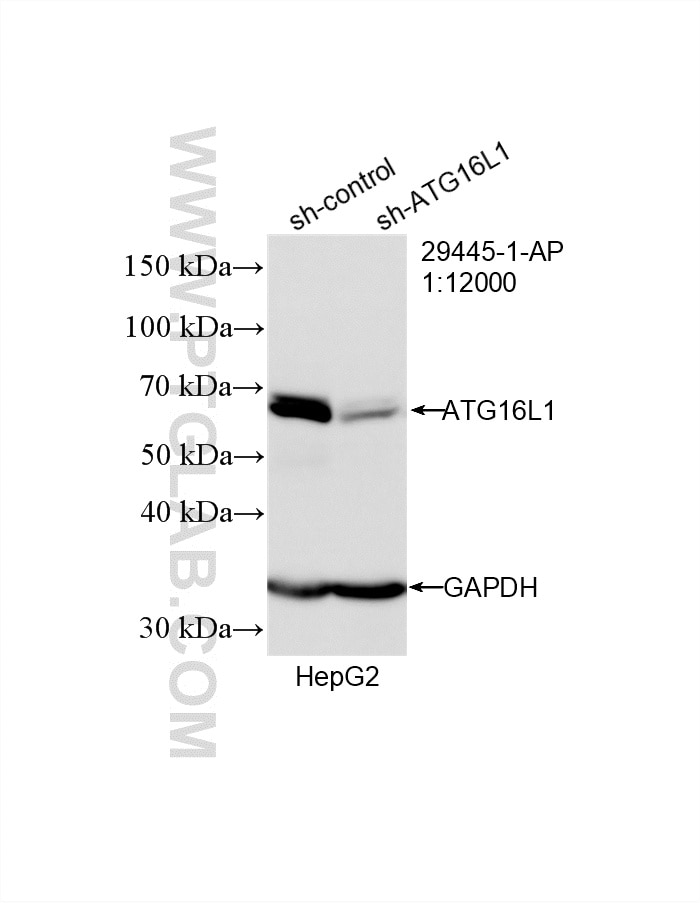
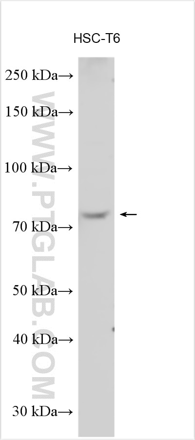
| Positive WB detected in | COLO 320 cells, HepG2 cells, HSC-T6 cells, HEK-293 cells, MCF-7 cells |
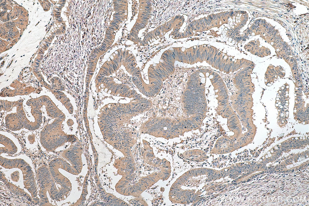
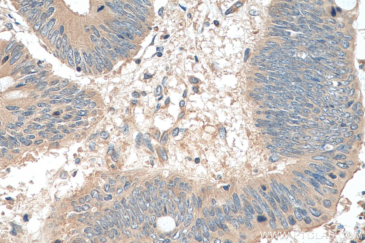
| Positive IHC detected in | human colon cancer tissue Note: suggested antigen retrieval with TE buffer pH 9.0; (*) Alternatively, antigen retrieval may be performed with citrate buffer pH 6.0 |
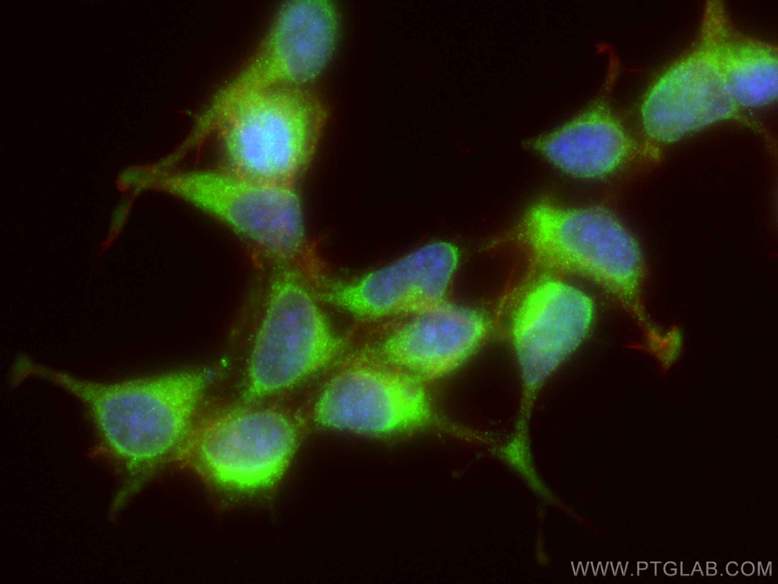
| Positive IF/ICC detected in | HEK-293 cells |
Recommended dilution
| Application | Dilution |
|---|---|
| Western Blot (WB) | WB : 1:1000-1:8000 |
| Immunohistochemistry (IHC) | IHC : 1:200-1:800 |
| Immunofluorescence (IF)/ICC | IF/ICC : 1:50-1:500 |
| It is recommended that this reagent should be titrated in each testing system to obtain optimal results. | |
| Sample-dependent, Check data in validation data gallery. | |
Published Applications
| WB | See 8 publications below |
| IHC | See 2 publications below |
| IF | See 3 publications below |
Product Information
29445-1-AP targets ATG16L1 in WB, IHC, IF/ICC, ELISA applications and shows reactivity with human, mouse, rat samples.
| Tested Reactivity | human, mouse, rat |
| Cited Reactivity | human, mouse, rat |
| Host / Isotype | Rabbit / IgG |
| Class | Polyclonal |
| Type | Antibody |
| Immunogen |
CatNo: Ag31318 Product name: Recombinant human ATG16L1 protein Source: e coli.-derived, PGEX-4T Tag: GST Domain: 1-230 aa of BC000061 Sequence: NKLLEKSDLHSVLAQKLQAEKHDVPNRHEISPGHDGTWNDNQLQEMAQLRIKHQEELTELHKKRGELAQLVIDLNNQMQRKDREMQMNEAKIAECLQTISDLETECLDLRTKLCDLERANQTLKDEYDALQITFTALEGKLRKTTEENQELVTRWMAEKAQEANRLNAENEKDSRRRQARLQKELAEAAKEPLPVEQDDDIEVIVDETSDHTEETSPVRAISRAATKRLS Predict reactive species |
| Full Name | ATG16 autophagy related 16-like 1 (S. cerevisiae) |
| Calculated Molecular Weight | 607 aa, 68 kDa |
| Observed Molecular Weight | 68-75 kDa |
| GenBank Accession Number | BC000061 |
| Gene Symbol | ATG16L1 |
| Gene ID (NCBI) | 55054 |
| RRID | AB_2918308 |
| Conjugate | Unconjugated |
| Form | Liquid |
| Purification Method | Antigen affinity purification |
| UNIPROT ID | Q676U5 |
| Storage Buffer | PBS with 0.02% sodium azide and 50% glycerol, pH 7.3. |
| Storage Conditions | Store at -20°C. Stable for one year after shipment. Aliquoting is unnecessary for -20oC storage. 20ul sizes contain 0.1% BSA. |
Background Information
Human ATG16L1 is a 607 amino acid protein (~68 kDa) comprising three major domains: the N‐terminal ATG5 binding domain (ATG5‐BD), the central coiled‐coil domain (CCD) and a predicted C‐terminal WD40‐domain. ATG16L1α and β (Atg16L1α, 66 kDa; and Atg16L1β, 68 kDa) are the major isoforms expressed in intestinal epithelium and macrophages , and all isoforms encode exon 9, which contains Thr 300. Atg16L1 mediates the cellular degradative process of autophagy and is considered a critical regulator of inflammation based on its genetic association with inflammatory bowel disease. ATG16L1 has been implicated in Crohn's disease. (PMID: 24553140, PMID: 22740627,PMID: 28685931)
Protocols
| Product Specific Protocols | |
|---|---|
| IF protocol for ATG16L1 antibody 29445-1-AP | Download protocol |
| IHC protocol for ATG16L1 antibody 29445-1-AP | Download protocol |
| WB protocol for ATG16L1 antibody 29445-1-AP | Download protocol |
| Standard Protocols | |
|---|---|
| Click here to view our Standard Protocols |
Publications
| Species | Application | Title |
|---|---|---|
J Nanobiotechnology Engineering exosomes derived from TNF-α preconditioned IPFP-MSCs enhance both yield and therapeutic efficacy for osteoarthritis | ||
Cell Signal Inhibition of XIST restrains paclitaxel resistance in breast cancer cells by targeting hsa-let-7d-5p/ATG16L1 through regulation of autophagy | ||
Food Funct EPA and DHA differentially coordinate the crosstalk between host and gut microbiota and block DSS-induced colitis in mice by a reinforced colonic mucus barrier. | ||
Anticancer Drugs Purvalanol A induces apoptosis and reverses cisplatin resistance in ovarian cancer | ||
Cell Signal GLDC attenuates liver ischemia-reperfusion injury by inhibiting macrophage recruitment and activation via PTBP1/P2RY6 | ||
Reviews
The reviews below have been submitted by verified Proteintech customers who received an incentive for providing their feedback.
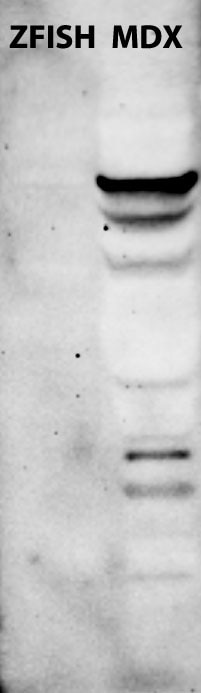
FH Boel (Verified Customer) (07-30-2025) | 25µg gastrocnemius muscle total protein extract from mdx mouse (dystrophin deficient mice that are used as a model for Duchenne muscular dystrophy) generated a strong band of full lenght protein plus multiple additional bands of lower molecular weight. 25µg of total protein extract from pooled wild type zebrafish 5 days post fertilization generated no protein bands.
 |