Anticorps Polyclonal de lapin anti-ATOH1
ATOH1 Polyclonal Antibody for WB, IHC, IF/ICC, FC (Intra), ELISA
Hôte / Isotype
Lapin / IgG
Réactivité testée
Humain, rat, souris et plus (1)
Applications
WB, IHC, IF/ICC, FC (Intra), chIP, ELISA
Conjugaison
Non conjugué
N° de cat : 21215-1-AP
Synonymes
Galerie de données de validation
 at dilution of 1:8000 incubated at room temperature for 1.5 hours.")
 at dilution of 1:3000 incubated at room temperature for 1.5 hours.")
 at dilution of 1:10000 incubated at room temperature for 1.5 hours.")
 at dilution of 1:600 incubated at room temperature for 1.5 hours.")
 at dilution of 1:2000 (under 20x lens). Heat mediated antigen retrieval with Tris-EDTA buffer (pH 9.0).")
 at dilution of 1:2000 (under 20x lens). Heat mediated antigen retrieval with Tris-EDTA buffer (pH 9.0).")
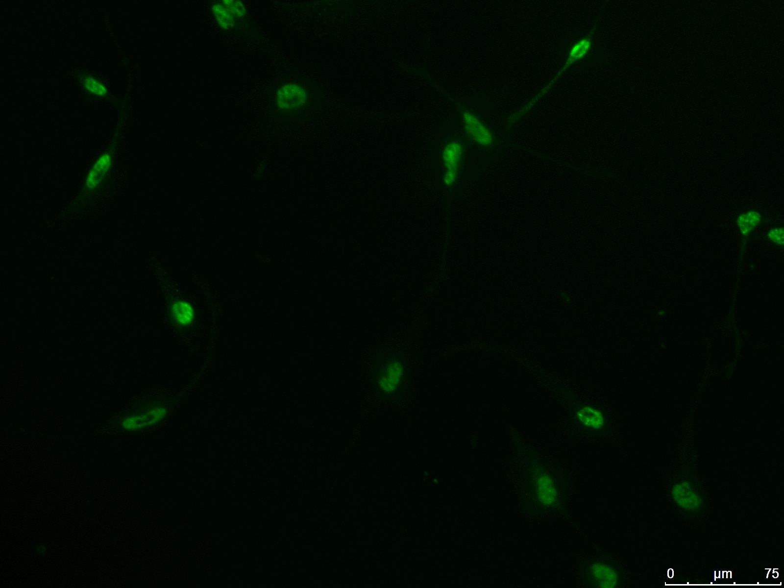
 fixed HepG2 cells using ATOH1 antibody (21215-1-AP) at dilution of 1:800 and CoraLite®488-Conjugated AffiniPure Goat Anti-Rabbit IgG(H+L).")
 fixed HepG2 cells using ATOH1 antibody (21215-1-AP) at dilution of 1:200 and CoraLite®594-Conjugated Goat Anti-Rabbit IgG(H+L) (SA00013-4), CL488-phalloidin (green).")
 fixed HepG2 cells using 21215-1-AP (ATOH1 antibody) at dilution of 1:100 and CoraLite488-Conjugated AffiniPure Goat Anti-Rabbit IgG(H+L).")
 and CoraLite®488-Conjugated AffiniPure Goat Anti-Rabbit IgG(H+L) at dilution 1:1000 (red), or 0.4 ug Isotype Control. Cells were fixed and permeabilized with Transcription Factor Staining Buffer Kit (PF00011).")
Applications testées
| Résultats positifs en WB | cellules Neuro-2a, cellules C6, cellules HepG2, tissu cérébral de rat, tissu cérébral de souris, tissu d'intestin grêle de souris |
| Résultats positifs en IHC | tissu de cancer du côlon humain, il est suggéré de démasquer l'antigène avec un tampon de TE buffer pH 9.0; (*) À défaut, 'le démasquage de l'antigène peut être 'effectué avec un tampon citrate pH 6,0. |
| Résultats positifs en IF/ICC | cellules HepG2, |
| Résultats positifs en FC (Intra) | cellules HepG2, |
Dilution recommandée
| Application | Dilution |
|---|---|
| Western Blot (WB) | WB : 1:1000-1:6000 |
| Immunohistochimie (IHC) | IHC : 1:1000-1:4000 |
| Immunofluorescence (IF)/ICC | IF/ICC : 1:400-1:1600 |
| Flow Cytometry (FC) (INTRA) | FC (INTRA) : 0.4 ug per 10^6 cells in a 100 µl suspension |
| It is recommended that this reagent should be titrated in each testing system to obtain optimal results. | |
| Sample-dependent, check data in validation data gallery | |
Applications publiées
| WB | See 23 publications below |
| IHC | See 4 publications below |
| IF | See 22 publications below |
| ChIP | See 1 publications below |
Informations sur le produit
21215-1-AP cible ATOH1 dans les applications de WB, IHC, IF/ICC, FC (Intra), chIP, ELISA et montre une réactivité avec des échantillons Humain, rat, souris
| Réactivité | Humain, rat, souris |
| Réactivité citée | rat, Humain, singe, souris |
| Hôte / Isotype | Lapin / IgG |
| Clonalité | Polyclonal |
| Type | Anticorps |
| Immunogène | ATOH1 Protéine recombinante Ag15615 |
| Nom complet | atonal homolog 1 (Drosophila) |
| Masse moléculaire calculée | 354 aa, 38 kDa |
| Poids moléculaire observé | 38-40 kDa |
| Numéro d’acquisition GenBank | BC069594 |
| Symbole du gène | ATOH1 |
| Identification du gène (NCBI) | 474 |
| Conjugaison | Non conjugué |
| Forme | Liquide |
| Méthode de purification | Purification par affinité contre l'antigène |
| Tampon de stockage | PBS with 0.02% sodium azide and 50% glycerol |
| Conditions de stockage | Stocker à -20°C. Stable pendant un an après l'expédition. L'aliquotage n'est pas nécessaire pour le stockage à -20oC Les 20ul contiennent 0,1% de BSA. |
Informations générales
ATOH1, also named as Protein atonal homolog 1 or BHLHA14, is a 364 amino acid protein, which contains 1 bHLH (basic helix-loop-helix) domain. ATOH1,as a transcriptional regulator in the nucleus activates E box-dependent transcription in collaboration with TCF3/E47, but the activity is completely antagonized by the negative regulator of neurogenesis HES1. ATOH1 plays a role in the differentiation of subsets of neural cells by activating E box-dependent transcription.
Protocole
| Product Specific Protocols | |
|---|---|
| WB protocol for ATOH1 antibody 21215-1-AP | Download protocol |
| IHC protocol for ATOH1 antibody 21215-1-AP | Download protocol |
| IF protocol for ATOH1 antibody 21215-1-AP | Download protocol |
| Standard Protocols | |
|---|---|
| Click here to view our Standard Protocols |
Publications
| Species | Application | Title |
|---|---|---|
Cell Res Decoding the spatiotemporal regulation of transcription factors during human spinal cord development | ||
Immunity IL-17RA-signaling in Lgr5+ intestinal stem cells induces expression of transcription factor ATOH1 to promote secretory cell lineage commitment. | ||
Nat Cell Biol Merkel cell polyomavirus activates LSD1-mediated blockade of non-canonical BAF to regulate transformation and tumorigenesis. | ||
J Clin Invest Sox2 haploinsufficiency primes regeneration and Wnt responsiveness in the mouse cochlea. | ||
Dev Cell Self-organized pattern formation in the developing mouse neural tube by a temporal relay of BMP signaling | ||
Proc Natl Acad Sci U S A Sufu- and Spop-mediated regulation of Gli2 is essential for the control of mammalian cochlear hair cell differentiation |
Avis
The reviews below have been submitted by verified Proteintech customers who received an incentive for providing their feedback.
FH Nicole (Verified Customer) (08-08-2024) | Works really well both in Western Blot and Immunofluorescence
|
FH Yin (Verified Customer) (12-20-2021) | It did not work.
|
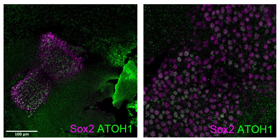
FH Stephen (Verified Customer) (11-04-2020) | ATOH1 antibody was tested on embryonic chicken tissue. Staining showed nuclear localisation in inner ear vestibular crista in sox2 positive prosensory region of 6 day old embryo
 |
FH Roland (Verified Customer) (09-17-2020) | We tested several ATOH1 antibodies and this is the only one which turned out to be specific as confirmed bei knockdown experiments.
|
FH Biren (Verified Customer) (08-31-2020) | I used human GBM stem cells as a negative control to validate the specificity of this antibody. The cells used were confirmed by RNAseq to not express ATOH1, yet all cells were positively stained at the previously used concentration of 1/500, indicating that this antibody is not specific.
 |
FH Erithelgi (Verified Customer) (09-25-2019) | The antibody detects a band of the expected size, in cells that induce GFP-ATOH1 under a DOX promoter. No bands detected in non-induced cells.
|
FH Nicole (Verified Customer) (07-23-2019) | Used to stain cultured stem cells for differentiation markers.
|

