Anticorps Polyclonal de lapin anti-ATP5I
ATP5I Polyclonal Antibody for WB, IHC, IF/ICC, ELISA
Hôte / Isotype
Lapin / IgG
Réactivité testée
Humain, rat, souris et plus (1)
Applications
WB, IHC, IF/ICC, ELISA
Conjugaison
Non conjugué
N° de cat : 16483-1-AP
Synonymes
Galerie de données de validation
Applications testées
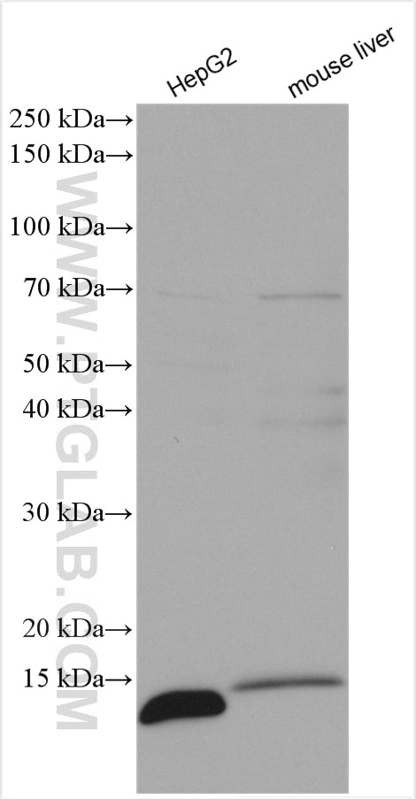
| Résultats positifs en WB | cellules HepG2, tissu hépatique de souris |
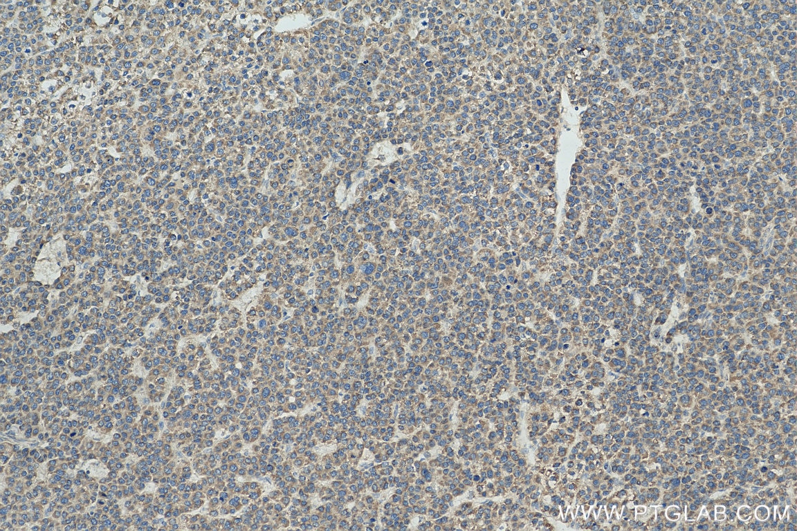
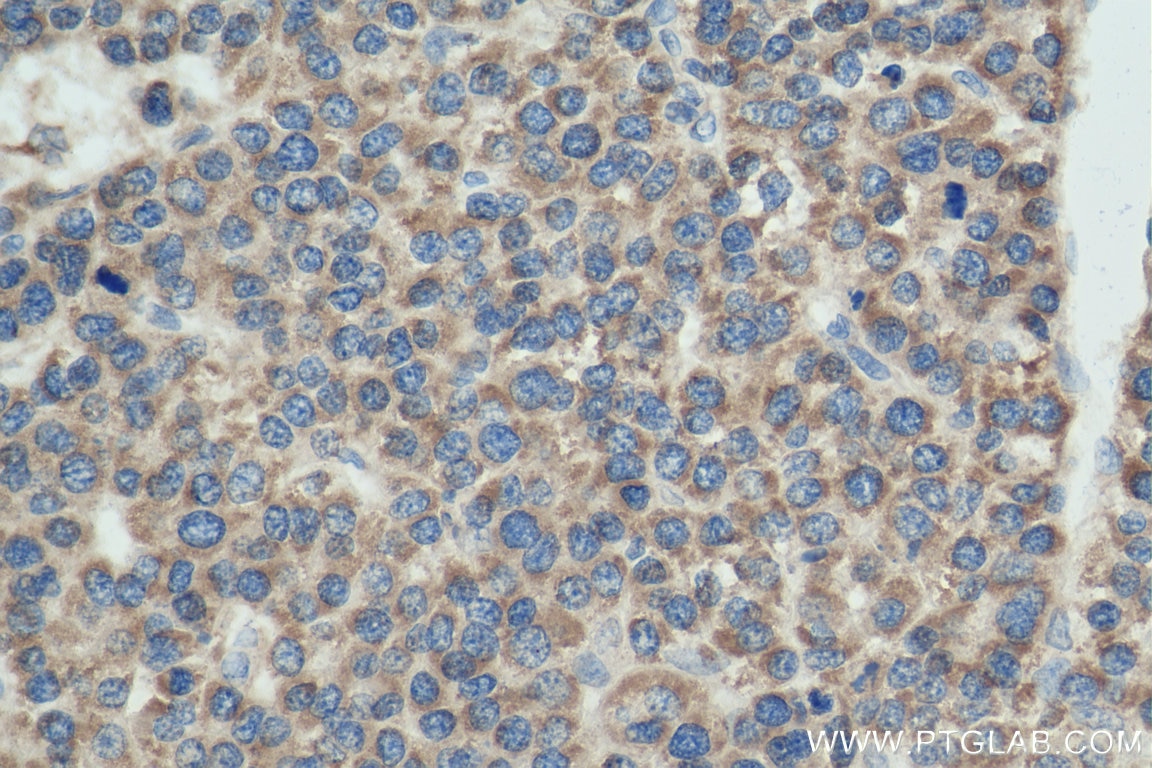
| Résultats positifs en IHC | tissu de cancer du foie humain, il est suggéré de démasquer l'antigène avec un tampon de TE buffer pH 9.0; (*) À défaut, 'le démasquage de l'antigène peut être 'effectué avec un tampon citrate pH 6,0. |
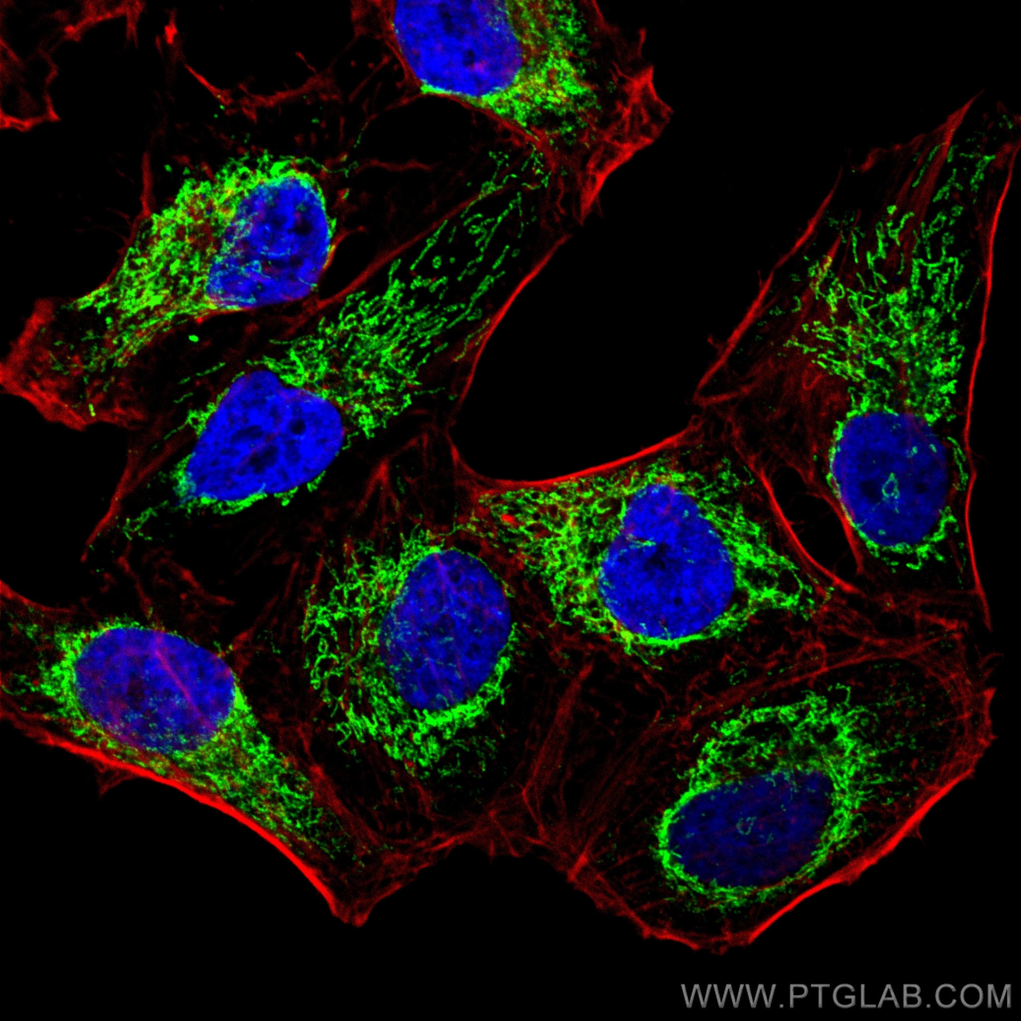
| Résultats positifs en IF/ICC | cellules HeLa, |
Dilution recommandée
| Application | Dilution |
|---|---|
| Western Blot (WB) | WB : 1:500-1:2000 |
| Immunohistochimie (IHC) | IHC : 1:50-1:500 |
| Immunofluorescence (IF)/ICC | IF/ICC : 1:50-1:500 |
| It is recommended that this reagent should be titrated in each testing system to obtain optimal results. | |
| Sample-dependent, check data in validation data gallery | |
Applications publiées
| WB | See 7 publications below |
| IF | See 4 publications below |
Informations sur le produit
16483-1-AP cible ATP5I dans les applications de WB, IHC, IF/ICC, ELISA et montre une réactivité avec des échantillons Humain, rat, souris
| Réactivité | Humain, rat, souris |
| Réactivité citée | Humain, poulet, souris |
| Hôte / Isotype | Lapin / IgG |
| Clonalité | Polyclonal |
| Type | Anticorps |
| Immunogène | ATP5I Protéine recombinante Ag9605 |
| Nom complet | ATP synthase, H+ transporting, mitochondrial F0 complex, subunit E |
| Masse moléculaire calculée | 69 aa, 8 kDa |
| Poids moléculaire observé | 8 kDa |
| Numéro d’acquisition GenBank | BC003679 |
| Symbole du gène | ATP5I |
| Identification du gène (NCBI) | 521 |
| Conjugaison | Non conjugué |
| Forme | Liquide |
| Méthode de purification | Purification par affinité contre l'antigène |
| Tampon de stockage | PBS with 0.02% sodium azide and 50% glycerol |
| Conditions de stockage | Stocker à -20°C. Stable pendant un an après l'expédition. L'aliquotage n'est pas nécessaire pour le stockage à -20oC Les 20ul contiennent 0,1% de BSA. |
Informations générales
ATP5I(ATP synthase subunit e) is also named as ATP5K and belongs to the ATPase e subunit family. The ATP5I gene encodes the e subunit of the mitochondrial ATP synthase Fo complex. Mitochondrial membrane ATP synthase(F1F0 ATP synthase or Complex V) produces ATP from ADP in the presence of a proton gradient across the membrane which is generated by electron transport complexes of the respiratory chain. Antisense ATP5I in a human HCC cell line inhibited cell growth suggesting that ATP5I acts through the MAP kinase pathway(PMID:11939412).
Protocole
| Product Specific Protocols | |
|---|---|
| WB protocol for ATP5I antibody 16483-1-AP | Download protocol |
| IHC protocol for ATP5I antibody 16483-1-AP | Download protocol |
| IF protocol for ATP5I antibody 16483-1-AP | Download protocol |
| Standard Protocols | |
|---|---|
| Click here to view our Standard Protocols |
Publications
| Species | Application | Title |
|---|---|---|
EBioMedicine Mitochonic Acid 5 (MA-5) Facilitates ATP Synthase Oligomerization and Cell Survival in Various Mitochondrial Diseases. | ||
Front Aging Neurosci Hippocampus-Based Mitochondrial Respiratory Function Decline Is Responsible for Perioperative Neurocognitive Disorders. | ||
Genet Sel Evol The mRNA-lncRNA landscape of multiple tissues uncovers key regulators and molecular pathways that underlie heterosis for feed intake and efficiency in laying chickens | ||
J Proteomics Profiling and identification of new proteins involved in brain ischemia using MALDI-imaging-mass-spectrometry. | ||
Biology (Basel) Mitochondrial Translation Occurs Preferentially in the Peri-Nuclear Mitochondrial Network of Cultured Human Cells. |
Avis
The reviews below have been submitted by verified Proteintech customers who received an incentive for providing their feedback.
FH Yan (Verified Customer) (01-28-2020) | It worked well when I performed western blot in embryonic tissue.
|


