- Phare
- Validé par KD/KO
Anticorps Polyclonal de lapin anti-C3/C3b/C3c
C3/C3b/C3c Polyclonal Antibody for WB, IHC, IF/ICC, IF-P, IP, ELISA
Hôte / Isotype
Lapin / IgG
Réactivité testée
Humain, rat et plus (3)
Applications
WB, IHC, IF/ICC, IF-P, IP, CoIP, ELISA
Conjugaison
Non conjugué
N° de cat : 21337-1-AP
Synonymes
Galerie de données de validation
 at dilution of 1:1000 incubated at room temperature for 1.5 hours.")
 at dilution of 1:10000 incubated at room temperature for 1.5 hours.")
 with human plasma lysate 4000 ug.")
 at dilution of 1:2000 (under 10x lens). Heat mediated antigen retrieval with Tris-EDTA buffer (pH 9.0).")
 at dilution of 1:2000 (under 40x lens). Heat mediated antigen retrieval with Tris-EDTA buffer (pH 9.0).")
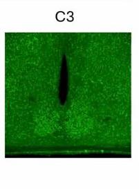
 at dilution of 1:200 (under 10x lens). Heat mediated antigen retrieval with Tris-EDTA buffer (pH 9.0).")
 at dilution of 1:200 (under 40x lens). Heat mediated antigen retrieval with Tris-EDTA buffer (pH 9.0).")
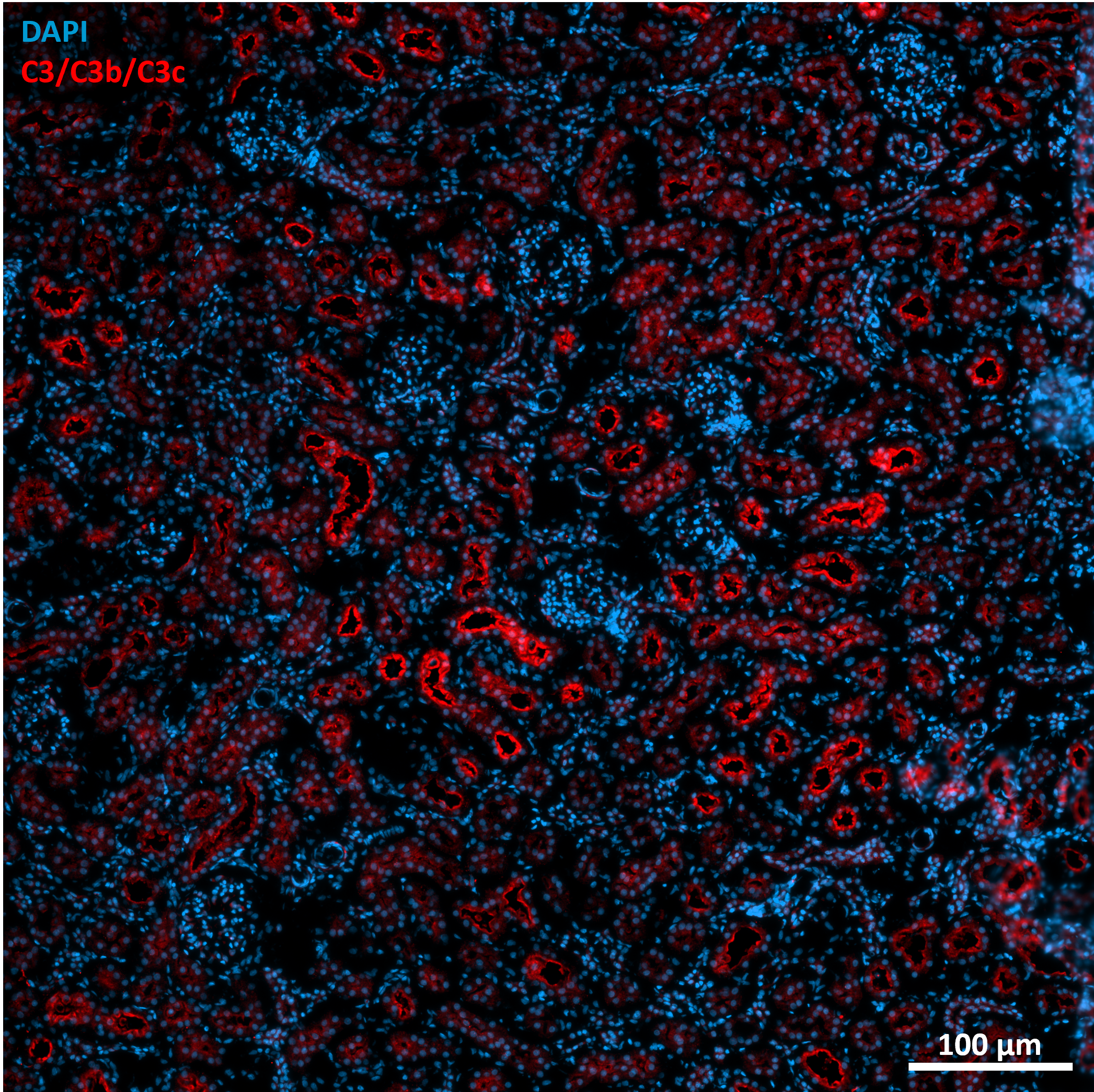
 fixed human liver cancer tissue using C3/C3b/C3c antibody (21337-1-AP) at dilution of 1:200 and CoraLite®488-Conjugated AffiniPure Goat Anti-Rabbit IgG(H+L).")
 fixed human liver cancer tissue using C3/C3b/C3c antibody (21337-1-AP) at dilution of 1:200 and CoraLite®488-Conjugated AffiniPure Goat Anti-Rabbit IgG(H+L).")
 fixed HepG2 cells using C3/C3b/C3c antibody (21337-1-AP) at dilution of 1:200 and CoraLite®488-Conjugated Goat Anti-Rabbit IgG(H+L) (SA00013-2).")
Applications testées
| Résultats positifs en WB | rat plasma, plasma humain |
| Résultats positifs en IP | tissu plasmatique humain |
| Résultats positifs en IHC | tissu hépatique humain, tissu de cancer du foie humain il est suggéré de démasquer l'antigène avec un tampon de TE buffer pH 9.0; (*) À défaut, 'le démasquage de l'antigène peut être 'effectué avec un tampon citrate pH 6,0. |
| Résultats positifs en IF-P | tissu de cancer du foie humain, |
| Résultats positifs en IF/ICC | cellules HepG2, |
Dilution recommandée
| Application | Dilution |
|---|---|
| Western Blot (WB) | WB : 1:5000-1:50000 |
| Immunoprécipitation (IP) | IP : 0.5-4.0 ug for 1.0-3.0 mg of total protein lysate |
| Immunohistochimie (IHC) | IHC : 1:1000-1:4000 |
| Immunofluorescence (IF)-P | IF-P : 1:50-1:500 |
| Immunofluorescence (IF)/ICC | IF/ICC : 1:50-1:500 |
| It is recommended that this reagent should be titrated in each testing system to obtain optimal results. | |
| Sample-dependent, check data in validation data gallery | |
Applications publiées
| KD/KO | See 3 publications below |
| WB | See 66 publications below |
| IHC | See 15 publications below |
| IF | See 54 publications below |
| CoIP | See 1 publications below |
Informations sur le produit
21337-1-AP cible C3/C3b/C3c dans les applications de WB, IHC, IF/ICC, IF-P, IP, CoIP, ELISA et montre une réactivité avec des échantillons Humain, rat
| Réactivité | Humain, rat |
| Réactivité citée | rat, bovin, Humain, porc, souris |
| Hôte / Isotype | Lapin / IgG |
| Clonalité | Polyclonal |
| Type | Anticorps |
| Immunogène | C3/C3b/C3c Protéine recombinante Ag15537 |
| Nom complet | complement component 3 |
| Masse moléculaire calculée | 1663 aa, 187 kDa |
| Poids moléculaire observé | 115 kDa |
| Numéro d’acquisition GenBank | BC150179 |
| Symbole du gène | C3 |
| Identification du gène (NCBI) | 718 |
| Conjugaison | Non conjugué |
| Forme | Liquide |
| Méthode de purification | Purification par affinité contre l'antigène |
| Tampon de stockage | PBS with 0.02% sodium azide and 50% glycerol |
| Conditions de stockage | Stocker à -20°C. Stable pendant un an après l'expédition. L'aliquotage n'est pas nécessaire pour le stockage à -20oC Les 20ul contiennent 0,1% de BSA. |
Informations générales
The complement system is an important effector that bridges the innate and adaptive immune systems (PMID: 20010915). The third component of complement, C3, plays a central role in activating the complement system. Its processing by C3 convertase is the central reaction in both classical and alternative complement pathways (PMID: 11414361). Human C3 (190-195 kDa), composed of α and β chains (115-120 and 75 kDa, respectively), is cleaved into C3a and C3b by C3 convertase. C3b is composed of the α' chain and β chain (PMID: 27210597). Factor I cleaves the α′ chain of C3b to 68 kDa and 43 kDa degradation products (iC3b) (PMID: 25395424; 14527961). This antibody raised against 1314-1663 aa of human C3 protein recognizes the C3 α chain (115-120 kDa), C3b α' chain (110-115 kDa), and C3c α' chain fragment 2 (43 kDa).
Protocole
| Product Specific Protocols | |
|---|---|
| WB protocol for C3/C3b/C3c antibody 21337-1-AP | Download protocol |
| IHC protocol for C3/C3b/C3c antibody 21337-1-AP | Download protocol |
| IF protocol for C3/C3b/C3c antibody 21337-1-AP | Download protocol |
| IP protocol for C3/C3b/C3c antibody 21337-1-AP | Download protocol |
| Standard Protocols | |
|---|---|
| Click here to view our Standard Protocols |
Publications
| Species | Application | Title |
|---|---|---|
Nat Cell Biol Autophagy enables microglia to engage amyloid plaques and prevents microglial senescence | ||
Brain Behav Immun PDE4 inhibition alleviates HMGB1/C1q/C3-mediated excessive phagocytic pruning of synapses by microglia and depressive-like behaviors in mice | ||
J Autoimmun TIGIT-Fc fusion protein alleviates murine lupus nephritis through the regulation of SPI-B-PAX5-XBP1 axis-mediated B-cell differentiation | ||
Gut Microbes Targeted modulation of intestinal barrier and mucosal immune-related microbiota attenuates IgA nephropathy progression | ||
Nat Commun Increased circulating levels of Factor H-Related Protein 4 are strongly associated with age-related macular degeneration. |
Avis
The reviews below have been submitted by verified Proteintech customers who received an incentive for providing their feedback.
FH Boyan (Verified Customer) (08-27-2025) | good for WB, two bands at ~190 and 130 kd, both of which are C3 forms
|
FH Marisa (Verified Customer) (07-31-2025) | We used the C3/C3b/C3c antibody in immunofluorescence and obtained a detectable signal, though some background was present. With slight optimization, it worked for visualizing complement activation by confocal microscopy.
 |
FH Marion (Verified Customer) (08-01-2024) | LPS induction model Fixative : Paraformaldehyde 4% Permeabilization: 10 min; RT; 0.2% Triton X-100 Blocking: 2 h; RT; 10% Normal donkey serum; 1% BSA Primary antibody: 1 night; 4°C Secondary antibody: ab150064 (Abcam); 1h30; RT Antibodies diluted in blocking solution
 |

