Anticorps Monoclonal anti-CD146/MCAM
CD146/MCAM Monoclonal Antibody for WB, IHC, IF/ICC, IF-P, ELISA
Hôte / Isotype
Mouse / IgG1
Réactivité testée
Humain
Applications
WB, IHC, IF/ICC, IF-P, ELISA
Conjugaison
Non conjugué
CloneNo.
4D8A9
N° de cat : 66153-1-Ig
Synonymes
Galerie de données de validation
Applications testées
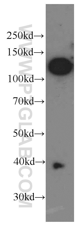
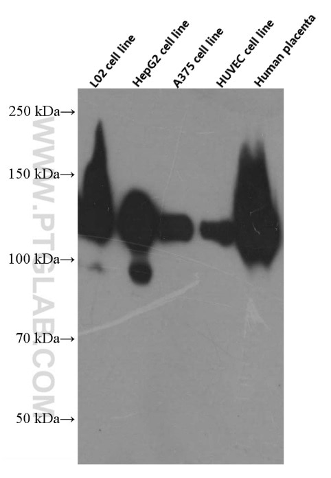
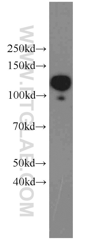
| Résultats positifs en WB | cellules HepG2, cellules A375, cellules HeLa, cellules HUVEC, cellules L02, tissu placentaire humain |
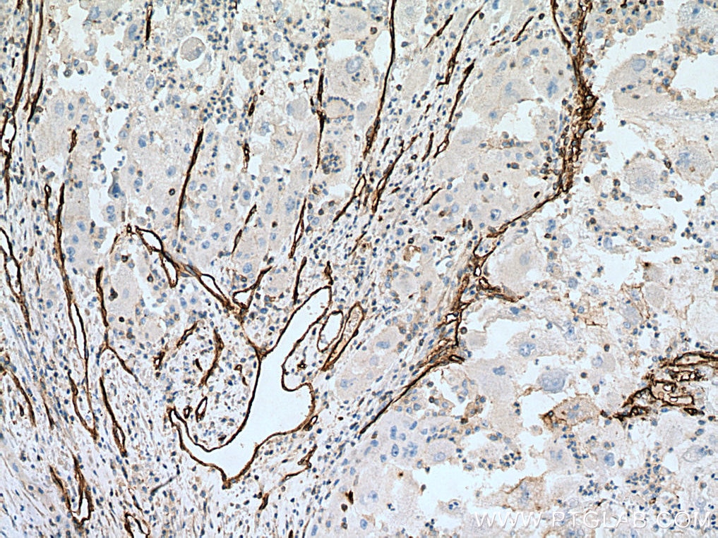
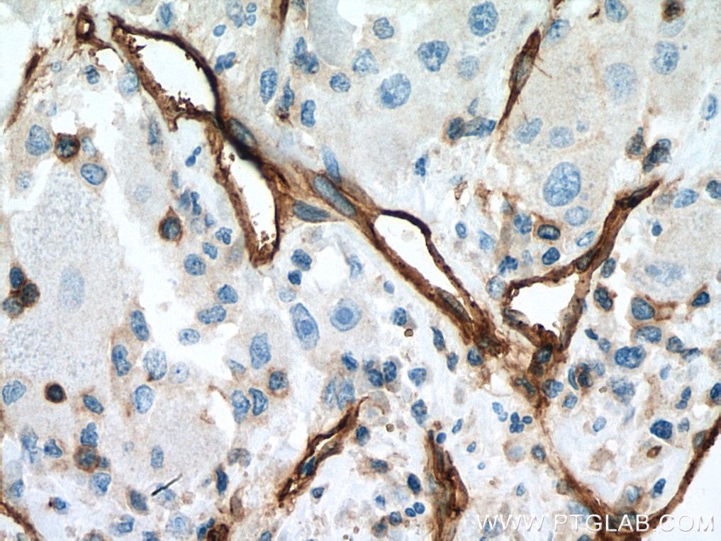
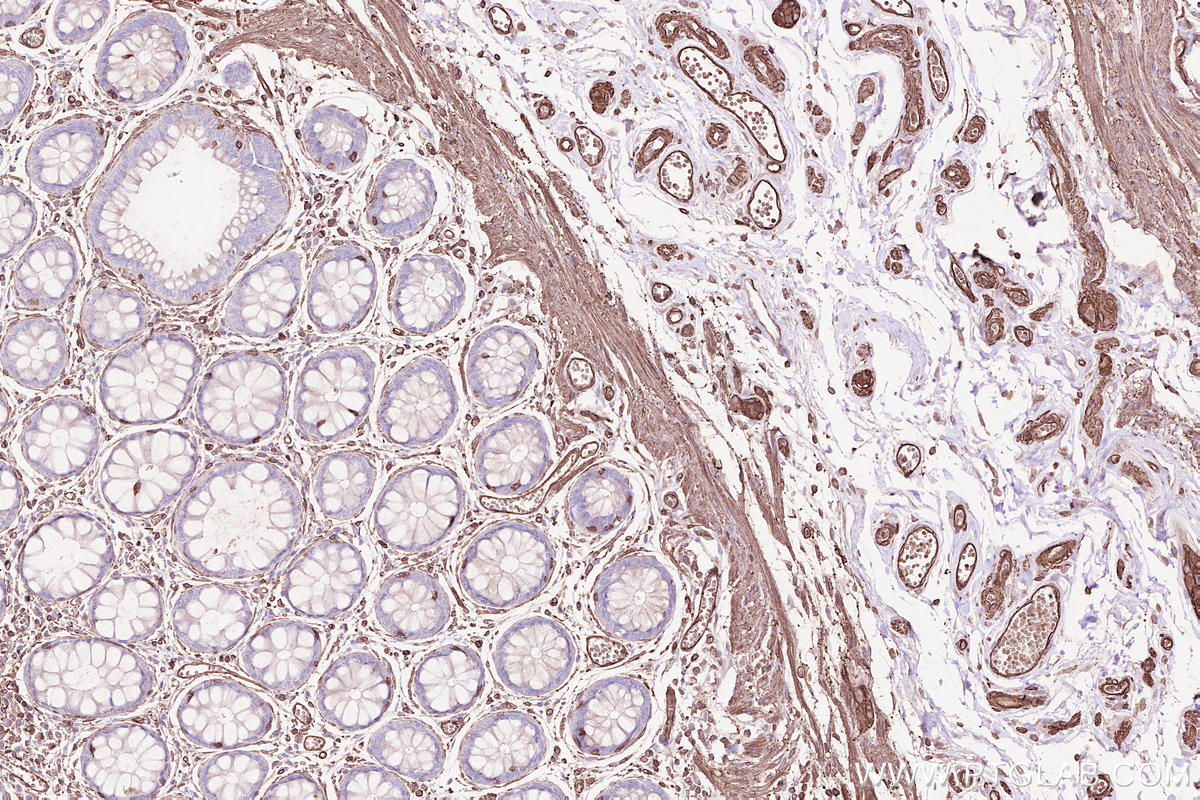
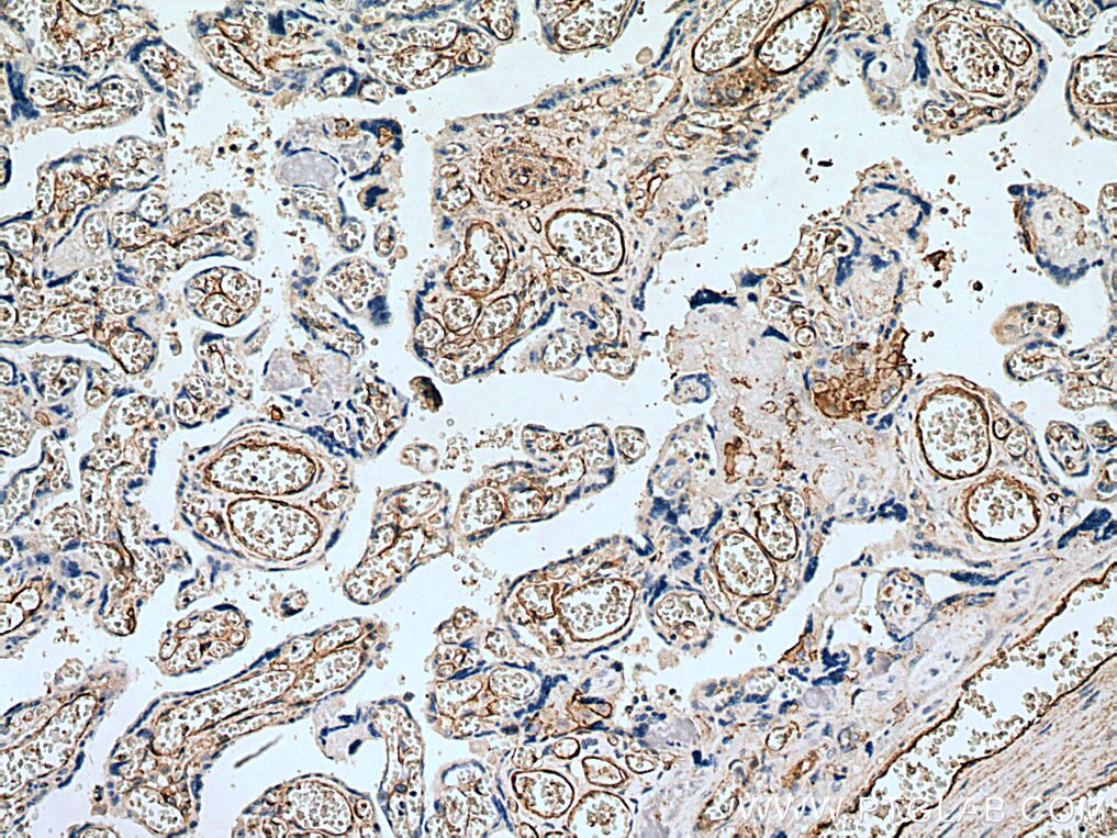
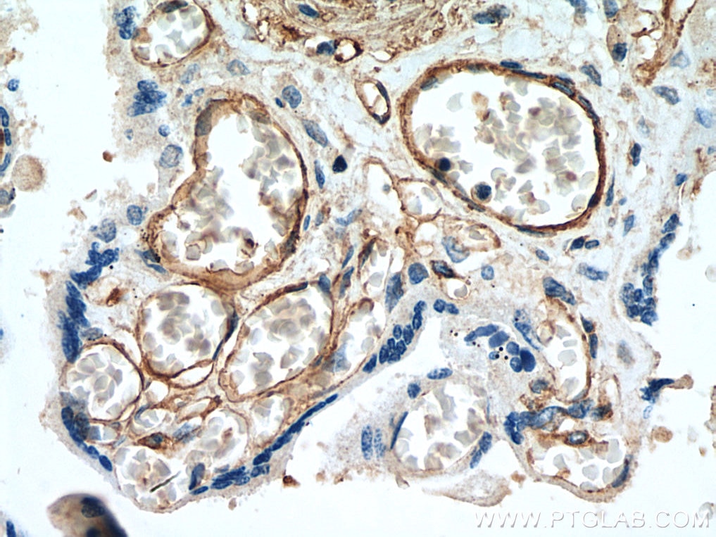
| Résultats positifs en IHC | tissu de cancer du foie humain, tissu placentaire humain il est suggéré de démasquer l'antigène avec un tampon de TE buffer pH 9.0; (*) À défaut, 'le démasquage de l'antigène peut être 'effectué avec un tampon citrate pH 6,0. |
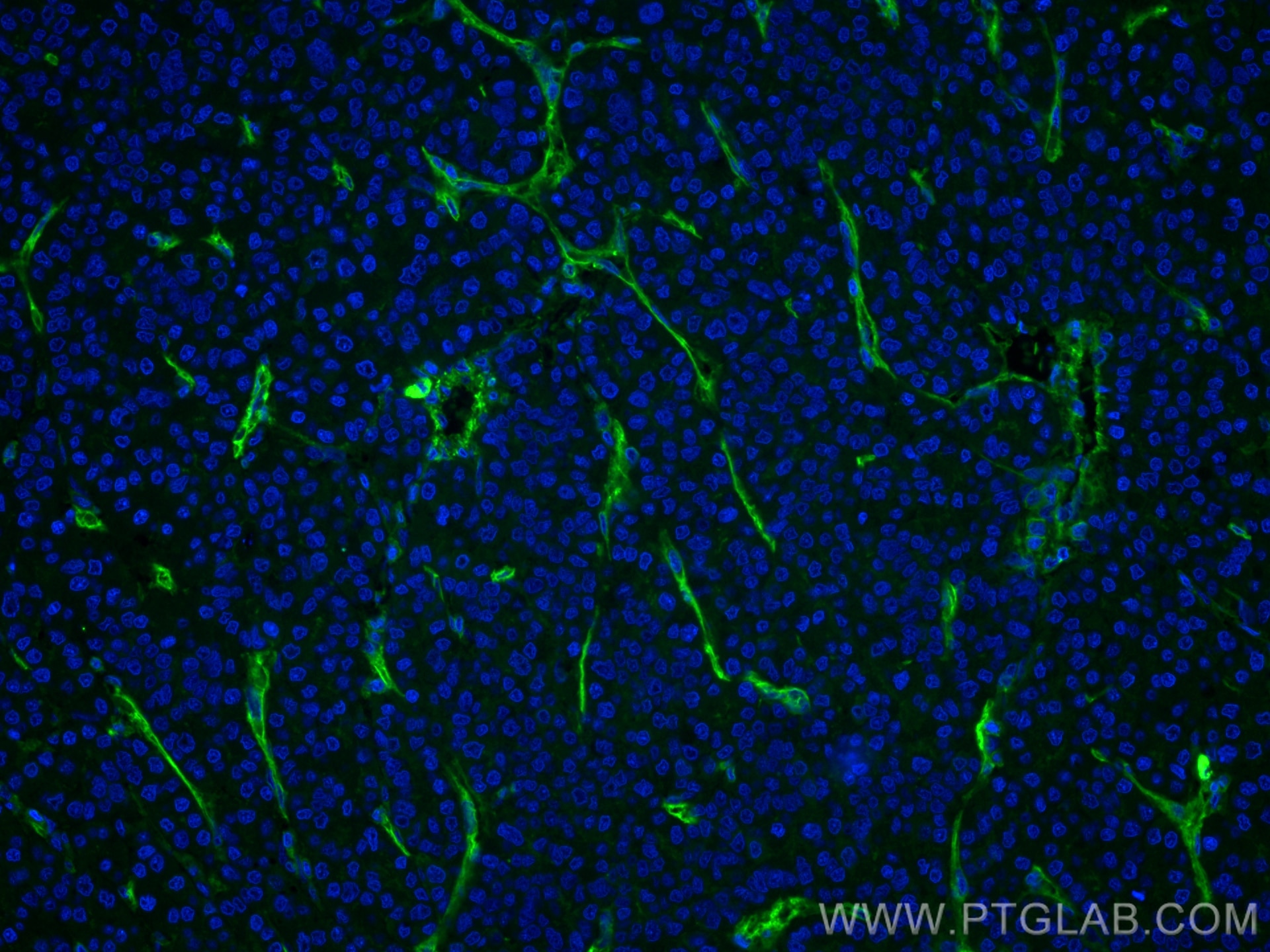
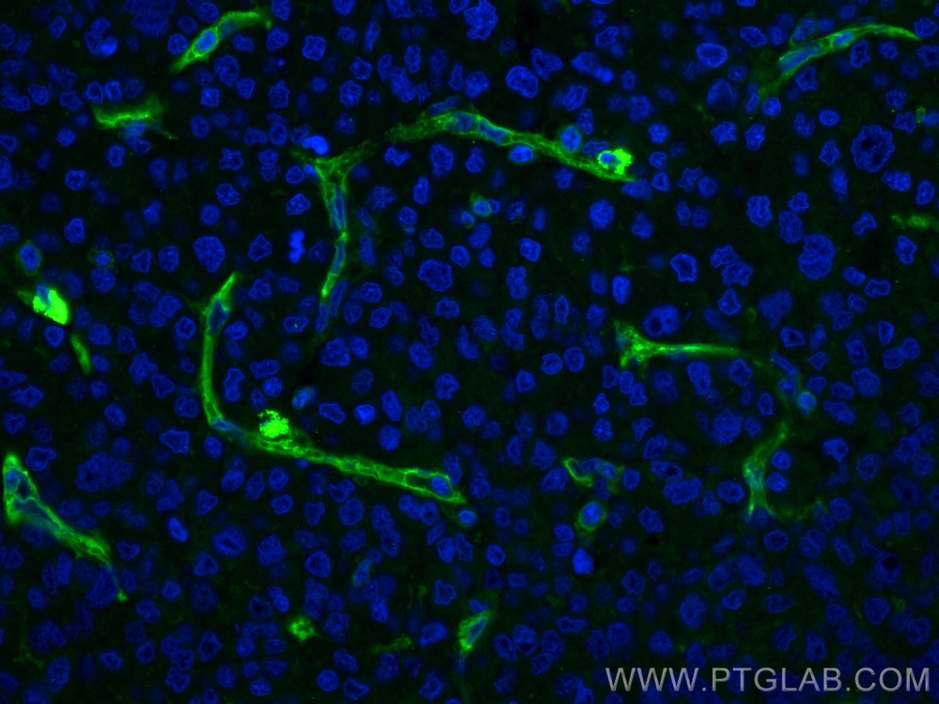
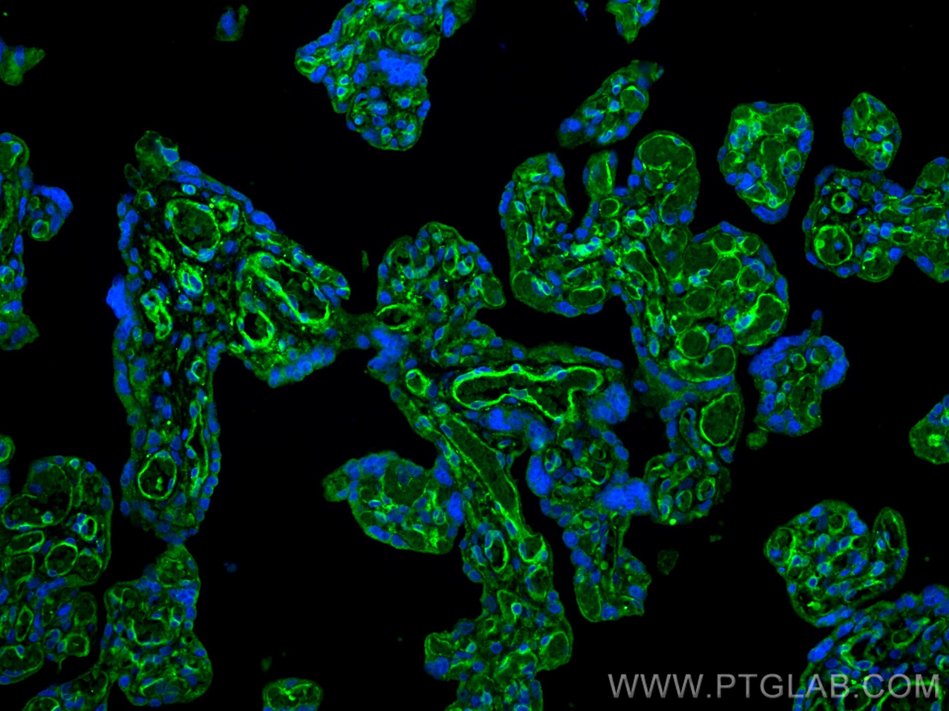
| Résultats positifs en IF-P | tissu de cancer du foie humain, tissu placentaire humain |
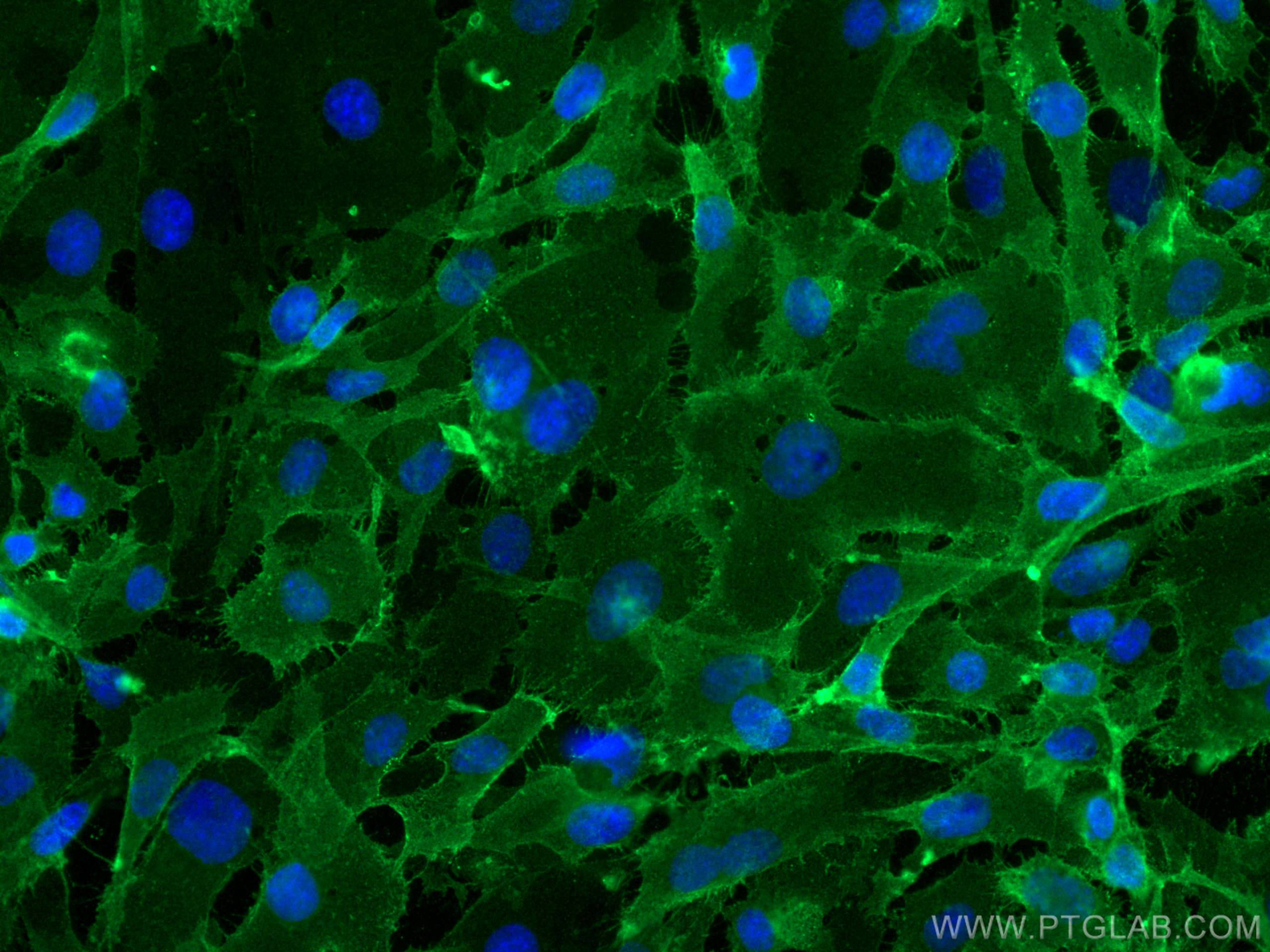
| Résultats positifs en IF/ICC | cellules HUVEC, |
Dilution recommandée
| Application | Dilution |
|---|---|
| Western Blot (WB) | WB : 1:2000-1:20000 |
| Immunohistochimie (IHC) | IHC : 1:1000-1:4000 |
| Immunofluorescence (IF)-P | IF-P : 1:1000-1:4000 |
| Immunofluorescence (IF)/ICC | IF/ICC : 1:250-1:1000 |
| It is recommended that this reagent should be titrated in each testing system to obtain optimal results. | |
| Sample-dependent, check data in validation data gallery | |
Applications publiées
| WB | See 1 publications below |
| IHC | See 1 publications below |
| FC | See 1 publications below |
Informations sur le produit
66153-1-Ig cible CD146/MCAM dans les applications de WB, IHC, IF/ICC, IF-P, ELISA et montre une réactivité avec des échantillons Humain
| Réactivité | Humain |
| Réactivité citée | Humain |
| Hôte / Isotype | Mouse / IgG1 |
| Clonalité | Monoclonal |
| Type | Anticorps |
| Immunogène | CD146/MCAM Protéine recombinante Ag11855 |
| Nom complet | melanoma cell adhesion molecule |
| Masse moléculaire calculée | 646 aa, 72 kDa |
| Poids moléculaire observé | 120 kDa |
| Numéro d’acquisition GenBank | BC056418 |
| Symbole du gène | CD146 |
| Identification du gène (NCBI) | 4162 |
| Conjugaison | Non conjugué |
| Forme | Liquide |
| Méthode de purification | Purification par protéine G |
| Tampon de stockage | PBS with 0.02% sodium azide and 50% glycerol |
| Conditions de stockage | Stocker à -20°C. Stable pendant un an après l'expédition. L'aliquotage n'est pas nécessaire pour le stockage à -20oC Les 20ul contiennent 0,1% de BSA. |
Informations générales
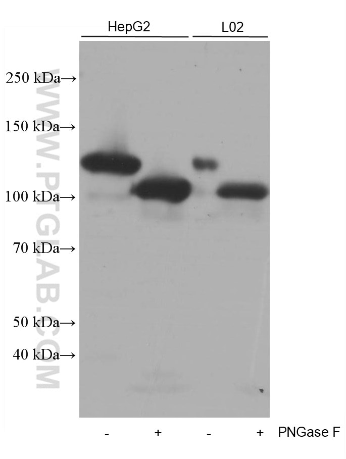
CD146, also known as melanoma cell adhesion molecule (MCAM) or MUC18, originally identified as a biomarker of melanoma progression, is a transmembrane glycoprotein of 113-130 kDa, belonging to the immunoglobulin (Ig) superfamily (PMID: 8378324; 25993332). Structurally, it consists of five Ig domains, a transmembrane domain, and a cytoplasmic region. In normal adult tissue, CD146 is primarily expressed by vascular endothelium and smooth muscle. CD146 is a key cell adhesion protein in vascular endothelial cell activity and angiogenesis, and has been used as marker of circulating endothelium cells (CECs) (PMID: 19356677). In addition to the membrane-anchored form of CD146, a soluble form of CD146 (sCD146, 105 kDa) has also been found in human plasma and in the supernatant of cultured human endothelial cells (PMID: 9462829; 19229070; 16374253; 14597988). This antibody detects a band at approximately 120 kDa that corresponds to the molecular weight of glycosylated CD146. Treatment of lysates of HepG2 cells and L02 cells with PNGase F, which removes oligosaccharides from N-linked glycoproteins, led to a down-shift of the detected band.
Protocole
| Product Specific Protocols | |
|---|---|
| WB protocol for CD146/MCAM antibody 66153-1-Ig | Download protocol |
| IHC protocol for CD146/MCAM antibody 66153-1-Ig | Download protocol |
| IF protocol for CD146/MCAM antibody 66153-1-Ig | Download protocol |
| Standard Protocols | |
|---|---|
| Click here to view our Standard Protocols |
Publications
| Species | Application | Title |
|---|---|---|
Exp Cell Res Nrf2 activation is involved in osteogenic differentiation of periodontal ligament stem cells under cyclic mechanical stretch. | ||
Cell Signal Glioma-derived small extracellular vesicles induce pericyte-phenotype transition of glioma stem cells under hypoxic conditions | ||
Ultrastruct Pathol Immunohistochemical and ultrastructural identification of telocytes in the infantile hemangioma |
Avis
The reviews below have been submitted by verified Proteintech customers who received an incentive for providing their feedback.
FH Boyan (Verified Customer) (02-05-2021) | Not sure whether it works, because I cannot detect anything in whole RPE1 cell lysates.
|
