- Phare
- Validé par KD/KO
Anticorps Polyclonal de lapin anti-CEP164
CEP164 Polyclonal Antibody for WB, IHC, IF/ICC, IP, ELISA
Hôte / Isotype
Lapin / IgG
Réactivité testée
canin, Humain et plus (1)
Applications
WB, IHC, IF/ICC, IP, ELISA
Conjugaison
Non conjugué
N° de cat : 22227-1-AP
Synonymes
Galerie de données de validation
 at dilution of 1:300 incubated at room temperature for 1.5 hours.")
 with RPE1 cells by Laboratory of Protein Dynamics and Signaling; Center for Cancer Research, National Cancer Institute.")
 at dilution of 1:50 (under 40x lens).")
 at dilution of 1:1000 (under 20x lens). Heat mediated antigen retrieval with Tris-EDTA buffer (pH 9.0).")
 at dilution of 1:50 (under 10x lens).")
 at dilution of 1:50 (under 40x lens).")
 at dilution of 1:50 (under 10x lens).")
 fixed HeLa cells using CEP164 antibody (22227-1-AP) at dilution of 1:400 and CoraLite®488-Conjugated AffiniPure Goat Anti-Rabbit IgG(H+L).")
 with HeLa cells (1.5% formaldehyde, 10 min RT) by Laboratory of Protein Dynamics and Signaling; Center for Cancer Research, National Cancer Institute.")
 fixed HeLa cells using CEP164 antibody (22227-1-AP) at dilution of 1:200 and CoraLite®488-Conjugated AffiniPure Goat Anti-Rabbit IgG(H+L).")
 fixed hTERT-RPE1 cells using CEP164 antibody (22227-1-AP) at dilution of 1:200 and CoraLite®488-Conjugated AffiniPure Goat Anti-Rabbit IgG(H+L).")
 fixed A549 cells using CEP164 antibody (22227-1-AP) at dilution of 1:400 and CoraLite®488-Conjugated AffiniPure Goat Anti-Rabbit IgG(H+L).")
 fixed PC-3 cells using CEP164 antibody (22227-1-AP) at dilution of 1:200 and CoraLite®488-Conjugated AffiniPure Goat Anti-Rabbit IgG(H+L).")
 fixed MDCK cells using 22227-1-AP (CEP164 antibody) at dilution of 1:50 and Alexa Fluor 488-conjugated AffiniPure Goat Anti-Rabbit IgG(H+L).")
Applications testées
| Résultats positifs en WB | cellules HEK-293 |
| Résultats positifs en IP | cellules RPE1 |
| Résultats positifs en IHC | tissu de côlon humain, tissu de cancer du col de l'utérus humain il est suggéré de démasquer l'antigène avec un tampon de TE buffer pH 9.0; (*) À défaut, 'le démasquage de l'antigène peut être 'effectué avec un tampon citrate pH 6,0. |
| Résultats positifs en IF/ICC | cellules HeLa, cellules A549, cellules hTERT-RPE1, cellules MDCK, cellules PC-3 |
Dilution recommandée
| Application | Dilution |
|---|---|
| Western Blot (WB) | WB : 1:200-1:1000 |
| Immunoprécipitation (IP) | IP : 0.5-4.0 ug for 1.0-3.0 mg of total protein lysate |
| Immunohistochimie (IHC) | IHC : 1:100-1:1000 |
| Immunofluorescence (IF)/ICC | IF/ICC : 1:200-1:800 |
| It is recommended that this reagent should be titrated in each testing system to obtain optimal results. | |
| Sample-dependent, check data in validation data gallery | |
Applications publiées
| KD/KO | See 2 publications below |
| WB | See 11 publications below |
| IHC | See 1 publications below |
| IF | See 58 publications below |
| IP | See 1 publications below |
Informations sur le produit
22227-1-AP cible CEP164 dans les applications de WB, IHC, IF/ICC, IP, ELISA et montre une réactivité avec des échantillons canin, Humain
| Réactivité | canin, Humain |
| Réactivité citée | Humain, souris |
| Hôte / Isotype | Lapin / IgG |
| Clonalité | Polyclonal |
| Type | Anticorps |
| Immunogène | CEP164 Protéine recombinante Ag17570 |
| Nom complet | centrosomal protein 164kDa |
| Masse moléculaire calculée | 1460 aa, 164 kDa |
| Poids moléculaire observé | 164 kDa |
| Numéro d’acquisition GenBank | BC000602 |
| Symbole du gène | CEP164 |
| Identification du gène (NCBI) | 22897 |
| Conjugaison | Non conjugué |
| Forme | Liquide |
| Méthode de purification | Purification par affinité contre l'antigène |
| Tampon de stockage | PBS with 0.02% sodium azide and 50% glycerol |
| Conditions de stockage | Stocker à -20°C. Stable pendant un an après l'expédition. L'aliquotage n'est pas nécessaire pour le stockage à -20oC Les 20ul contiennent 0,1% de BSA. |
Informations générales
CEP164, also called KIAA1052 or NPHP15, is a 1460 amino acid protein containing 1 WW domain. CEP164 localizes in the microtubule organizing center and is expressed in several cell lines. CEP164 plays a role in microtubule organization and/or maintenance for the formation of primary cilia, a microtubule-based structure that protrudes from the surface of epithelial cells. CEP164 plays a critical role in the G2/M checkpoint and nuclear divisions. The expression of CEP164 is normally limited to the mother centriole, and CEP164 can be used as a useful marker for mother centriole.
Protocole
| Product Specific Protocols | |
|---|---|
| WB protocol for CEP164 antibody 22227-1-AP | Download protocol |
| IHC protocol for CEP164 antibody 22227-1-AP | Download protocol |
| IF protocol for CEP164 antibody 22227-1-AP | Download protocol |
| Standard Protocols | |
|---|---|
| Click here to view our Standard Protocols |
Publications
| Species | Application | Title |
|---|---|---|
Cell Res NudCL2 is an autophagy receptor that mediates selective autophagic degradation of CP110 at mother centrioles to promote ciliogenesis. | ||
Nat Commun M-Phase Phosphoprotein 9 regulates ciliogenesis by modulating CP110-CEP97 complex localization at the mother centriole. | ||
Nat Commun Sub-centrosomal mapping identifies augmin-γTuRC as part of a centriole-stabilizing scaffold. | ||
Nat Commun DNA replication licensing factor Cdc6 and Plk4 kinase antagonistically regulate centrosome duplication via Sas-6. | ||
EMBO J ANKRD26 recruits PIDD1 to centriolar distal appendages to activate the PIDDosome following centrosome amplification. |
Avis
The reviews below have been submitted by verified Proteintech customers who received an incentive for providing their feedback.
FH Ludovic (Verified Customer) (02-27-2024) | A fabulous antibody to identify the distal appendage of the primary cilium. Never seen an antibody that works so well!
|
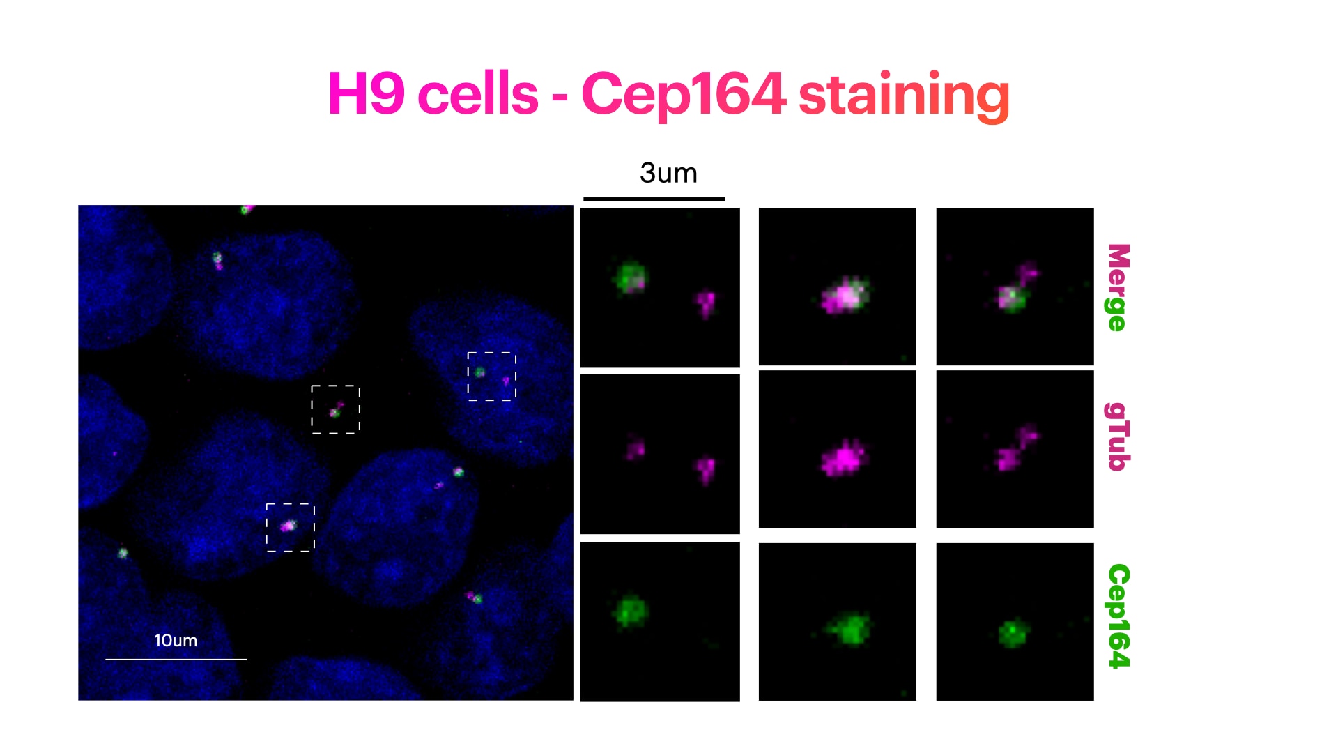
FH Elisa (Verified Customer) (04-04-2023) | H9 cells stained for Hoechst (DNA marker, in green), Cep164 (mother centriole distal appendage marker, in green) and g-Tubulin (pericentriolar matrix marker, in magenta). H9 cells were fixed in cold methanol for 10' at -20C. Cells were then rehydrated with PBS for 5'. Membrane permeabilization was then performed with 0.1% Triton + 0.1% Tween +0.01%SDS in PBS for 5'. Cells were finally incubated with blocking buffer (5% BSA+ 0.1% Tween in PBS) for 30' at RT. Primary antibody was diluted in blocking buffer 1:300 and incubated for 1h at room temperature. Alexa-488-Anti-rabbit was used as secondary antibody (1:600 dilution) (1h at room temperature).
 |
FH Pierrick (Verified Customer) (10-24-2019) | Great antibody for immunofluorescence on human cells.Works well in WB
|

