Anticorps Polyclonal de lapin anti-CEP250/CNAP1
CEP250/CNAP1 Polyclonal Antibody for WB, IHC, IF/ICC, IP, ELISA
Hôte / Isotype
Lapin / IgG
Réactivité testée
Humain et plus (1)
Applications
WB, IHC, IF/ICC, IP, ELISA
Conjugaison
Non conjugué
N° de cat : 14498-1-AP
Synonymes
Galerie de données de validation
 at dilution of 1:1500 incubated at room temperature for 1.5 hours.")
 with HEK-293 cells lysate 1800 ug.")
.")
.")
 fixed hTERT-RPE1 cells using CEP250/CNAP1 antibody (14498-1-AP) at dilution of 1:100 and Cy3-conjugated Affinipure Goat Anti-Rabbit IgG(H+L), SCLT1 antibody (14875-1-AP, Magenta), CoraLite®488 ARL13B antibody (CL488-17711, green).")
Applications testées
| Résultats positifs en WB | cellules A431, cellules HEK-293T, cellules HeLa |
| Résultats positifs en IP | cellules HEK-293, |
| Résultats positifs en IHC | tissu placentaire humain, il est suggéré de démasquer l'antigène avec un tampon de TE buffer pH 9.0; (*) À défaut, 'le démasquage de l'antigène peut être 'effectué avec un tampon citrate pH 6,0. |
| Résultats positifs en IF/ICC | cellules hTERT-RPE1, |
Dilution recommandée
| Application | Dilution |
|---|---|
| Western Blot (WB) | WB : 1:500-1:3000 |
| Immunoprécipitation (IP) | IP : 0.5-4.0 ug for 1.0-3.0 mg of total protein lysate |
| Immunohistochimie (IHC) | IHC : 1:50-1:500 |
| Immunofluorescence (IF)/ICC | IF/ICC : 1:50-1:500 |
| It is recommended that this reagent should be titrated in each testing system to obtain optimal results. | |
| Sample-dependent, check data in validation data gallery | |
Applications publiées
| WB | See 5 publications below |
| IHC | See 1 publications below |
| IF | See 31 publications below |
Informations sur le produit
14498-1-AP cible CEP250/CNAP1 dans les applications de WB, IHC, IF/ICC, IP, ELISA et montre une réactivité avec des échantillons Humain
| Réactivité | Humain |
| Réactivité citée | Humain, souris |
| Hôte / Isotype | Lapin / IgG |
| Clonalité | Polyclonal |
| Type | Anticorps |
| Immunogène | CEP250/CNAP1 Protéine recombinante Ag5925 |
| Nom complet | centrosomal protein 250kDa |
| Masse moléculaire calculée | 281 kDa |
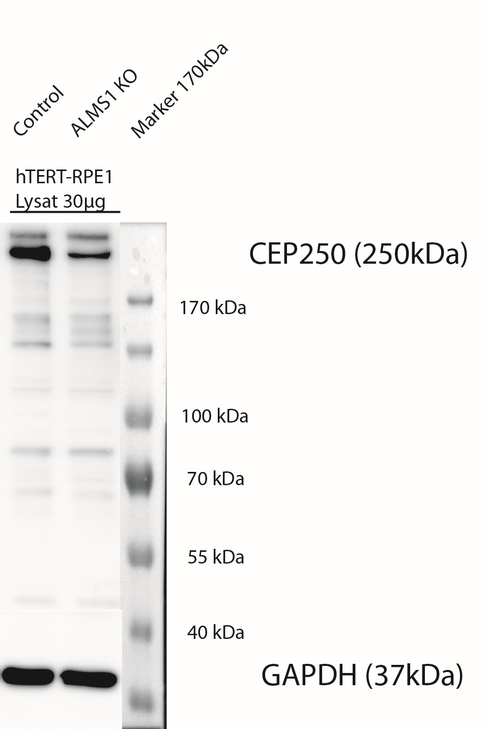
| Poids moléculaire observé | 250 kDa |
| Numéro d’acquisition GenBank | BC001433 |
| Symbole du gène | CEP250/CNAP1 |
| Identification du gène (NCBI) | 11190 |
| Conjugaison | Non conjugué |
| Forme | Liquide |
| Méthode de purification | Purification par affinité contre l'antigène |
| Tampon de stockage | PBS with 0.02% sodium azide and 50% glycerol |
| Conditions de stockage | Stocker à -20°C. Stable pendant un an après l'expédition. L'aliquotage n'est pas nécessaire pour le stockage à -20oC Les 20ul contiennent 0,1% de BSA. |
Informations générales
CEP250, also known as C-Nap1, is a 250 kDa coiled-coil protein that localizes to the proximal ends of mother and daughter centrioles. It is required for centriole-centriole cohesion during interphase of the cell cycle. It dissociates from the centrosomes when parental centrioles separate at the beginning of mitosis. The protein associates with and is phosphorylated by NIMA-related kinase 2, which is also associated with the centrosome.
Protocole
| Product Specific Protocols | |
|---|---|
| WB protocol for CEP250/CNAP1 antibody 14498-1-AP | Download protocol |
| IHC protocol for CEP250/CNAP1 antibody 14498-1-AP | Download protocol |
| IF protocol for CEP250/CNAP1 antibody 14498-1-AP | Download protocol |
| IP protocol for CEP250/CNAP1 antibody 14498-1-AP | Download protocol |
| Standard Protocols | |
|---|---|
| Click here to view our Standard Protocols |
Publications
| Species | Application | Title |
|---|---|---|
Science A liquid-like spindle domain promotes acentrosomal spindle assembly in mammalian oocytes. | ||
Nat Commun Microtubule asters anchored by FSD1 control axoneme assembly and ciliogenesis. | ||
Nat Commun Cytoplasmic E2f4 forms organizing centres for initiation of centriole amplification during multiciliogenesis. | ||
PLoS Biol Stable centrosomal roots disentangle to allow interphase centriole independence. | ||
Proc Natl Acad Sci U S A Dishevelled is a NEK2 kinase substrate controlling dynamics of centrosomal linker proteins. | ||
J Cell Biol Uncoordinated centrosome cycle underlies the instability of non-diploid somatic cells in mammals. |
Avis
The reviews below have been submitted by verified Proteintech customers who received an incentive for providing their feedback.
FH Karsten (Verified Customer) (12-22-2022) | WB: 1:600 dilution in 5% Milch in 1xTBST over nigth at 4°C, 30µg Protein loaded: result show prominent band at a hight of 250 kDa. unspecific bands also visible, but not prominent. worked fine IFS: 1:100, MetOH (-20°) fixation for 5 min at RT , Permeabilisation with 0.3% PBST (Triton) for 5 min at RT, Blocking with 1% BSA in PBS for 1h at RT. Antibody dilution 1:100 in 1%BSA in PBS 1h RT, sek Ak 1h at RT- nice basal body stainings at cilia in hTERT-RPE1 cells, that were starved for 3 days.
 |
FH Hairuo (Verified Customer) (12-16-2019) | Used in IF in neonatal mouse testis cry-sections.Works well.
|

