Anticorps Polyclonal de lapin anti-Claudin 3
Claudin 3 Polyclonal Antibody for IP, IHC, ELISA
Hôte / Isotype
Lapin / IgG
Réactivité testée
Humain, rat, souris et plus (1)
Applications
IHC, IF, IP, ELISA
Conjugaison
Non conjugué
N° de cat : 16456-1-AP
Synonymes
Galerie de données de validation
Applications testées
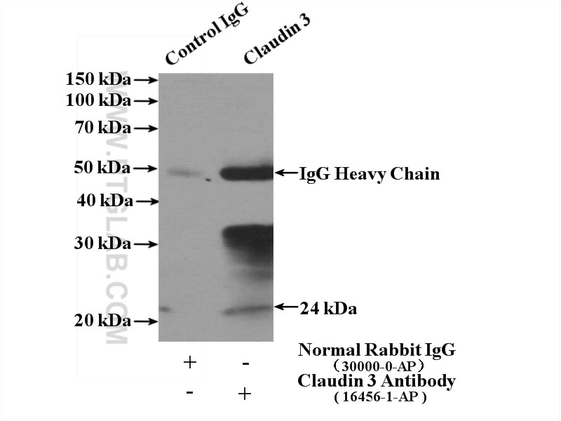
| Résultats positifs en IP | tissu de gros intestin de souris |
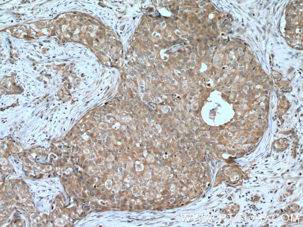
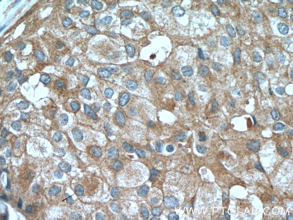
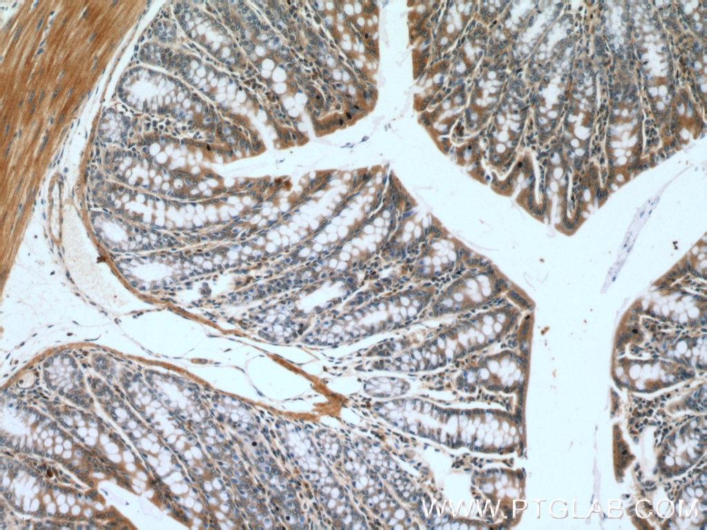
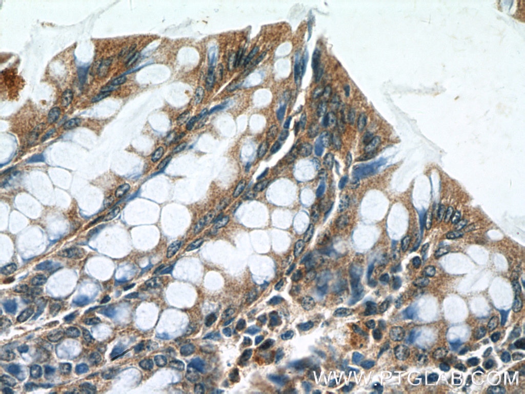
| Résultats positifs en IHC | tissu de cancer du sein humain, tissu de côlon de souris il est suggéré de démasquer l'antigène avec un tampon de TE buffer pH 9.0; (*) À défaut, 'le démasquage de l'antigène peut être 'effectué avec un tampon citrate pH 6,0. |
Dilution recommandée
| Application | Dilution |
|---|---|
| Immunoprécipitation (IP) | IP : 0.5-4.0 ug for 1.0-3.0 mg of total protein lysate |
| Immunohistochimie (IHC) | IHC : 1:50-1:500 |
| It is recommended that this reagent should be titrated in each testing system to obtain optimal results. | |
| Sample-dependent, check data in validation data gallery | |
Applications publiées
| IHC | See 13 publications below |
| IF | See 15 publications below |
Informations sur le produit
16456-1-AP cible Claudin 3 dans les applications de IHC, IF, IP, ELISA et montre une réactivité avec des échantillons Humain, rat, souris
| Réactivité | Humain, rat, souris |
| Réactivité citée | rat, Humain, porc, souris |
| Hôte / Isotype | Lapin / IgG |
| Clonalité | Polyclonal |
| Type | Anticorps |
| Immunogène | Claudin 3 Protéine recombinante Ag9411 |
| Nom complet | claudin 3 |
| Masse moléculaire calculée | 220 aa, 23 kDa |
| Poids moléculaire observé | 20-24 kDa |
| Numéro d’acquisition GenBank | BC016056 |
| Symbole du gène | Claudin 3 |
| Identification du gène (NCBI) | 1365 |
| Conjugaison | Non conjugué |
| Forme | Liquide |
| Méthode de purification | Purification par affinité contre l'antigène |
| Tampon de stockage | PBS with 0.02% sodium azide and 50% glycerol |
| Conditions de stockage | Stocker à -20°C. Stable pendant un an après l'expédition. L'aliquotage n'est pas nécessaire pour le stockage à -20oC Les 20ul contiennent 0,1% de BSA. |
Informations générales
Claudins are a family of proteins that are the most important components of the tight junctions, where they establish the paracellular barrier that controls the flow of molecules in the intercellular space between the cells of an epithelium. 23 claudins have been identified. They are small (20-27 kilodalton (kDa)) proteins with very similar structure. They have four transmembrane domains, with the N-terminus and the C-terminus in the cytoplasm.
Protocole
| Product Specific Protocols | |
|---|---|
| IHC protocol for Claudin 3 antibody 16456-1-AP | Download protocol |
| IP protocol for Claudin 3 antibody 16456-1-AP | Download protocol |
| Standard Protocols | |
|---|---|
| Click here to view our Standard Protocols |
Publications
| Species | Application | Title |
|---|---|---|
J Hazard Mater Insight into perfluorooctanoic acid-induced impairment of mouse embryo implantation via single-cell RNA-seq | ||
Aging Cell Advanced maternal age causes premature placental senescence and malformation via dysregulated α-Klotho expression in trophoblasts. | ||
EMBO Mol Med Deficiency in intestinal epithelial O-GlcNAcylation predisposes to gut inflammation. | ||
Sci China Life Sci Longitudinal gut fungal alterations and potential fungal biomarkers for the progression of primary liver disease | ||
J Hazard Mater Dual effects of JNK activation in blood-milk barrier damage induced by zinc oxide nanoparticles. | ||
Environ Int Translocation of transition metal oxide nanoparticles to breast milk and offspring: The necessity of bridging mother-offspring-integration toxicological assessments. |
Avis
The reviews below have been submitted by verified Proteintech customers who received an incentive for providing their feedback.
FH Priya (Verified Customer) (07-31-2023) | Used for Caco2 cells and mice tissue
|
FH Priya (Verified Customer) (06-21-2023) | Used this antibody for Caco2 cells andmice tissue
|
